Last additions - Retina Vascular Occlusions
|

Bilateral Branch Retinal Artery Occlusion - Intraarterial (hollenhorst) plaques visible543 views67 year old with acute visual field loss in each eye. Vision is 20/25 right eye 20/32 left eye. Images show intra-arterial plaques in each eye. The right eye on the optic nerve and the left eye in a branch retinal artery.Feb 06, 2017
|
|

Bilateral Branch Retinal Artery Occlusion - Intraarterial (hollenhorst) plaques visible491 views67 year old with acute visual field loss in each eye. Vision is 20/25 right eye 20/32 left eye. Images show intra-arterial plaques in each eye. The right eye on the optic nerve and the left eye in a branch retinal artery.Feb 06, 2017
|
|
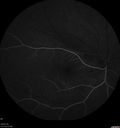
Bilateral Branch Retinal Artery Occlusion - Intraarterial (hollenhorst) plaques visible507 views67 year old with acute visual field loss in each eye. Vision is 20/25 right eye 20/32 left eye. Images show intra-arterial plaques in each eye. The right eye on the optic nerve and the left eye in a branch retinal artery.Feb 06, 2017
|
|

Bilateral Branch Retinal Artery Occlusion - Intraarterial (hollenhorst) plaques visible497 views67 year old with acute visual field loss in each eye. Vision is 20/25 right eye 20/32 left eye. Images show intra-arterial plaques in each eye. The right eye on the optic nerve and the left eye in a branch retinal artery.Feb 06, 2017
|
|

Bilateral Branch Retinal Artery Occlusion - Intraarterial (hollenhorst) plaques visible531 views67 year old with acute visual field loss in each eye. Vision is 20/25 right eye 20/32 left eye. Images show intra-arterial plaques in each eye. The right eye on the optic nerve and the left eye in a branch retinal artery.Feb 06, 2017
|
|

Bilateral Branch Retinal Artery Occlusion - Intraarterial (hollenhorst) plaques visible485 views67 year old with acute visual field loss in each eye. Vision is 20/25 right eye 20/32 left eye. Images show intra-arterial plaques in each eye. The right eye on the optic nerve and the left eye in a branch retinal artery.Feb 06, 2017
|
|

Bilateral Branch Retinal Artery Occlusion - Intraarterial (hollenhorst) plaques visible477 views67 year old with acute visual field loss in each eye. Vision is 20/25 right eye 20/32 left eye. Images show intra-arterial plaques in each eye. The right eye on the optic nerve and the left eye in a branch retinal artery.Feb 06, 2017
|
|

Bilateral Branch Retinal Artery Occlusion - Intraarterial (hollenhorst) plaques visible509 views67 year old with acute visual field loss in each eye. Vision is 20/25 right eye 20/32 left eye. Images show intra-arterial plaques in each eye. The right eye on the optic nerve and the left eye in a branch retinal artery.Feb 06, 2017
|
|

Bilateral Branch Retinal Artery Occlusion - Intraarterial (hollenhorst) plaques visible497 views67 year old with acute visual field loss in each eye. Vision is 20/25 right eye 20/32 left eye. Images show intra-arterial plaques in each eye. The right eye on the optic nerve and the left eye in a branch retinal artery.Feb 06, 2017
|
|

Bilateral Branch Retinal Artery Occlusion - Intraarterial (hollenhorst) plaques visible492 views67 year old with acute visual field loss in each eye. Vision is 20/25 right eye 20/32 left eye. Images show intra-arterial plaques in each eye. The right eye on the optic nerve and the left eye in a branch retinal artery.Feb 06, 2017
|
|

Bilateral Branch Retinal Artery Occlusion - Intraarterial (hollenhorst) plaques visible531 views67 year old with acute visual field loss in each eye. Vision is 20/25 right eye 20/32 left eye. Images show intra-arterial plaques in each eye. The right eye on the optic nerve and the left eye in a branch retinal artery.Feb 06, 2017
|
|

Bilateral Branch Retinal Artery Occlusion - Intraarterial (hollenhorst) plaques visible562 views67 year old with acute visual field loss in each eye. Vision is 20/25 right eye 20/32 left eye. Images show intra-arterial plaques in each eye. The right eye on the optic nerve and the left eye in a branch retinal artery.Feb 06, 2017
|
|

Bilateral Branch Retinal Artery Occlusion - Intraarterial (hollenhorst) plaques visible595 views67 year old with acute visual field loss in each eye. Vision is 20/25 right eye 20/32 left eye. Images show intra-arterial plaques in each eye. The right eye on the optic nerve and the left eye in a branch retinal artery.Feb 06, 2017
|
|

Bilateral Branch Retinal Artery Occlusion - Intraarterial (hollenhorst) plaques visible653 views67 year old with acute visual field loss in each eye. Vision is 20/25 right eye 20/32 left eye. Images show intra-arterial plaques in each eye. The right eye on the optic nerve and the left eye in a branch retinal artery.Feb 06, 2017
|
|

Central Retinal Artery Occlusion - MAP761 viewsProgression from acute to atrophy - decreasing retinal thickenssNov 28, 2016
|
|

Branch Retinal Vein Occlusion508 views84 year old man - 2 months vision loss - VA 20/40 - 20/20 with one injection of EyleaSep 29, 2016
|
|

Branch Retinal Vein Occlusion469 views84 year old man - 2 months vision loss - VA 20/40 - 20/20 with one injection of EyleaSep 29, 2016
|
|

Branch Retinal Vein Occlusion509 views84 year old man - 2 months vision loss - VA 20/40 - 20/20 with one injection of EyleaSep 29, 2016
|
|

Branch Retinal Vein Occlusion466 views84 year old man - 2 months vision loss - VA 20/40 - 20/20 with one injection of EyleaSep 29, 2016
|
|

Branch Retinal Vein Occlusion495 views84 year old man - 2 months vision loss - VA 20/40 - 20/20 with one injection of EyleaSep 29, 2016
|
|

Branch Retinal Vein Occlusion485 views84 year old man - 2 months vision loss - VA 20/40 - 20/20 with one injection of EyleaSep 29, 2016
|
|

Branch Retinal Vein Occlusion447 views84 year old man - 2 months vision loss - VA 20/40 - 20/20 with one injection of EyleaSep 29, 2016
|
|

Branch Retinal Vein Occlusion470 views84 year old man - 2 months vision loss - VA 20/40 - 20/20 with one injection of EyleaSep 29, 2016
|
|

Branch Retinal Vein Occlusion486 views84 year old man - 2 months vision loss - VA 20/40 - 20/20 with one injection of EyleaSep 29, 2016
|
|

Branch Retinal Vein Occlusion478 views84 year old man - 2 months vision loss - VA 20/40 - 20/20 with one injection of EyleaSep 29, 2016
|
|

Branch Retinal Vein Occlusion452 views84 year old man - 2 months vision loss - VA 20/40 - 20/20 with one injection of EyleaSep 29, 2016
|
|

Branch Retinal Vein Occlusion502 views84 year old man - 2 months vision loss - VA 20/40 - 20/20 with one injection of EyleaSep 29, 2016
|
|

Branch Retinal Vein Occlusion474 views84 year old man - 2 months vision loss - VA 20/40 - 20/20 with one injection of EyleaSep 29, 2016
|
|

Branch Retinal Vein Occlusion537 views84 year old man - 2 months vision loss - VA 20/40 - 20/20 with one injection of EyleaSep 29, 2016
|
|

Branch Retinal Vein Occlusion508 views84 year old man - 2 months vision loss - VA 20/40 - 20/20 with one injection of EyleaSep 29, 2016
|
|

Branch Retinal Vein Occlusion519 views84 year old man - 2 months vision loss - VA 20/40 - 20/20 with one injection of EyleaSep 29, 2016
|
|

Branch Retinal Vein Occlusion521 views84 year old man - 2 months vision loss - VA 20/40 - 20/20 with one injection of EyleaSep 29, 2016
|
|

Branch Retinal Vein Occlusion548 views84 year old man - 2 months vision loss - VA 20/40 - 20/20 with one injection of EyleaSep 29, 2016
|
|

Branch Retinal Artery Occlusion581 viewsAcute Inferotemporal Occlusion Right Eye - FA shows delayed circulation, OCT shows edema of superficial layers of retinaSep 29, 2016
|
|

Branch Retinal Artery Occlusion533 viewsAcute Inferotemporal Occlusion Right Eye - FA shows delayed circulation, OCT shows edema of superficial layers of retinaSep 29, 2016
|
|

Branch Retinal Artery Occlusion493 viewsAcute Inferotemporal Occlusion Right Eye - FA shows delayed circulation, OCT shows edema of superficial layers of retinaSep 29, 2016
|
|

Branch Retinal Artery Occlusion515 viewsAcute Inferotemporal Occlusion Right Eye - FA shows delayed circulation, OCT shows edema of superficial layers of retinaSep 29, 2016
|
|

Branch Retinal Artery Occlusion527 viewsAcute Inferotemporal Occlusion Right Eye - FA shows delayed circulation, OCT shows edema of superficial layers of retinaSep 29, 2016
|
|

Branch Retinal Artery Occlusion520 viewsAcute Inferotemporal Occlusion Right Eye - FA shows delayed circulation, OCT shows edema of superficial layers of retinaSep 29, 2016
|
|

Branch Retinal Artery Occlusion507 viewsAcute Inferotemporal Occlusion Right Eye - FA shows delayed circulation, OCT shows edema of superficial layers of retinaSep 29, 2016
|
|

Branch Retinal Artery Occlusion520 viewsAcute Inferotemporal Occlusion Right Eye - FA shows delayed circulation, OCT shows edema of superficial layers of retinaSep 29, 2016
|
|

Branch Retinal Artery Occlusion527 viewsAcute Inferotemporal Occlusion Right Eye - FA shows delayed circulation, OCT shows edema of superficial layers of retinaSep 29, 2016
|
|

Branch Retinal Artery Occlusion587 viewsAcute Inferotemporal Occlusion Right Eye - FA shows delayed circulation, OCT shows edema of superficial layers of retinaSep 29, 2016
|
|

Branch Retinal Artery Occlusion521 viewsAcute Inferotemporal Occlusion Right Eye - FA shows delayed circulation, OCT shows edema of superficial layers of retinaSep 29, 2016
|
|

Branch Retinal Artery Occlusion470 viewsAcute Inferotemporal Occlusion Right Eye - FA shows delayed circulation, OCT shows edema of superficial layers of retinaSep 29, 2016
|
|

Branch Retinal Artery Occlusion446 viewsAcute Inferotemporal Occlusion Right Eye - FA shows delayed circulation, OCT shows edema of superficial layers of retinaSep 29, 2016
|
|

Branch Retinal Artery Occlusion544 viewsAcute Inferotemporal Occlusion Right Eye - FA shows delayed circulation, OCT shows edema of superficial layers of retinaSep 29, 2016
|
|

Branch Retinal Artery Occlusion590 viewsAcute Inferotemporal Occlusion Right Eye - FA shows delayed circulation, OCT shows edema of superficial layers of retinaSep 29, 2016
|
|

Branch Retinal Artery Occlusion499 viewsAcute Inferotemporal Occlusion Right Eye - FA shows delayed circulation, OCT shows edema of superficial layers of retinaSep 29, 2016
|
|

Branch Retinal Artery Occlusion543 viewsAcute Inferotemporal Occlusion Right Eye - FA shows delayed circulation, OCT shows edema of superficial layers of retinaSep 29, 2016
|
|

Branch Retinal Artery Occlusion564 viewsAcute Inferotemporal Occlusion Right Eye - FA shows delayed circulation, OCT shows edema of superficial layers of retinaSep 29, 2016
|
|

Branch Retinal Artery Occlusion534 viewsAcute Inferotemporal Occlusion Right Eye - FA shows delayed circulation, OCT shows edema of superficial layers of retinaSep 29, 2016
|
|

Branch Retinal Artery Occlusion583 viewsAcute Inferotemporal Occlusion Right Eye - FA shows delayed circulation, OCT shows edema of superficial layers of retinaSep 29, 2016
|
|

Branch Retinal Artery Occlusion557 viewsAcute Inferotemporal Occlusion Right Eye - FA shows delayed circulation, OCT shows edema of superficial layers of retinaSep 29, 2016
|
|

Branch Retinal Artery Occlusion614 viewsAcute Inferotemporal Occlusion Right Eye - FA shows delayed circulation, OCT shows edema of superficial layers of retinaSep 29, 2016
|
|

Acute Branch Retinal Artery Occlusion Left Eye661 viewsThis 84 year old woman had an old BRVO in the right eye and saw a shadow for 2 days in the left eye. She has a superotemporal BRAO. The photos show retinal whitening, the OCT shows inner retinal edema and the FA shows poor circulation.Nov 19, 2015
|
|

Acute Branch Retinal Artery Occlusion Left Eye551 viewsThis 84 year old woman had an old BRVO in the right eye and saw a shadow for 2 days in the left eye. She has a superotemporal BRAO. The photos show retinal whitening, the OCT shows inner retinal edema and the FA shows poor circulation.Nov 19, 2015
|
|

Acute Branch Retinal Artery Occlusion Left Eye537 viewsThis 84 year old woman had an old BRVO in the right eye and saw a shadow for 2 days in the left eye. She has a superotemporal BRAO. The photos show retinal whitening, the OCT shows inner retinal edema and the FA shows poor circulation.Nov 19, 2015
|
|

Acute Branch Retinal Artery Occlusion Left Eye556 viewsThis 84 year old woman had an old BRVO in the right eye and saw a shadow for 2 days in the left eye. She has a superotemporal BRAO. The photos show retinal whitening, the OCT shows inner retinal edema and the FA shows poor circulation.Nov 19, 2015
|
|

Acute Branch Retinal Artery Occlusion Left Eye549 viewsThis 84 year old woman had an old BRVO in the right eye and saw a shadow for 2 days in the left eye. She has a superotemporal BRAO. The photos show retinal whitening, the OCT shows inner retinal edema and the FA shows poor circulation.Nov 19, 2015
|
|

Acute Branch Retinal Artery Occlusion Left Eye528 viewsThis 84 year old woman had an old BRVO in the right eye and saw a shadow for 2 days in the left eye. She has a superotemporal BRAO. The photos show retinal whitening, the OCT shows inner retinal edema and the FA shows poor circulation.Nov 19, 2015
|
|

Acute Branch Retinal Artery Occlusion Left Eye530 viewsThis 84 year old woman had an old BRVO in the right eye and saw a shadow for 2 days in the left eye. She has a superotemporal BRAO. The photos show retinal whitening, the OCT shows inner retinal edema and the FA shows poor circulation.Nov 19, 2015
|
|

Acute Branch Retinal Artery Occlusion Left Eye508 viewsThis 84 year old woman had an old BRVO in the right eye and saw a shadow for 2 days in the left eye. She has a superotemporal BRAO. The photos show retinal whitening, the OCT shows inner retinal edema and the FA shows poor circulation.Nov 19, 2015
|
|

Acute Branch Retinal Artery Occlusion Left Eye555 viewsThis 84 year old woman had an old BRVO in the right eye and saw a shadow for 2 days in the left eye. She has a superotemporal BRAO. The photos show retinal whitening, the OCT shows inner retinal edema and the FA shows poor circulation.Nov 19, 2015
|
|

Acute Branch Retinal Artery Occlusion Left Eye671 viewsThis 84 year old woman had an old BRVO in the right eye and saw a shadow for 2 days in the left eye. She has a superotemporal BRAO. The photos show retinal whitening, the OCT shows inner retinal edema and the FA shows poor circulation.Nov 19, 2015
|
|

Acute Branch Retinal Artery Occlusion Left Eye546 viewsThis 84 year old woman had an old BRVO in the right eye and saw a shadow for 2 days in the left eye. She has a superotemporal BRAO. The photos show retinal whitening, the OCT shows inner retinal edema and the FA shows poor circulation.Nov 19, 2015
|
|

Acute Branch Retinal Artery Occlusion Left Eye673 viewsThis 84 year old woman had an old BRVO in the right eye and saw a shadow for 2 days in the left eye. She has a superotemporal BRAO. The photos show retinal whitening, the OCT shows inner retinal edema and the FA shows poor circulation.Nov 19, 2015
|
|

Acute Branch Retinal Artery Occlusion Left Eye637 viewsThis 84 year old woman had an old BRVO in the right eye and saw a shadow for 2 days in the left eye. She has a superotemporal BRAO. The photos show retinal whitening, the OCT shows inner retinal edema and the FA shows poor circulation.Nov 19, 2015
|
|

Acute Branch Retinal Artery Occlusion Left Eye661 viewsThis 84 year old woman had an old BRVO in the right eye and saw a shadow for 2 days in the left eye. She has a superotemporal BRAO. The photos show retinal whitening, the OCT shows inner retinal edema and the FA shows poor circulation.Nov 19, 2015
|
|

Acute Branch Retinal Artery Occlusion Left Eye624 viewsThis 84 year old woman had an old BRVO in the right eye and saw a shadow for 2 days in the left eye. She has a superotemporal BRAO. The photos show retinal whitening, the OCT shows inner retinal edema and the FA shows poor circulation.Nov 19, 2015
|
|

Acute Branch Retinal Artery Occlusion Left Eye581 viewsThis 84 year old woman had an old BRVO in the right eye and saw a shadow for 2 days in the left eye. She has a superotemporal BRAO. The photos show retinal whitening, the OCT shows inner retinal edema and the FA shows poor circulation.Nov 19, 2015
|
|

Acute Branch Retinal Artery Occlusion Left Eye508 viewsThis 84 year old woman had an old BRVO in the right eye and saw a shadow for 2 days in the left eye. She has a superotemporal BRAO. The photos show retinal whitening, the OCT shows inner retinal edema and the FA shows poor circulation.Nov 19, 2015
|
|

Acute Branch Retinal Artery Occlusion Left Eye436 viewsThis 84 year old woman had an old BRVO in the right eye and saw a shadow for 2 days in the left eye. She has a superotemporal BRAO. The photos show retinal whitening, the OCT shows inner retinal edema and the FA shows poor circulation.Nov 19, 2015
|
|

Acute Branch Retinal Artery Occlusion Left Eye493 viewsThis 84 year old woman had an old BRVO in the right eye and saw a shadow for 2 days in the left eye. She has a superotemporal BRAO. The photos show retinal whitening, the OCT shows inner retinal edema and the FA shows poor circulation.Nov 19, 2015
|
|

Acute Branch Retinal Artery Occlusion Left Eye574 viewsThis 84 year old woman had an old BRVO in the right eye and saw a shadow for 2 days in the left eye. She has a superotemporal BRAO. The photos show retinal whitening, the OCT shows inner retinal edema and the FA shows poor circulation.Nov 19, 2015
|
|

Acute Branch Retinal Artery Occlusion Left Eye507 viewsThis 84 year old woman had an old BRVO in the right eye and saw a shadow for 2 days in the left eye. She has a superotemporal BRAO. The photos show retinal whitening, the OCT shows inner retinal edema and the FA shows poor circulation.Nov 19, 2015
|
|

Acute Branch Retinal Artery Occlusion Left Eye502 viewsThis 84 year old woman had an old BRVO in the right eye and saw a shadow for 2 days in the left eye. She has a superotemporal BRAO. The photos show retinal whitening, the OCT shows inner retinal edema and the FA shows poor circulation.Nov 19, 2015
|
|

Acute Branch Retinal Artery Occlusion Left Eye483 viewsThis 84 year old woman had an old BRVO in the right eye and saw a shadow for 2 days in the left eye. She has a superotemporal BRAO. The photos show retinal whitening, the OCT shows inner retinal edema and the FA shows poor circulation.Nov 19, 2015
|
|

Acute Branch Retinal Artery Occlusion Left Eye649 viewsThis 84 year old woman had an old BRVO in the right eye and saw a shadow for 2 days in the left eye. She has a superotemporal BRAO. The photos show retinal whitening, the OCT shows inner retinal edema and the FA shows poor circulation.Nov 19, 2015
|
|

Acute Branch Retinal Artery Occlusion Left Eye656 viewsThis 84 year old woman had an old BRVO in the right eye and saw a shadow for 2 days in the left eye. She has a superotemporal BRAO. The photos show retinal whitening, the OCT shows inner retinal edema and the FA shows poor circulation.Nov 19, 2015
|
|

Acute Branch Retinal Artery Occlusion Left Eye675 viewsThis 84 year old woman had an old BRVO in the right eye and saw a shadow for 2 days in the left eye. She has a superotemporal BRAO. The photos show retinal whitening, the OCT shows inner retinal edema and the FA shows poor circulation.Nov 19, 2015
|
|

Acute Branch Retinal Artery Occlusion Left Eye689 viewsThis 84 year old woman had an old BRVO in the right eye and saw a shadow for 2 days in the left eye. She has a superotemporal BRAO. The photos show retinal whitening, the OCT shows inner retinal edema and the FA shows poor circulation.Nov 19, 2015
|
|

Acute Branch Retinal Artery Occlusion Left Eye618 viewsThis 84 year old woman had an old BRVO in the right eye and saw a shadow for 2 days in the left eye. She has a superotemporal BRAO. The photos show retinal whitening, the OCT shows inner retinal edema and the FA shows poor circulation.Nov 19, 2015
|
|

Acute Branch Retinal Artery Occlusion Left Eye743 viewsThis 84 year old woman had an old BRVO in the right eye and saw a shadow for 2 days in the left eye. She has a superotemporal BRAO. The photos show retinal whitening, the OCT shows inner retinal edema and the FA shows poor circulation.Nov 19, 2015
|
|
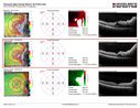
Mtehylenetetrahydrofolate reductase c677T homozygous CRVO pregnant woman (MTHFR)479 viewsVision improved from 20/50 to 20/16 in one month following ozurdex injections. 38 year old woman with only one eye. The left eye was removed as a child for retinoblastoma.Oct 05, 2015
|
|
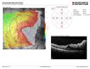
Mtehylenetetrahydrofolate reductase c677T homozygous CRVO pregnant woman (MTHFR)505 viewsVision improved from 20/50 to 20/16 in one month following ozurdex injections. 38 year old woman with only one eye. The left eye was removed as a child for retinoblastoma.Oct 05, 2015
|
|
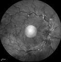
Mtehylenetetrahydrofolate reductase c677T homozygous CRVO pregnant woman (MTHFR)427 viewsVision improved from 20/50 to 20/16 in one month following ozurdex injections. 38 year old woman with only one eye. The left eye was removed as a child for retinoblastoma.Oct 05, 2015
|
|
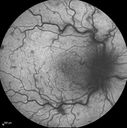
Mtehylenetetrahydrofolate reductase c677T homozygous CRVO pregnant woman (MTHFR)454 viewsVision improved from 20/50 to 20/16 in one month following ozurdex injections. 38 year old woman with only one eye. The left eye was removed as a child for retinoblastoma.Oct 05, 2015
|
|
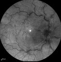
Mtehylenetetrahydrofolate reductase c677T homozygous CRVO pregnant woman (MTHFR)445 viewsVision improved from 20/50 to 20/16 in one month following ozurdex injections. 38 year old woman with only one eye. The left eye was removed as a child for retinoblastoma.Oct 05, 2015
|
|
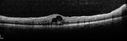
Mtehylenetetrahydrofolate reductase c677T homozygous CRVO pregnant woman (MTHFR)442 viewsVision improved from 20/50 to 20/16 in one month following ozurdex injections. 38 year old woman with only one eye. The left eye was removed as a child for retinoblastoma.Oct 05, 2015
|
|
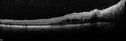
Mtehylenetetrahydrofolate reductase c677T homozygous CRVO pregnant woman (MTHFR)428 viewsVision improved from 20/50 to 20/16 in one month following ozurdex injections. 38 year old woman with only one eye. The left eye was removed as a child for retinoblastoma.Oct 05, 2015
|
|
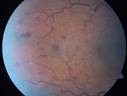
Mtehylenetetrahydrofolate reductase c677T homozygous CRVO pregnant woman (MTHFR)440 viewsVision improved from 20/50 to 20/16 in one month following ozurdex injections. 38 year old woman with only one eye. The left eye was removed as a child for retinoblastoma.Oct 05, 2015
|
|
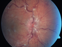
Mtehylenetetrahydrofolate reductase c677T homozygous CRVO pregnant woman (MTHFR)471 viewsVision improved from 20/50 to 20/16 in one month following ozurdex injections. 38 year old woman with only one eye. The left eye was removed as a child for retinoblastoma.Oct 05, 2015
|
|
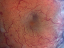
Mtehylenetetrahydrofolate reductase c677T homozygous CRVO pregnant woman (MTHFR)454 viewsVision improved from 20/50 to 20/16 in one month following ozurdex injections. 38 year old woman with only one eye. The left eye was removed as a child for retinoblastoma.Oct 05, 2015
|
|

Mtehylenetetrahydrofolate reductase c677T homozygous CRVO pregnant woman (MTHFR)494 viewsVision improved from 20/50 to 20/16 in one month following ozurdex injections. 38 year old woman with only one eye. The left eye was removed as a child for retinoblastoma.Oct 05, 2015
|
|

Non macular BRVO with good vision (Fellow eye has severe BRVO)585 viewsSep 22, 2015
|
|

Non macular BRVO with good vision (Fellow eye has severe BRVO)535 viewsSep 22, 2015
|
|

Non macular BRVO with good vision (Fellow eye has severe BRVO)601 viewsSep 22, 2015
|
|

Ischemic BRVO with non-perfusion (nonperfusion) - recovered from severe vitreous hemorrhage537 viewsSep 22, 2015
|
|
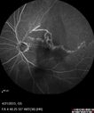
Ischemic BRVO with non-perfusion (nonperfusion) - recovered from severe vitreous hemorrhage577 viewsSep 22, 2015
|
|
| 1845 files on 19 page(s) |
 |
|
|
|
|
12 | |
|
|
|
|


