Last additions - Retina Vascular Occlusions
|

Ischemic BRVO with non-perfusion (nonperfusion) - recovered from severe vitreous hemorrhage517 viewsSep 22, 2015
|
|

Ischemic BRVO with non-perfusion (nonperfusion) - recovered from severe vitreous hemorrhage491 viewsSep 22, 2015
|
|

Ischemic BRVO with non-perfusion (nonperfusion) - recovered from severe vitreous hemorrhage473 viewsVascular remodelingSep 22, 2015
|
|

Ischemic BRVO with non-perfusion (nonperfusion) - recovered from severe vitreous hemorrhage478 viewsSep 22, 2015
|
|

Ischemic BRVO with non-perfusion (nonperfusion) - recovered from severe vitreous hemorrhage505 viewsSep 22, 2015
|
|

Ischemic BRVO with non-perfusion (nonperfusion) - recovered from severe vitreous hemorrhage530 viewsSep 22, 2015
|
|

Ischemic BRVO with non-perfusion (nonperfusion) - recovered from severe vitreous hemorrhage484 viewsSep 22, 2015
|
|

Ischemic BRVO with non-perfusion (nonperfusion) - recovered from severe vitreous hemorrhage507 viewsSep 22, 2015
|
|

Ischemic BRVO with non-perfusion (nonperfusion) - recovered from severe vitreous hemorrhage526 viewsSep 22, 2015
|
|

Minimal macular changes on SD OCT483 viewsSuperficial and deep capillary plexus visible because of collateral vesselsSep 22, 2015
|
|

Ischemic BRVO with non-perfusion (nonperfusion) - recovered from severe vitreous hemorrhage539 viewsSep 22, 2015
|
|

Normal SD OCT672 viewsSep 22, 2015
|
|

Normal506 viewsSep 22, 2015
|
|
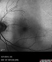
FAF shows increased FAF superiorly where the retina has been damaged because of loss of chromophores600 viewsSep 22, 2015
|
|

Normal FAF519 viewsSep 22, 2015
|
|
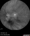
480 viewsSep 22, 2015
|
|
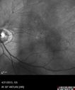
499 viewsSep 22, 2015
|
|
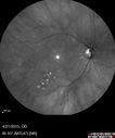
521 viewsSep 22, 2015
|
|
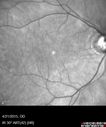
465 viewsSep 22, 2015
|
|

Ghost vessels with BRVO872 viewsSep 22, 2015
|
|

531 viewsSep 22, 2015
|
|

512 viewsSep 22, 2015
|
|

544 viewsSep 22, 2015
|
|

590 viewsSep 22, 2015
|
|

BRAO and PAMM675 viewsGray paracentral lesion from BRAO is a PAMM lesionOct 11, 2014
|
|

BRAO and PAMM1033 viewsLayers of retina are thickened showing ischemia to the middle retina (deep retinal capillary plexus)Oct 11, 2014
|
|

BRAO and PAMM615 viewsOct 11, 2014
|
|

BRAO and PAMM636 viewsOct 11, 2014
|
|

BRAO and PAMM705 viewsOct 11, 2014
|
|

BRAO and PAMM625 viewsOct 11, 2014
|
|

BRAO and PAMM676 viewsOct 11, 2014
|
|

BRAO and PAMM602 viewsOct 11, 2014
|
|

BRAO and PAMM527 viewsOct 11, 2014
|
|

BRAO and PAMM464 viewsOct 11, 2014
|
|

BRAO and PAMM579 viewsOct 11, 2014
|
|

BRAO and PAMM529 viewsOct 11, 2014
|
|

BRAO and PAMM685 viewsOct 11, 2014
|
|

Choroidal Nevus651 viewsOct 11, 2014
|
|

BRAO and PAMM630 viewsOct 11, 2014
|
|

BRAO and PAMM652 viewsOct 11, 2014
|
|

BRAO and PAMM672 viewsOct 11, 2014
|
|

BRAO and PAMM627 viewsOct 11, 2014
|
|

BRAO and PAMM831 viewsOct 11, 2014
|
|

Retinal Artery Occlusion1196 viewsPatient comes in for eval on artery occlusions in both eyes. VA is 20/400, right eye and NLP, left eye. Fundus photos show paniretinal scars in the right eye with arterial narrowing and the left eye has arterial narrowing as well.Sep 23, 2014
|
|
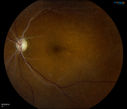
Retinal Artery Occlusion1706 viewsPatient comes in for eval on artery occlusions in both eyes. VA is 20/400, right eye and NLP, left eye. Fundus photos show paniretinal scars in the right eye with arterial narrowing and the left eye has arterial narrowing as well.Sep 23, 2014
|
|

Retinal Artery Occlusion1033 viewsPatient comes in for eval on artery occlusions in both eyes. VA is 20/400, right eye and NLP, left eye. Fundus photos show paniretinal scars in the right eye with arterial narrowing and the left eye has arterial narrowing as well.Sep 23, 2014
|
|

Retinal Artery Occlusion594 viewsPatient comes in for eval on artery occlusions in both eyes. VA is 20/400, right eye and NLP, left eye. Fundus photos show paniretinal scars in the right eye with arterial narrowing and the left eye has arterial narrowing as well.Sep 23, 2014
|
|

Retinal Vein Occlusion415 viewsPatient comes in with loss of vision, left eye. VA is count fingers in the left eye. Fundus photos show large vein occlusion with macular edema in the left eye. Patient proceeded with Avastin Injection..Sep 23, 2014
|
|

Retinal Vein Occlusion399 viewsPatient comes in with loss of vision, left eye. VA is count fingers in the left eye. Fundus photos show large vein occlusion with macular edema in the left eye. Patient proceeded with Avastin Injection..Sep 23, 2014
|
|

Branch Retinal Vein Occlusion with NVE (Neovascularization Elsewhere)737 viewsPre and post laser therapy - NV has partially regressedSep 06, 2014
|
|

Branch Retinal Vein Occlusion with NVE (Neovascularization Elsewhere)583 viewsSep 06, 2014
|
|

Branch Retinal Vein Occlusion with NVE (Neovascularization Elsewhere)654 viewsOne month post laserSep 06, 2014
|
|

Branch Retinal Vein Occlusion with NVE (Neovascularization Elsewhere)545 viewsSep 06, 2014
|
|
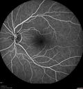
Branch Retinal Vein Occlusion with NVE (Neovascularization Elsewhere)523 viewsSep 06, 2014
|
|

Branch Retinal Vein Occlusion with NVE (Neovascularization Elsewhere)479 viewsSep 06, 2014
|
|
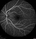
Branch Retinal Vein Occlusion with NVE (Neovascularization Elsewhere)481 viewsSep 06, 2014
|
|

Branch Retinal Vein Occlusion with NVE (Neovascularization Elsewhere)556 viewsLate Leakage on FASep 06, 2014
|
|

Branch Retinal Vein Occlusion with NVE (Neovascularization Elsewhere)472 viewsLate Leakage on FASep 06, 2014
|
|

558 viewsLate Leakage on FASep 06, 2014
|
|

Branch Retinal Vein Occlusion with NVE (Neovascularization Elsewhere)545 viewsLate Leakage on FASep 06, 2014
|
|

Branch Retinal Vein Occlusion with NVE (Neovascularization Elsewhere)674 viewsLate Leakage on FA - NV is adjacent to non-perfusionSep 06, 2014
|
|

Branch Retinal Vein Occlusion with NVE (Neovascularization Elsewhere)680 viewsLate Leakage on FA - NV is adjacent to non-perfusionSep 06, 2014
|
|

Branch Retinal Vein Occlusion with NVE (Neovascularization Elsewhere)559 viewsLate Leakage on FA - NV is adjacent to non-perfusionSep 06, 2014
|
|

Branch Retinal Vein Occlusion with NVE (Neovascularization Elsewhere)757 viewsLate Leakage on FA - NV is adjacent to non-perfusionSep 06, 2014
|
|

Branch Retinal Vein Occlusion with NVE (Neovascularization Elsewhere)689 viewsLate Leakage on FA - NV is adjacent to non-perfusionSep 06, 2014
|
|

Branch Retinal Vein Occlusion with NVE (Neovascularization Elsewhere)568 viewsLate Leakage on FA - NV is adjacent to non-perfusionSep 06, 2014
|
|

Branch Retinal Vein Occlusion with NVE (Neovascularization Elsewhere)470 viewsSep 06, 2014
|
|

Branch Retinal Vein Occlusion with NVE (Neovascularization Elsewhere)508 viewsSep 06, 2014
|
|

Branch Retinal Vein Occlusion with NVE (Neovascularization Elsewhere)500 viewsSep 06, 2014
|
|

Branch Retinal Vein Occlusion with NVE (Neovascularization Elsewhere)494 viewsSep 06, 2014
|
|

Branch Retinal Vein Occlusion with NVE (Neovascularization Elsewhere)469 viewsSep 06, 2014
|
|

Branch Retinal Vein Occlusion with NVE (Neovascularization Elsewhere)496 viewsSep 06, 2014
|
|

Branch Retinal Vein Occlusion with NVE (Neovascularization Elsewhere)576 viewsSep 06, 2014
|
|
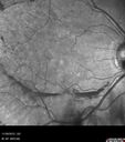
Branch Retinal Vein Occlusion with NVE (Neovascularization Elsewhere)541 viewsSep 06, 2014
|
|

Branch Retinal Vein Occlusion with NVE (Neovascularization Elsewhere)542 viewsSep 06, 2014
|
|

Branch Retinal Vein Occlusion with NVE (Neovascularization Elsewhere)582 viewsSD-OCT of NVE - shows it to be pre-retinalSep 06, 2014
|
|

Branch Retinal Vein Occlusion with NVE (Neovascularization Elsewhere)532 viewsSD-OCT of NVE - shows it to be pre-retinalSep 06, 2014
|
|

Branch Retinal Vein Occlusion with NVE (Neovascularization Elsewhere)485 viewsSD-OCT of NVE - shows it to be pre-retinalSep 06, 2014
|
|

Branch Retinal Vein Occlusion with NVE (Neovascularization Elsewhere)545 viewsSep 06, 2014
|
|

Branch Retinal Vein Occlusion with NVE (Neovascularization Elsewhere)683 viewsSep 06, 2014
|
|

Branch Retinal Vein Occlusion with NVE (Neovascularization Elsewhere)716 viewsSep 06, 2014
|
|

Branch Retinal Vein Occlusion with NVE (Neovascularization Elsewhere)741 viewsSep 06, 2014
|
|

Branch Retinal Vein Occlusion with NVE (Neovascularization Elsewhere)656 viewsSep 06, 2014
|
|

Branch Retinal Vein Occlusion with NVE (Neovascularization Elsewhere)705 viewsSep 06, 2014
|
|

Branch Retinal Vein Occlusion with NVE (Neovascularization Elsewhere)831 viewsSep 06, 2014
|
|

Central Retinal Artery Occlusion1105 viewsCentral Retinal Artery Occlusion with cilio artery perfusion. May 15, 2014
|
|

FA: CRAO838 viewsCRAO with cilio artery perfusionMay 15, 2014
|
|

Stasis Retinopathy, Central Retinal Vein Occlusion723 viewsYoung male who complains of decreased vision in the left eye. VA is 20/20 in the left eye. Fundus photo shows mild central retinal vein occlusion in the left eye. Mar 28, 2014
|
|
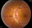
Stasis Retinopathy, Central Retinal Vein Occlusion1272 viewsYoung male who complains of decreased vision in the left eye. VA is 20/20 in the left eye. Fundus photo shows mild central retinal vein occlusion in the left eye. Mar 28, 2014
|
|

Stasis Retinopathy, Central Retinal Vein Occlusion568 viewsYoung male who complains of decreased vision in the left eye. VA is 20/20 in the left eye. Fundus photo shows mild central retinal vein occlusion in the left eye. Mar 28, 2014
|
|

Central Retinal Vein Occlusion1356 viewsPatient comes in with mild blurred vision in the right eye. Fundus exam shows CRVO with scattered retinal hemorrhages in the right eye.Mar 27, 2014
|
|

telangiectasia905 views20/20 pt with blurry area in vaMar 07, 2014
|
|

Branch Retinal Vein Occlusion with worse macular edema from hypertension - 1 month later - edema is less557 viewsVision back to 20/25 with resolved edema (no need for other therapy with better blood pressure control)Feb 18, 2014
|
|

Branch Retinal Vein Occlusion with worse macular edema from hypertension - FA shows Edema Right Eye571 viewsFeb 18, 2014
|
|

Branch Retinal Vein Occlusion with worse macular edema from hypertension - FA shows Edema Right Eye475 viewsFeb 18, 2014
|
|

Branch Retinal Vein Occlusion with worse macular edema from hypertension - FA shows Edema Right Eye472 viewsFeb 18, 2014
|
|

Branch Retinal Vein Occlusion with worse macular edema from hypertension - FA shows Edema Right Eye467 viewsFeb 18, 2014
|
|

Branch Retinal Vein Occlusion with worse macular edema from hypertension - FA shows Edema Right Eye493 viewsFeb 18, 2014
|
|

Branch Retinal Vein Occlusion with worse macular edema from hypertension - FA shows Edema Right Eye564 viewsFeb 18, 2014
|
|
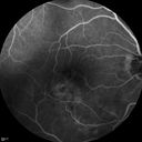
Branch Retinal Vein Occlusion with worse macular edema from hypertension - FA shows Edema Right Eye543 viewsFeb 18, 2014
|
|
| 1845 files on 19 page(s) |
 |
|
|
|
|
13 | |
|
|
|
|


