Last additions - Retina Vascular Occlusions
|

Central Retinal Vein Occlusion - No macular edema - Pre-foveal opercula424 views62 year old female with 20/20 vision. She had been seeing a spot out of the left eye for 4 years and then noticed splotches in her vision. She never devleoped edema or NVFeb 27, 2019
|
|

Central Retinal Vein Occlusion - No macular edema - Pre-foveal opercula415 views62 year old female with 20/20 vision. She had been seeing a spot out of the left eye for 4 years and then noticed splotches in her vision. She never devleoped edema or NVFeb 27, 2019
|
|

Central Retinal Vein Occlusion - No macular edema - Pre-foveal opercula419 views62 year old female with 20/20 vision. She had been seeing a spot out of the left eye for 4 years and then noticed splotches in her vision. She never devleoped edema or NVFeb 27, 2019
|
|

Central Retinal Vein Occlusion - No macular edema - Pre-foveal opercula415 views62 year old female with 20/20 vision. She had been seeing a spot out of the left eye for 4 years and then noticed splotches in her vision. She never devleoped edema or NVFeb 27, 2019
|
|

Central Retinal Vein Occlusion - No macular edema - Pre-foveal opercula443 views62 year old female with 20/20 vision. She had been seeing a spot out of the left eye for 4 years and then noticed splotches in her vision. She never devleoped edema or NVFeb 27, 2019
|
|

Central Retinal Vein Occlusion - No macular edema - Pre-foveal opercula421 views62 year old female with 20/20 vision. She had been seeing a spot out of the left eye for 4 years and then noticed splotches in her vision. She never devleoped edema or NVFeb 27, 2019
|
|

Central Retinal Vein Occlusion - No macular edema - Pre-foveal opercula405 views62 year old female with 20/20 vision. She had been seeing a spot out of the left eye for 4 years and then noticed splotches in her vision. She never devleoped edema or NVFeb 27, 2019
|
|

Central Retinal Vein Occlusion - No macular edema - Pre-foveal opercula534 views62 year old female with 20/20 vision. She had been seeing a spot out of the left eye for 4 years and then noticed splotches in her vision. She never devleoped edema or NVFeb 27, 2019
|
|

Central Retinal Vein Occlusion - No macular edema - Pre-foveal opercula438 views62 year old female with 20/20 vision. She had been seeing a spot out of the left eye for 4 years and then noticed splotches in her vision. She never devleoped edema or NVFeb 27, 2019
|
|

Central Retinal Vein Occlusion - No macular edema - Pre-foveal opercula409 views62 year old female with 20/20 vision. She had been seeing a spot out of the left eye for 4 years and then noticed splotches in her vision. She never devleoped edema or NVFeb 27, 2019
|
|

Central Retinal Vein Occlusion - No macular edema - Pre-foveal opercula455 views62 year old female with 20/20 vision. She had been seeing a spot out of the left eye for 4 years and then noticed splotches in her vision. She never devleoped edema or NVFeb 27, 2019
|
|

Central Retinal Vein Occlusion - No macular edema - Pre-foveal opercula431 views62 year old female with 20/20 vision. She had been seeing a spot out of the left eye for 4 years and then noticed splotches in her vision. She never devleoped edema or NVFeb 27, 2019
|
|

Central Retinal Vein Occlusion - No macular edema - Pre-foveal opercula425 views62 year old female with 20/20 vision. She had been seeing a spot out of the left eye for 4 years and then noticed splotches in her vision. She never devleoped edema or NVFeb 27, 2019
|
|

Central Retinal Vein Occlusion - No macular edema - Pre-foveal opercula434 views62 year old female with 20/20 vision. She had been seeing a spot out of the left eye for 4 years and then noticed splotches in her vision. She never devleoped edema or NVFeb 27, 2019
|
|

Central Retinal Vein Occlusion - No macular edema - Pre-foveal opercula450 views62 year old female with 20/20 vision. She had been seeing a spot out of the left eye for 4 years and then noticed splotches in her vision. She never devleoped edema or NVFeb 27, 2019
|
|

Central Retinal Vein Occlusion - No macular edema - Pre-foveal opercula467 views62 year old female with 20/20 vision. She had been seeing a spot out of the left eye for 4 years and then noticed splotches in her vision. She never devleoped edema or NVFeb 27, 2019
|
|

Central Retinal Vein Occlusion - No macular edema - Pre-foveal opercula420 views62 year old female with 20/20 vision. She had been seeing a spot out of the left eye for 4 years and then noticed splotches in her vision. She never devleoped edema or NVFeb 27, 2019
|
|

Central Retinal Vein Occlusion - No macular edema - Pre-foveal opercula393 views62 year old female with 20/20 vision. She had been seeing a spot out of the left eye for 4 years and then noticed splotches in her vision. She never devleoped edema or NVFeb 27, 2019
|
|

Central Retinal Vein Occlusion - No macular edema - Pre-foveal opercula410 views62 year old female with 20/20 vision. She had been seeing a spot out of the left eye for 4 years and then noticed splotches in her vision. She never devleoped edema or NVFeb 27, 2019
|
|

Central Retinal Vein Occlusion - No macular edema - Pre-foveal opercula436 views62 year old female with 20/20 vision. She had been seeing a spot out of the left eye for 4 years and then noticed splotches in her vision. She never devleoped edema or NVFeb 27, 2019
|
|

Central Retinal Vein Occlusion - No macular edema - Pre-foveal opercula441 views62 year old female with 20/20 vision. She had been seeing a spot out of the left eye for 4 years and then noticed splotches in her vision. She never devleoped edema or NVFeb 27, 2019
|
|

Central Retinal Vein Occlusion - No macular edema - Pre-foveal opercula497 views62 year old female with 20/20 vision. She had been seeing a spot out of the left eye for 4 years and then noticed splotches in her vision. She never devleoped edema or NVFeb 27, 2019
|
|

Central Retinal Vein Occlusion - No macular edema - Pre-foveal opercula452 views62 year old female with 20/20 vision. She had been seeing a spot out of the left eye for 4 years and then noticed splotches in her vision. She never devleoped edema or NVFeb 27, 2019
|
|

Central Retinal Vein Occlusion - No macular edema - Pre-foveal opercula389 views62 year old female with 20/20 vision. She had been seeing a spot out of the left eye for 4 years and then noticed splotches in her vision. She never devleoped edema or NVFeb 27, 2019
|
|

Central Retinal Vein Occlusion - No macular edema - Pre-foveal opercula439 views62 year old female with 20/20 vision. She had been seeing a spot out of the left eye for 4 years and then noticed splotches in her vision. She never devleoped edema or NVFeb 27, 2019
|
|

Central Retinal Artery Occlusion - Diabetic Retinopathy - NO cherry red spot448 views58 year old man with sudden vision loss in the right eye. The vision had been 20/40 and dropped to 20/200 the day prior to presentation.
VA 20/200 OD, 20/20 OS
FA shows CRAO. No cherry red spot.Feb 27, 2019
|
|

Central Retinal Artery Occlusion - Diabetic Retinopathy - NO cherry red spot536 views58 year old man with sudden vision loss in the right eye. The vision had been 20/40 and dropped to 20/200 the day prior to presentation.
VA 20/200 OD, 20/20 OS
FA shows CRAO. No cherry red spot.Feb 27, 2019
|
|

Central Retinal Artery Occlusion - Diabetic Retinopathy - NO cherry red spot474 views58 year old man with sudden vision loss in the right eye. The vision had been 20/40 and dropped to 20/200 the day prior to presentation.
VA 20/200 OD, 20/20 OS
FA shows CRAO. No cherry red spot.Feb 27, 2019
|
|

Central Retinal Artery Occlusion - Diabetic Retinopathy - NO cherry red spot530 views58 year old man with sudden vision loss in the right eye. The vision had been 20/40 and dropped to 20/200 the day prior to presentation.
VA 20/200 OD, 20/20 OS
FA shows CRAO. No cherry red spot.Feb 27, 2019
|
|
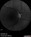
Central Retinal Artery Occlusion - Diabetic Retinopathy - NO cherry red spot493 views58 year old man with sudden vision loss in the right eye. The vision had been 20/40 and dropped to 20/200 the day prior to presentation.
VA 20/200 OD, 20/20 OS
FA shows CRAO. No cherry red spot.Feb 27, 2019
|
|

Central Retinal Artery Occlusion - Diabetic Retinopathy - NO cherry red spot550 views58 year old man with sudden vision loss in the right eye. The vision had been 20/40 and dropped to 20/200 the day prior to presentation.
VA 20/200 OD, 20/20 OS
FA shows CRAO. No cherry red spot.Feb 27, 2019
|
|

Central Retinal Artery Occlusion - Diabetic Retinopathy - NO cherry red spot484 views58 year old man with sudden vision loss in the right eye. The vision had been 20/40 and dropped to 20/200 the day prior to presentation.
VA 20/200 OD, 20/20 OS
FA shows CRAO. No cherry red spot.Feb 27, 2019
|
|

Central Retinal Artery Occlusion - Diabetic Retinopathy - NO cherry red spot524 views58 year old man with sudden vision loss in the right eye. The vision had been 20/40 and dropped to 20/200 the day prior to presentation.
VA 20/200 OD, 20/20 OS
FA shows CRAO. No cherry red spot.Feb 27, 2019
|
|

Central Retinal Artery Occlusion - Diabetic Retinopathy - NO cherry red spot442 views58 year old man with sudden vision loss in the right eye. The vision had been 20/40 and dropped to 20/200 the day prior to presentation.
VA 20/200 OD, 20/20 OS
FA shows CRAO. No cherry red spot.Feb 27, 2019
|
|

Central Retinal Artery Occlusion - Diabetic Retinopathy - NO cherry red spot562 views58 year old man with sudden vision loss in the right eye. The vision had been 20/40 and dropped to 20/200 the day prior to presentation.
VA 20/200 OD, 20/20 OS
FA shows CRAO. No cherry red spot.Feb 27, 2019
|
|

Central Retinal Artery Occlusion - Diabetic Retinopathy - NO cherry red spot573 views58 year old man with sudden vision loss in the right eye. The vision had been 20/40 and dropped to 20/200 the day prior to presentation.
VA 20/200 OD, 20/20 OS
FA shows CRAO. No cherry red spot.Feb 27, 2019
|
|

Central Retinal Artery Occlusion - Diabetic Retinopathy - NO cherry red spot620 views58 year old man with sudden vision loss in the right eye. The vision had been 20/40 and dropped to 20/200 the day prior to presentation.
VA 20/200 OD, 20/20 OS
FA shows CRAO. No cherry red spot.Feb 27, 2019
|
|

Twig Old Macular Branch Retinal Vein Occlusion - Venous Venous Collaterals500 views88 year old man with 20/25 vision in his left and only eye. There is vascular remodeling consistent with an old twig BRVO.Feb 14, 2019
|
|

Twig Old Macular Branch Retinal Vein Occlusion - Venous Venous Collaterals513 views88 year old man with 20/25 vision in his left and only eye. There is vascular remodeling consistent with an old twig BRVO.Feb 14, 2019
|
|

Twig Old Macular Branch Retinal Vein Occlusion - Venous Venous Collaterals471 views88 year old man with 20/25 vision in his left and only eye. There is vascular remodeling consistent with an old twig BRVO.Feb 14, 2019
|
|

Twig Old Macular Branch Retinal Vein Occlusion - Venous Venous Collaterals533 views88 year old man with 20/25 vision in his left and only eye. There is vascular remodeling consistent with an old twig BRVO.Feb 14, 2019
|
|

Twig Old Macular Branch Retinal Vein Occlusion - Venous Venous Collaterals440 views88 year old man with 20/25 vision in his left and only eye. There is vascular remodeling consistent with an old twig BRVO.Feb 14, 2019
|
|

Twig Old Macular Branch Retinal Vein Occlusion - Venous Venous Collaterals472 views88 year old man with 20/25 vision in his left and only eye. There is vascular remodeling consistent with an old twig BRVO.Feb 14, 2019
|
|

Twig Old Macular Branch Retinal Vein Occlusion - Venous Venous Collaterals735 views88 year old man with 20/25 vision in his left and only eye. There is vascular remodeling consistent with an old twig BRVO.Feb 14, 2019
|
|

Ischemic CRVO OD and Arterial Macroaneurysm OS456 views83 year old female post PPV, Laser, Ahmed valve for CRVO with vitreous heme and NVG in the right eye. VA was 20/160 3 months following vitrectomy. Left eye has MA which was not treated and involuted post-hemeJan 25, 2019
|
|

Ischemic CRVO OD and Arterial Macroaneurysm OS551 views83 year old female post PPV, Laser, Ahmed valve for CRVO with vitreous heme and NVG in the right eye. VA was 20/160 3 months following vitrectomy. Left eye has MA which was not treated and involuted post-hemeJan 25, 2019
|
|

Ischemic CRVO OD and Arterial Macroaneurysm OS420 views83 year old female post PPV, Laser, Ahmed valve for CRVO with vitreous heme and NVG in the right eye. VA was 20/160 3 months following vitrectomy. Left eye has MA which was not treated and involuted post-hemeJan 25, 2019
|
|

Ischemic CRVO OD and Arterial Macroaneurysm OS485 views83 year old female post PPV, Laser, Ahmed valve for CRVO with vitreous heme and NVG in the right eye. VA was 20/160 3 months following vitrectomy. Left eye has MA which was not treated and involuted post-hemeJan 25, 2019
|
|

Ischemic CRVO OD and Arterial Macroaneurysm OS500 views83 year old female post PPV, Laser, Ahmed valve for CRVO with vitreous heme and NVG in the right eye. VA was 20/160 3 months following vitrectomy. Left eye has MA which was not treated and involuted post-hemeJan 25, 2019
|
|

CRVO 25 year old man - Heterozygous Factor V Leiden415 views20/100 initial vision. Improved to 20/30 with Lucentis which continued for 3 years, treat and extend, and stopped.Dec 18, 2018
|
|

CRVO 25 year old man - Heterozygous Factor V Leiden482 views20/100 initial vision. Improved to 20/30 with Lucentis which continued for 3 years, treat and extend, and stopped.Dec 18, 2018
|
|

CRVO 25 year old man - Heterozygous Factor V Leiden446 views20/100 initial vision. Improved to 20/30 with Lucentis which continued for 3 years, treat and extend, and stopped.Dec 18, 2018
|
|

CRVO 25 year old man - Heterozygous Factor V Leiden451 views20/100 initial vision. Improved to 20/30 with Lucentis which continued for 3 years, treat and extend, and stopped.Dec 18, 2018
|
|

CRVO 25 year old man - Heterozygous Factor V Leiden414 views20/100 initial vision. Improved to 20/30 with Lucentis which continued for 3 years, treat and extend, and stopped.Dec 18, 2018
|
|

CRVO 25 year old man - Heterozygous Factor V Leiden393 views20/100 initial vision. Improved to 20/30 with Lucentis which continued for 3 years, treat and extend, and stopped.Dec 18, 2018
|
|

CRVO 25 year old man - Heterozygous Factor V Leiden413 views20/100 initial vision. Improved to 20/30 with Lucentis which continued for 3 years, treat and extend, and stopped.Dec 18, 2018
|
|

CRVO 25 year old man - Heterozygous Factor V Leiden469 views20/100 initial vision. Improved to 20/30 with Lucentis which continued for 3 years, treat and extend, and stopped.Dec 18, 2018
|
|

CRVO 25 year old man - Heterozygous Factor V Leiden419 views20/100 initial vision. Improved to 20/30 with Lucentis which continued for 3 years, treat and extend, and stopped.Dec 18, 2018
|
|

CRVO 25 year old man - Heterozygous Factor V Leiden428 views20/100 initial vision. Improved to 20/30 with Lucentis which continued for 3 years, treat and extend, and stopped.Dec 18, 2018
|
|

CRVO 25 year old man - Heterozygous Factor V Leiden459 views20/100 initial vision. Improved to 20/30 with Lucentis which continued for 3 years, treat and extend, and stopped.Dec 18, 2018
|
|

CRVO 25 year old man - Heterozygous Factor V Leiden438 views20/100 initial vision. Improved to 20/30 with Lucentis which continued for 3 years, treat and extend, and stopped.Dec 18, 2018
|
|

CRVO 25 year old man - Heterozygous Factor V Leiden435 views20/100 initial vision. Improved to 20/30 with Lucentis which continued for 3 years, treat and extend, and stopped.Dec 18, 2018
|
|

CRVO 25 year old man - Heterozygous Factor V Leiden419 views20/100 initial vision. Improved to 20/30 with Lucentis which continued for 3 years, treat and extend, and stopped.Dec 18, 2018
|
|

CRVO 25 year old man - Heterozygous Factor V Leiden426 views20/100 initial vision. Improved to 20/30 with Lucentis which continued for 3 years, treat and extend, and stopped.Dec 18, 2018
|
|

CRVO 25 year old man - Heterozygous Factor V Leiden426 views20/100 initial vision. Improved to 20/30 with Lucentis which continued for 3 years, treat and extend, and stopped.Dec 18, 2018
|
|

CRVO 25 year old man - Heterozygous Factor V Leiden435 views20/100 initial vision. Improved to 20/30 with Lucentis which continued for 3 years, treat and extend, and stopped.Dec 18, 2018
|
|

CRVO 25 year old man - Heterozygous Factor V Leiden437 views20/100 initial vision. Improved to 20/30 with Lucentis which continued for 3 years, treat and extend, and stopped.Dec 18, 2018
|
|

CRVO 25 year old man - Heterozygous Factor V Leiden519 views20/100 initial vision. Improved to 20/30 with Lucentis which continued for 3 years, treat and extend, and stopped.Dec 18, 2018
|
|

CRVO 25 year old man - Heterozygous Factor V Leiden435 views20/100 initial vision. Improved to 20/30 with Lucentis which continued for 3 years, treat and extend, and stopped.Dec 18, 2018
|
|

CRVO 25 year old man - Heterozygous Factor V Leiden472 views20/100 initial vision. Improved to 20/30 with Lucentis which continued for 3 years, treat and extend, and stopped.Dec 18, 2018
|
|

CRVO 25 year old man - Heterozygous Factor V Leiden417 views20/100 initial vision. Improved to 20/30 with Lucentis which continued for 3 years, treat and extend, and stopped.Dec 18, 2018
|
|

CRVO 25 year old man - Heterozygous Factor V Leiden484 views20/100 initial vision. Improved to 20/30 with Lucentis which continued for 3 years, treat and extend, and stopped.Dec 18, 2018
|
|

CRVO 25 year old man - Heterozygous Factor V Leiden429 views20/100 initial vision. Improved to 20/30 with Lucentis which continued for 3 years, treat and extend, and stopped.Dec 18, 2018
|
|

CRVO 25 year old man - Heterozygous Factor V Leiden548 views20/100 initial vision. Improved to 20/30 with Lucentis which continued for 3 years, treat and extend, and stopped.Dec 18, 2018
|
|

Central Retinal Artery Occlusion then Neovascular Glaucoma498 views81 year old male with sudden vision loss. 2 months later developed NVGJul 27, 2018
|
|

Central Retinal Artery Occlusion then Neovascular Glaucoma567 views81 year old male with sudden vision loss. 2 months later developed NVGJul 27, 2018
|
|

Central Retinal Artery Occlusion then Neovascular Glaucoma468 views81 year old male with sudden vision loss. 2 months later developed NVGJul 27, 2018
|
|

Central Retinal Artery Occlusion then Neovascular Glaucoma429 views81 year old male with sudden vision loss. 2 months later developed NVGJul 27, 2018
|
|

Central Retinal Artery Occlusion then Neovascular Glaucoma415 views81 year old male with sudden vision loss. 2 months later developed NVGJul 27, 2018
|
|

Central Retinal Artery Occlusion then Neovascular Glaucoma511 views81 year old male with sudden vision loss. 2 months later developed NVGJul 27, 2018
|
|

Central Retinal Artery Occlusion then Neovascular Glaucoma545 views81 year old male with sudden vision loss. 2 months later developed NVGJul 27, 2018
|
|

Central Retinal Artery Occlusion then Neovascular Glaucoma438 views81 year old male with sudden vision loss. 2 months later developed NVGJul 27, 2018
|
|

Central Retinal Artery Occlusion then Neovascular Glaucoma501 views81 year old male with sudden vision loss. 2 months later developed NVGJul 27, 2018
|
|

Central Retinal Artery Occlusion then Neovascular Glaucoma496 views81 year old male with sudden vision loss. 2 months later developed NVGJul 27, 2018
|
|

DRIL - Disorganization Retinal Inner Layers842 viewsFollowing treatment for BRVO vision stayed at 3/200 despite resolution of edemaJul 05, 2018
|
|

DRIL - Disorganization Retinal Inner Layers452 viewsFollowing treatment for BRVO vision stayed at 3/200 despite resolution of edemaJul 05, 2018
|
|

DRIL - Disorganization Retinal Inner Layers442 viewsFollowing treatment for BRVO vision stayed at 3/200 despite resolution of edemaJul 05, 2018
|
|

DRIL - Disorganization Retinal Inner Layers443 viewsFollowing treatment for BRVO vision stayed at 3/200 despite resolution of edemaJul 05, 2018
|
|

PAMM with Follow-up581 viewsMay 17, 2018
|
|

Cardiac Catheterization induced Cilioretinal Artery Occlusion - Paracentral Acute Middle Maculopathy482 views69 year old female with sudden vision loss on completing cardiac catheterization. The SD OCT shows a PAMM lesion and the lesion looks gray on color imaging. There is delayed filling of the cilioretinal artery.Mar 06, 2018
|
|
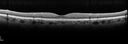
Cardiac Catheterization induced Cilioretinal Artery Occlusion - Paracentral Acute Middle Maculopathy425 views69 year old female with sudden vision loss on completing cardiac catheterization. The SD OCT shows a PAMM lesion and the lesion looks gray on color imaging. There is delayed filling of the cilioretinal artery.Mar 06, 2018
|
|
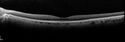
Cardiac Catheterization induced Cilioretinal Artery Occlusion - Paracentral Acute Middle Maculopathy457 views69 year old female with sudden vision loss on completing cardiac catheterization. The SD OCT shows a PAMM lesion and the lesion looks gray on color imaging. There is delayed filling of the cilioretinal artery.Mar 06, 2018
|
|
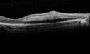
Cardiac Catheterization induced Cilioretinal Artery Occlusion - Paracentral Acute Middle Maculopathy488 views69 year old female with sudden vision loss on completing cardiac catheterization. The SD OCT shows a PAMM lesion and the lesion looks gray on color imaging. There is delayed filling of the cilioretinal artery.Mar 06, 2018
|
|
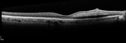
Cardiac Catheterization induced Cilioretinal Artery Occlusion - Paracentral Acute Middle Maculopathy428 views69 year old female with sudden vision loss on completing cardiac catheterization. The SD OCT shows a PAMM lesion and the lesion looks gray on color imaging. There is delayed filling of the cilioretinal artery.Mar 06, 2018
|
|
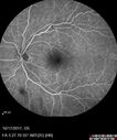
Cardiac Catheterization induced Cilioretinal Artery Occlusion - Paracentral Acute Middle Maculopathy421 views69 year old female with sudden vision loss on completing cardiac catheterization. The SD OCT shows a PAMM lesion and the lesion looks gray on color imaging. There is delayed filling of the cilioretinal artery.Mar 06, 2018
|
|

Cardiac Catheterization induced Cilioretinal Artery Occlusion - Paracentral Acute Middle Maculopathy464 views69 year old female with sudden vision loss on completing cardiac catheterization. The SD OCT shows a PAMM lesion and the lesion looks gray on color imaging. There is delayed filling of the cilioretinal artery.Mar 06, 2018
|
|

Cardiac Catheterization induced Cilioretinal Artery Occlusion - Paracentral Acute Middle Maculopathy416 views69 year old female with sudden vision loss on completing cardiac catheterization. The SD OCT shows a PAMM lesion and the lesion looks gray on color imaging. There is delayed filling of the cilioretinal artery.Mar 06, 2018
|
|

Cardiac Catheterization induced Cilioretinal Artery Occlusion - Paracentral Acute Middle Maculopathy482 views69 year old female with sudden vision loss on completing cardiac catheterization. The SD OCT shows a PAMM lesion and the lesion looks gray on color imaging. There is delayed filling of the cilioretinal artery.Mar 06, 2018
|
|

Cardiac Catheterization induced Cilioretinal Artery Occlusion - Paracentral Acute Middle Maculopathy424 views69 year old female with sudden vision loss on completing cardiac catheterization. The SD OCT shows a PAMM lesion and the lesion looks gray on color imaging. There is delayed filling of the cilioretinal artery.Mar 06, 2018
|
|

Cardiac Catheterization induced Cilioretinal Artery Occlusion - Paracentral Acute Middle Maculopathy416 views69 year old female with sudden vision loss on completing cardiac catheterization. The SD OCT shows a PAMM lesion and the lesion looks gray on color imaging. There is delayed filling of the cilioretinal artery.Mar 06, 2018
|
|
| 1798 files on 18 page(s) |
 |
|
|
|
|
9 | |
|
|
|
|


