Last additions - Age-Related Macular Degeneration - Wet
|

Wet AMD 1 month after Eylea - macula is dry and vision is better505 viewsSep 22, 2015
|
|

New wet AMD OS with subretinal hemorrhage and recent vision loss, Scar OD451 viewsSep 22, 2015
|
|

New wet AMD OS with subretinal hemorrhage and recent vision loss, Scar OD435 viewsSep 22, 2015
|
|

New wet AMD OS with subretinal hemorrhage and recent vision loss, Scar OD460 viewsSep 22, 2015
|
|

New wet AMD OS with subretinal hemorrhage and recent vision loss, Scar OD464 viewsSep 22, 2015
|
|

New wet AMD OS with subretinal hemorrhage and recent vision loss, Scar OD422 viewsSep 22, 2015
|
|

New wet AMD OS with subretinal hemorrhage and recent vision loss, Scar OD404 viewsSep 22, 2015
|
|

New wet AMD OS with subretinal hemorrhage and recent vision loss, Scar OD445 viewsSep 22, 2015
|
|

New wet AMD OS with subretinal hemorrhage and recent vision loss, Scar OD466 viewsSep 22, 2015
|
|

New wet AMD OS with subretinal hemorrhage and recent vision loss, Scar OD453 viewsSep 22, 2015
|
|

New wet AMD OS with subretinal hemorrhage and recent vision loss, Scar OD446 viewsSep 22, 2015
|
|
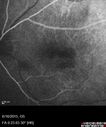
New wet AMD OS with subretinal hemorrhage and recent vision loss, Scar OD478 viewsSep 22, 2015
|
|

New wet AMD OS with subretinal hemorrhage and recent vision loss, Scar OD497 viewsSep 22, 2015
|
|

New wet AMD OS with subretinal hemorrhage and recent vision loss, Scar OD516 viewsSep 22, 2015
|
|

New wet AMD OS with subretinal hemorrhage and recent vision loss, Scar OD513 viewsSep 22, 2015
|
|

New wet AMD OS with subretinal hemorrhage and recent vision loss, Scar OD521 viewsSep 22, 2015
|
|
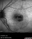
New wet AMD OS with subretinal hemorrhage and recent vision loss, Scar OD449 viewsSep 22, 2015
|
|

New wet AMD OS with subretinal hemorrhage and recent vision loss, Scar OD466 viewsSep 22, 2015
|
|

New wet AMD OS with subretinal hemorrhage and recent vision loss, Scar OD486 viewsSep 22, 2015
|
|

New wet AMD OS with subretinal hemorrhage and recent vision loss, Scar OD504 viewsSep 22, 2015
|
|

New wet AMD OS with subretinal hemorrhage and recent vision loss, Scar OD551 viewsSep 22, 2015
|
|

New wet AMD OS with subretinal hemorrhage and recent vision loss, Scar OD645 viewsSep 22, 2015
|
|

OCT shows lamellar macular hole left eye612 viewsWet AMD in right and better eye failed extend to 2 months but was able to stop therapy once he started on systemic Avastin for cancer. The last image shows the macula dry 9 months without local therapy. The left eye has a lamellar macular hole.Apr 02, 2015
|
|

OCT shows lamellar macular hole left eye511 viewsWet AMD in right and better eye failed extend to 2 months but was able to stop therapy once he started on systemic Avastin for cancer. The last image shows the macula dry 9 months without local therapy. The left eye has a lamellar macular hole.Apr 02, 2015
|
|

OCT shows lamellar macular hole left eye532 viewsWet AMD in right and better eye failed extend to 2 months but was able to stop therapy once he started on systemic Avastin for cancer. The last image shows the macula dry 9 months without local therapy. The left eye has a lamellar macular hole.Apr 02, 2015
|
|

OCT shows lamellar macular hole right eye - macula is DRY after 9 months without local treatment - VA 20/40507 viewsWet AMD in right and better eye failed extend to 2 months but was able to stop therapy once he started on systemic Avastin for cancer. The last image shows the macula dry 9 months without local therapy. The left eye has a lamellar macular hole.Apr 02, 2015
|
|

Lamellar macular hole459 viewsWet AMD in right and better eye failed extend to 2 months but was able to stop therapy once he started on systemic Avastin for cancer. The last image shows the macula dry 9 months without local therapy. The left eye has a lamellar macular hole.Apr 02, 2015
|
|

Macula is Dry469 viewsWet AMD in right and better eye failed extend to 2 months but was able to stop therapy once he started on systemic Avastin for cancer. The last image shows the macula dry 9 months without local therapy. The left eye has a lamellar macular hole.Apr 02, 2015
|
|

Wet AMD OD and Lamellar Macular hole OS420 viewsWet AMD in right and better eye failed extend to 2 months but was able to stop therapy once he started on systemic Avastin for cancer. The last image shows the macula dry 9 months without local therapy. The left eye has a lamellar macular hole.Apr 02, 2015
|
|

Wet AMD OD and Lamellar Macular hole OS455 viewsWet AMD in right and better eye failed extend to 2 months but was able to stop therapy once he started on systemic Avastin for cancer. The last image shows the macula dry 9 months without local therapy. The left eye has a lamellar macular hole.Apr 02, 2015
|
|

Wet AMD OD and Lamellar Macular hole OS466 viewsWet AMD in right and better eye failed extend to 2 months but was able to stop therapy once he started on systemic Avastin for cancer. The last image shows the macula dry 9 months without local therapy. The left eye has a lamellar macular hole.Apr 02, 2015
|
|

Wet AMD OD and Lamellar Macular hole OS412 viewsWet AMD in right and better eye failed extend to 2 months but was able to stop therapy once he started on systemic Avastin for cancer. The last image shows the macula dry 9 months without local therapy. The left eye has a lamellar macular hole.Apr 02, 2015
|
|

Wet AMD OD and Lamellar Macular hole OS495 viewsWet AMD in right and better eye failed extend to 2 months but was able to stop therapy once he started on systemic Avastin for cancer. The last image shows the macula dry 9 months without local therapy. The left eye has a lamellar macular hole.Apr 02, 2015
|
|

Wet AMD OD and Lamellar Macular hole OS415 viewsWet AMD in right and better eye failed extend to 2 months but was able to stop therapy once he started on systemic Avastin for cancer. The last image shows the macula dry 9 months without local therapy. The left eye has a lamellar macular hole.Apr 02, 2015
|
|

Wet AMD OD and Lamellar Macular hole OS410 viewsWet AMD in right and better eye failed extend to 2 months but was able to stop therapy once he started on systemic Avastin for cancer. The last image shows the macula dry 9 months without local therapy. The left eye has a lamellar macular hole.Apr 02, 2015
|
|

Wet AMD OD and Lamellar Macular hole OS418 viewsWet AMD in right and better eye failed extend to 2 months but was able to stop therapy once he started on systemic Avastin for cancer. The last image shows the macula dry 9 months without local therapy. The left eye has a lamellar macular hole.Apr 02, 2015
|
|

Wet AMD OD and Lamellar Macular hole OS417 viewsWet AMD in right and better eye failed extend to 2 months but was able to stop therapy once he started on systemic Avastin for cancer. The last image shows the macula dry 9 months without local therapy. The left eye has a lamellar macular hole.Apr 02, 2015
|
|

Wet AMD OD and Lamellar Macular hole OS450 viewsWet AMD in right and better eye failed extend to 2 months but was able to stop therapy once he started on systemic Avastin for cancer. The last image shows the macula dry 9 months without local therapy. The left eye has a lamellar macular hole.Apr 02, 2015
|
|

Wet AMD OD and Lamellar Macular hole OS426 viewsWet AMD in right and better eye failed extend to 2 months but was able to stop therapy once he started on systemic Avastin for cancer. The last image shows the macula dry 9 months without local therapy. The left eye has a lamellar macular hole.Apr 02, 2015
|
|

Wet AMD OD and Lamellar Macular hole OS481 viewsWet AMD in right and better eye failed extend to 2 months but was able to stop therapy once he started on systemic Avastin for cancer. The last image shows the macula dry 9 months without local therapy. The left eye has a lamellar macular hole.Apr 02, 2015
|
|

Wet AMD OD and Lamellar Macular hole OS476 viewsWet AMD in right and better eye failed extend to 2 months but was able to stop therapy once he started on systemic Avastin for cancer. The last image shows the macula dry 9 months without local therapy. The left eye has a lamellar macular hole.Apr 02, 2015
|
|

Wet AMD OD and Lamellar Macular hole OS486 viewsWet AMD in right and better eye failed extend to 2 months but was able to stop therapy once he started on systemic Avastin for cancer. The last image shows the macula dry 9 months without local therapy. The left eye has a lamellar macular hole.Apr 02, 2015
|
|

Wet AMD OD and Lamellar Macular hole OS493 viewsWet AMD in right and better eye failed extend to 2 months but was able to stop therapy once he started on systemic Avastin for cancer. The last image shows the macula dry 9 months without local therapy. The left eye has a lamellar macular hole.Apr 02, 2015
|
|

Wet AMD OD and Lamellar Macular hole OS436 viewsWet AMD in right and better eye failed extend to 2 months but was able to stop therapy once he started on systemic Avastin for cancer. The last image shows the macula dry 9 months without local therapy. The left eye has a lamellar macular hole.Apr 02, 2015
|
|

Wet AMD OD and Lamellar Macular hole OS427 viewsWet AMD in right and better eye failed extend to 2 months but was able to stop therapy once he started on systemic Avastin for cancer. The last image shows the macula dry 9 months without local therapy. The left eye has a lamellar macular hole.Apr 02, 2015
|
|

Wet AMD OD and Lamellar Macular hole OS447 viewsWet AMD in right and better eye failed extend to 2 months but was able to stop therapy once he started on systemic Avastin for cancer. The last image shows the macula dry 9 months without local therapy. The left eye has a lamellar macular hole.Apr 02, 2015
|
|

Wet AMD OD and Lamellar Macular hole OS490 viewsWet AMD in right and better eye failed extend to 2 months but was able to stop therapy once he started on systemic Avastin for cancer. The last image shows the macula dry 9 months without local therapy. The left eye has a lamellar macular hole.Apr 02, 2015
|
|

Wet AMD OD and Lamellar Macular hole OS443 viewsWet AMD in right and better eye failed extend to 2 months but was able to stop therapy once he started on systemic Avastin for cancer. The last image shows the macula dry 9 months without local therapy. The left eye has a lamellar macular hole.Apr 02, 2015
|
|

Wet AMD OD and Lamellar Macular hole OS433 viewsWet AMD in right and better eye failed extend to 2 months but was able to stop therapy once he started on systemic Avastin for cancer. The last image shows the macula dry 9 months without local therapy. The left eye has a lamellar macular hole.Apr 02, 2015
|
|

Wet AMD OD and Lamellar Macular hole OS437 viewsWet AMD in right and better eye failed extend to 2 months but was able to stop therapy once he started on systemic Avastin for cancer. The last image shows the macula dry 9 months without local therapy. The left eye has a lamellar macular hole.Apr 02, 2015
|
|

Wet AMD OD and Lamellar Macular hole OS455 viewsWet AMD in right and better eye failed extend to 2 months but was able to stop therapy once he started on systemic Avastin for cancer. The last image shows the macula dry 9 months without local therapy. The left eye has a lamellar macular hole.Apr 02, 2015
|
|

Wet AMD OD and Lamellar Macular hole OS543 viewsWet AMD in right and better eye failed extend to 2 months but was able to stop therapy once he started on systemic Avastin for cancer. The last image shows the macula dry 9 months without local therapy. The left eye has a lamellar macular hole.Apr 02, 2015
|
|

Wet AMD OD and Lamellar Macular hole OS562 viewsWet AMD in right and better eye failed extend to 2 months but was able to stop therapy once he started on systemic Avastin for cancer. The last image shows the macula dry 9 months without local therapy. The left eye has a lamellar macular hole.Apr 02, 2015
|
|

Polypoidal Choroidal Vasculopathy with Pulsating ICG polyp left eye878 viewsOct 11, 2014
|
|
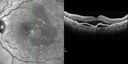
Polypoidal Choroidal Vasculopathy with Pulsating ICG polyp left eye714 viewsOct 11, 2014
|
|
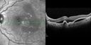
Polypoidal Choroidal Vasculopathy with Pulsating ICG polyp left eye633 viewsOct 11, 2014
|
|

Polypoidal Choroidal Vasculopathy with Pulsating ICG polyp left eye634 viewsOct 11, 2014
|
|

Polypoidal Choroidal Vasculopathy with Pulsating ICG polyp left eye581 viewsOct 11, 2014
|
|

Polypoidal Choroidal Vasculopathy with Pulsating ICG polyp left eye583 viewsOct 11, 2014
|
|

Polypoidal Choroidal Vasculopathy with Pulsating ICG polyp left eye574 viewsOct 11, 2014
|
|

Polypoidal Choroidal Vasculopathy with Pulsating ICG polyp left eye755 viewsOct 11, 2014
|
|
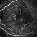
Polypoidal Choroidal Vasculopathy with Pulsating ICG polyp left eye748 viewsOct 11, 2014
|
|
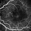
Polypoidal Choroidal Vasculopathy with Pulsating ICG polyp left eye639 viewsOct 11, 2014
|
|
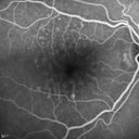
Polypoidal Choroidal Vasculopathy with Pulsating ICG polyp left eye669 viewsOct 11, 2014
|
|
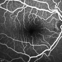
Polypoidal Choroidal Vasculopathy with Pulsating ICG polyp left eye613 viewsOct 11, 2014
|
|
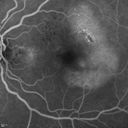
Polypoidal Choroidal Vasculopathy with Pulsating ICG polyp left eye658 viewsOct 11, 2014
|
|
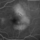
Polypoidal Choroidal Vasculopathy with Pulsating ICG polyp left eye612 viewsOct 11, 2014
|
|
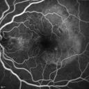
Polypoidal Choroidal Vasculopathy with Pulsating ICG polyp left eye560 viewsOct 11, 2014
|
|
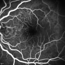
Polypoidal Choroidal Vasculopathy with Pulsating ICG polyp left eye579 viewsOct 11, 2014
|
|
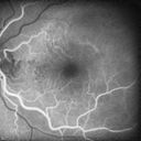
Polypoidal Choroidal Vasculopathy with Pulsating ICG polyp left eye575 viewsOct 11, 2014
|
|
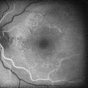
Polypoidal Choroidal Vasculopathy with Pulsating ICG polyp left eye559 viewsOct 11, 2014
|
|
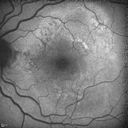
Polypoidal Choroidal Vasculopathy with Pulsating ICG polyp left eye611 viewsOct 11, 2014
|
|

Polypoidal Choroidal Vasculopathy with Pulsating ICG polyp left eye745 viewsOct 11, 2014
|
|

Polypoidal Choroidal Vasculopathy with Pulsating ICG polyp left eye695 viewsOct 11, 2014
|
|

Polypoidal Choroidal Vasculopathy with Pulsating ICG polyp left eye734 viewsOct 11, 2014
|
|

Polypoidal Choroidal Vasculopathy with Pulsating ICG polyp left eye597 viewsOct 11, 2014
|
|

Polypoidal Choroidal Vasculopathy with Pulsating ICG polyp left eye697 viewsOct 11, 2014
|
|

Polypoidal Choroidal Vasculopathy with Pulsating ICG polyp left eye658 viewsOct 11, 2014
|
|
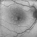
Polypoidal Choroidal Vasculopathy with Pulsating ICG polyp left eye568 viewsOct 11, 2014
|
|
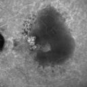
Polypoidal Choroidal Vasculopathy with Pulsating ICG polyp left eye635 viewsOct 11, 2014
|
|
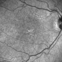
Polypoidal Choroidal Vasculopathy with Pulsating ICG polyp left eye569 viewsOct 11, 2014
|
|
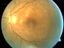
Polypoidal Choroidal Vasculopathy with Pulsating ICG polyp left eye562 viewsOct 11, 2014
|
|

Polypoidal Choroidal Vasculopathy with Pulsating ICG polyp left eye642 viewsOct 11, 2014
|
|
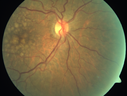
Polypoidal Choroidal Vasculopathy with Pulsating ICG polyp left eye570 viewsOct 11, 2014
|
|
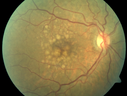
Polypoidal Choroidal Vasculopathy with Pulsating ICG polyp left eye691 viewsOct 11, 2014
|
|

Retinal Hemorrhage363 viewsFundus photo shows retinal hemorrhage located inferior, temporal to the macula in the left eye. Sep 16, 2014
|
|

Macular Degeneration497 viewsPatient complains of spot in the right eye. VA 20/400 in the right eye. Fundus photo shows macular degeneration changes with small layer of hemorrhage on the fovea in the right eye.Mar 28, 2014
|
|

Cartwheel CNVM - Classic - wet AMD - evidence of possible old CSR - ICG is normal - shows nothing553 views75 y.o Decreased Vision right eye for one year. VA OD: sc20/125+2 OS 20/25. Autofluorescence and SD OCT show evidence of old serous retinal detachment superior to the fovea suggesting a history of CSR. The macular lesion on FA shows a classic cartwheel appearanceNov 01, 2013
|
|

Cartwheel CNVM - Classic - wet AMD - evidence of possible old CSR - ICG is normal - shows nothing516 views75 y.o Decreased Vision right eye for one year. VA OD: sc20/125+2 OS 20/25. Autofluorescence and SD OCT show evidence of old serous retinal detachment superior to the fovea suggesting a history of CSR. The macular lesion on FA shows a classic cartwheel appearanceNov 01, 2013
|
|

Cartwheel CNVM - Classic - wet AMD - evidence of possible old CSR - ICG is normal - shows nothing549 views75 y.o Decreased Vision right eye for one year. VA OD: sc20/125+2 OS 20/25. Autofluorescence and SD OCT show evidence of old serous retinal detachment superior to the fovea suggesting a history of CSR. The macular lesion on FA shows a classic cartwheel appearanceNov 01, 2013
|
|
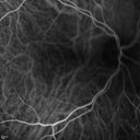
Cartwheel CNVM - Classic - wet AMD - evidence of possible old CSR - ICG is normal - shows nothing634 views75 y.o Decreased Vision right eye for one year. VA OD: sc20/125+2 OS 20/25. Autofluorescence and SD OCT show evidence of old serous retinal detachment superior to the fovea suggesting a history of CSR. The macular lesion on FA shows a classic cartwheel appearanceNov 01, 2013
|
|
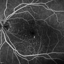
Cartwheel CNVM - Classic - wet AMD - evidence of possible old CSR - hydrophilic drusen stain early in FA582 views75 y.o Decreased Vision right eye for one year. VA OD: sc20/125+2 OS 20/25. Autofluorescence and SD OCT show evidence of old serous retinal detachment superior to the fovea suggesting a history of CSR. The macular lesion on FA shows a classic cartwheel appearanceNov 01, 2013
|
|
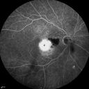
Cartwheel CNVM - Classic - wet AMD - evidence of possible old CSR - FA shows cartwheel shaped CNVM524 views75 y.o Decreased Vision right eye for one year. VA OD: sc20/125+2 OS 20/25. Autofluorescence and SD OCT show evidence of old serous retinal detachment superior to the fovea suggesting a history of CSR. The macular lesion on FA shows a classic cartwheel appearanceNov 01, 2013
|
|

Cartwheel CNVM - Classic - wet AMD - evidence of possible old CSR - FA shows cartwheel shaped CNVM710 views75 y.o Decreased Vision right eye for one year. VA OD: sc20/125+2 OS 20/25. Autofluorescence and SD OCT show evidence of old serous retinal detachment superior to the fovea suggesting a history of CSR. The macular lesion on FA shows a classic cartwheel appearanceNov 01, 2013
|
|

Cartwheel CNVM - Classic - wet AMD - evidence of possible old CSR - FA shows cartwheel shaped CNVM732 views75 y.o Decreased Vision right eye for one year. VA OD: sc20/125+2 OS 20/25. Autofluorescence and SD OCT show evidence of old serous retinal detachment superior to the fovea suggesting a history of CSR. The macular lesion on FA shows a classic cartwheel appearanceNov 01, 2013
|
|

Cartwheel CNVM - Classic - wet AMD - evidence of possible old CSR - FA shows cartwheel shaped CNVM714 views75 y.o Decreased Vision right eye for one year. VA OD: sc20/125+2 OS 20/25. Autofluorescence and SD OCT show evidence of old serous retinal detachment superior to the fovea suggesting a history of CSR. The macular lesion on FA shows a classic cartwheel appearanceNov 01, 2013
|
|

Cartwheel CNVM - Classic - wet AMD - evidence of possible old CSR583 views75 y.o Decreased Vision right eye for one year. VA OD: sc20/125+2 OS 20/25. Autofluorescence and SD OCT show evidence of old serous retinal detachment superior to the fovea suggesting a history of CSR. The macular lesion on FA shows a classic cartwheel appearanceNov 01, 2013
|
|
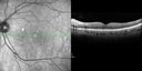
Cartwheel CNVM - Classic - wet AMD - evidence of possible old CSR557 views75 y.o Decreased Vision right eye for one year. VA OD: sc20/125+2 OS 20/25. Autofluorescence and SD OCT show evidence of old serous retinal detachment superior to the fovea suggesting a history of CSR. The macular lesion on FA shows a classic cartwheel appearanceNov 01, 2013
|
|

Cartwheel CNVM - Classic - wet AMD - evidence of possible old CSR868 views75 y.o Decreased Vision right eye for one year. VA OD: sc20/125+2 OS 20/25. Autofluorescence and SD OCT show evidence of old serous retinal detachment superior to the fovea suggesting a history of CSR. The macular lesion on FA shows a classic cartwheel appearanceNov 01, 2013
|
|

Cartwheel CNVM - Classic - wet AMD - evidence of possible old CSR - Autofluorescence shows brigh spot superior to subfoveal lesion897 views75 y.o Decreased Vision right eye for one year. VA OD: sc20/125+2 OS 20/25. Autofluorescence and SD OCT show evidence of old serous retinal detachment superior to the fovea suggesting a history of CSR. The macular lesion on FA shows a classic cartwheel appearanceNov 01, 2013
|
|
| 1175 files on 12 page(s) |
 |
|
|
|
|
|
8 | |
|
|
|


