Last additions - Macular Telangiectasis
|

415 viewsMay 04, 2020
|
|

423 viewsMay 04, 2020
|
|

405 viewsMay 04, 2020
|
|

423 viewsMay 04, 2020
|
|

Macular Telangiectasia - Mild440 views56 year old female with good vision and mild macular telangiectasia. VA 20/40 OD 20/16 OSDec 30, 2019
|
|

Macular Telangiectasia - Mild503 views56 year old female with good vision and mild macular telangiectasia. VA 20/40 OD 20/16 OSDec 30, 2019
|
|

Macular Telangiectasia - Mild403 views56 year old female with good vision and mild macular telangiectasia. VA 20/40 OD 20/16 OSDec 30, 2019
|
|

Macular Telangiectasia - Mild433 views56 year old female with good vision and mild macular telangiectasia. VA 20/40 OD 20/16 OSDec 30, 2019
|
|

Macular Telangiectasia - Mild400 views56 year old female with good vision and mild macular telangiectasia. VA 20/40 OD 20/16 OSDec 30, 2019
|
|

Macular Telangiectasia - Mild434 views56 year old female with good vision and mild macular telangiectasia. VA 20/40 OD 20/16 OSDec 30, 2019
|
|

Macular Telangiectasia - Mild493 views56 year old female with good vision and mild macular telangiectasia. VA 20/40 OD 20/16 OSDec 30, 2019
|
|

Macular Telangiectasia - Mild409 views56 year old female with good vision and mild macular telangiectasia. VA 20/40 OD 20/16 OSDec 30, 2019
|
|

Macular Telangiectasia - Mild421 views56 year old female with good vision and mild macular telangiectasia. VA 20/40 OD 20/16 OSDec 30, 2019
|
|

Macular Telangiectasia - Mild386 views56 year old female with good vision and mild macular telangiectasia. VA 20/40 OD 20/16 OSDec 30, 2019
|
|

Macular Telangiectasia - Mild465 views56 year old female with good vision and mild macular telangiectasia. VA 20/40 OD 20/16 OSDec 30, 2019
|
|

Macular Telangiectasia - Mild482 views56 year old female with good vision and mild macular telangiectasia. VA 20/40 OD 20/16 OSDec 30, 2019
|
|

Macular Telangiectasia - Mild438 views56 year old female with good vision and mild macular telangiectasia. VA 20/40 OD 20/16 OSDec 30, 2019
|
|

Macular Telangiectasia - Mild420 views56 year old female with good vision and mild macular telangiectasia. VA 20/40 OD 20/16 OSDec 30, 2019
|
|

Macular Telangiectasis and Vision loss following vitrectomy with membrane peel445 views76 year old man with MacTel who had membrane peel in the right eye 10 years ago with permanent vision loss.
VA OD: sc20/200-4 and VA OS: sc20/25-1Aug 07, 2019
|
|

Macular Telangiectasis and Vision loss following vitrectomy with membrane peel468 views76 year old man with MacTel who had membrane peel in the right eye 10 years ago with permanent vision loss.
VA OD: sc20/200-4 and VA OS: sc20/25-1Aug 07, 2019
|
|

Macular Telangiectasis and Vision loss following vitrectomy with membrane peel473 views76 year old man with MacTel who had membrane peel in the right eye 10 years ago with permanent vision loss.
VA OD: sc20/200-4 and VA OS: sc20/25-1Aug 07, 2019
|
|

Macular Telangiectasis and Vision loss following vitrectomy with membrane peel444 views76 year old man with MacTel who had membrane peel in the right eye 10 years ago with permanent vision loss.
VA OD: sc20/200-4 and VA OS: sc20/25-1Aug 07, 2019
|
|

Macular Telangiectasis and Vision loss following vitrectomy with membrane peel469 views76 year old man with MacTel who had membrane peel in the right eye 10 years ago with permanent vision loss.
VA OD: sc20/200-4 and VA OS: sc20/25-1Aug 07, 2019
|
|

Macular Telangiectasis and Vision loss following vitrectomy with membrane peel506 views76 year old man with MacTel who had membrane peel in the right eye 10 years ago with permanent vision loss.
VA OD: sc20/200-4 and VA OS: sc20/25-1Aug 07, 2019
|
|

Macular Telangiectasis and Vision loss following vitrectomy with membrane peel488 views76 year old man with MacTel who had membrane peel in the right eye 10 years ago with permanent vision loss.
VA OD: sc20/200-4 and VA OS: sc20/25-1Aug 07, 2019
|
|

Macular Telangiectasis and Vision loss following vitrectomy with membrane peel501 views76 year old man with MacTel who had membrane peel in the right eye 10 years ago with permanent vision loss.
VA OD: sc20/200-4 and VA OS: sc20/25-1Aug 07, 2019
|
|

Macular Telangiectasis and Vision loss following vitrectomy with membrane peel473 views76 year old man with MacTel who had membrane peel in the right eye 10 years ago with permanent vision loss.
VA OD: sc20/200-4 and VA OS: sc20/25-1Aug 07, 2019
|
|

Macular Telangiectasis and Vision loss following vitrectomy with membrane peel543 views76 year old man with MacTel who had membrane peel in the right eye 10 years ago with permanent vision loss.
VA OD: sc20/200-4 and VA OS: sc20/25-1Aug 07, 2019
|
|

Macular Telangiectasis and Vision loss following vitrectomy with membrane peel457 views76 year old man with MacTel who had membrane peel in the right eye 10 years ago with permanent vision loss.
VA OD: sc20/200-4 and VA OS: sc20/25-1Aug 07, 2019
|
|

Macular Telangiectasis and Vision loss following vitrectomy with membrane peel444 views76 year old man with MacTel who had membrane peel in the right eye 10 years ago with permanent vision loss.
VA OD: sc20/200-4 and VA OS: sc20/25-1Aug 07, 2019
|
|

Macular Telangiectasis and Vision loss following vitrectomy with membrane peel526 views76 year old man with MacTel who had membrane peel in the right eye 10 years ago with permanent vision loss.
VA OD: sc20/200-4 and VA OS: sc20/25-1Aug 07, 2019
|
|

Macular Telangiectasis and Vision loss following vitrectomy with membrane peel494 views76 year old man with MacTel who had membrane peel in the right eye 10 years ago with permanent vision loss.
VA OD: sc20/200-4 and VA OS: sc20/25-1Aug 07, 2019
|
|

Macular Telangiectasis - Mild - Retinal Crystals422 views58 year old man with long term mild vision loss. Considering Cataract surgery. VA 20/50 OD and 20/63 OS - Moderate cataract are presentJun 02, 2019
|
|

Macular Telangiectasis - Mild - Retinal Crystals485 views58 year old man with long term mild vision loss. Considering Cataract surgery. VA 20/50 OD and 20/63 OS - Moderate cataract are presentJun 02, 2019
|
|

Macular Telangiectasis - Mild - Retinal Crystals456 views58 year old man with long term mild vision loss. Considering Cataract surgery. VA 20/50 OD and 20/63 OS - Moderate cataract are presentJun 02, 2019
|
|

Macular Telangiectasis - Mild - Retinal Crystals424 views58 year old man with long term mild vision loss. Considering Cataract surgery. VA 20/50 OD and 20/63 OS - Moderate cataract are presentJun 02, 2019
|
|

Macular Telangiectasis - Mild - Retinal Crystals449 views58 year old man with long term mild vision loss. Considering Cataract surgery. VA 20/50 OD and 20/63 OS - Moderate cataract are presentJun 02, 2019
|
|

Macular Telangiectasis - Mild - Retinal Crystals437 views58 year old man with long term mild vision loss. Considering Cataract surgery. VA 20/50 OD and 20/63 OS - Moderate cataract are presentJun 02, 2019
|
|

Macular Telangiectasis - Mild - Retinal Crystals458 views58 year old man with long term mild vision loss. Considering Cataract surgery. VA 20/50 OD and 20/63 OS - Moderate cataract are presentJun 02, 2019
|
|

Macular Telangiectasis - Mild - Retinal Crystals479 views58 year old man with long term mild vision loss. Considering Cataract surgery. VA 20/50 OD and 20/63 OS - Moderate cataract are presentJun 02, 2019
|
|

Macular Telangiectasis - Mild - Retinal Crystals427 views58 year old man with long term mild vision loss. Considering Cataract surgery. VA 20/50 OD and 20/63 OS - Moderate cataract are presentJun 02, 2019
|
|

Macular Telangiectasis - Mild - Retinal Crystals428 views58 year old man with long term mild vision loss. Considering Cataract surgery. VA 20/50 OD and 20/63 OS - Moderate cataract are presentJun 02, 2019
|
|

Macular Telangiectasis - Very Mild413 views52 year old female with normal vision and suspicious OCT referred. Multimodal imaging shows very mild mactel (loss of macular pigment, ring on multicolor image and very mild OCT findings)Jan 28, 2019
|
|

Macular Telangiectasis - Very Mild547 views52 year old female with normal vision and suspicious OCT referred. Multimodal imaging shows very mild mactel (loss of macular pigment, ring on multicolor image and very mild OCT findings)Jan 28, 2019
|
|
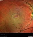
Macular Telangiectasis - Very Mild408 views52 year old female with normal vision and suspicious OCT referred. Multimodal imaging shows very mild mactel (loss of macular pigment, ring on multicolor image and very mild OCT findings)Jan 28, 2019
|
|

Macular Telangiectasis - Very Mild436 views52 year old female with normal vision and suspicious OCT referred. Multimodal imaging shows very mild mactel (loss of macular pigment, ring on multicolor image and very mild OCT findings)Jan 28, 2019
|
|

Macular Telangiectasis - Very Mild401 views52 year old female with normal vision and suspicious OCT referred. Multimodal imaging shows very mild mactel (loss of macular pigment, ring on multicolor image and very mild OCT findings)Jan 28, 2019
|
|

Macular Telangiectasis - Very Mild487 views52 year old female with normal vision and suspicious OCT referred. Multimodal imaging shows very mild mactel (loss of macular pigment, ring on multicolor image and very mild OCT findings)Jan 28, 2019
|
|
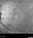
Macular Telangiectasis - Very Mild470 views52 year old female with normal vision and suspicious OCT referred. Multimodal imaging shows very mild mactel (loss of macular pigment, ring on multicolor image and very mild OCT findings)Jan 28, 2019
|
|

Macular Telangiectasis - Very Mild480 views52 year old female with normal vision and suspicious OCT referred. Multimodal imaging shows very mild mactel (loss of macular pigment, ring on multicolor image and very mild OCT findings)Jan 28, 2019
|
|
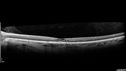
Macular Telangiectasis - Very Mild428 views52 year old female with normal vision and suspicious OCT referred. Multimodal imaging shows very mild mactel (loss of macular pigment, ring on multicolor image and very mild OCT findings)Jan 28, 2019
|
|

Macular Telangiectasis - Very Mild424 views52 year old female with normal vision and suspicious OCT referred. Multimodal imaging shows very mild mactel (loss of macular pigment, ring on multicolor image and very mild OCT findings)Jan 28, 2019
|
|

Macular Telangiectasis - Very Mild426 views52 year old female with normal vision and suspicious OCT referred. Multimodal imaging shows very mild mactel (loss of macular pigment, ring on multicolor image and very mild OCT findings)Jan 28, 2019
|
|
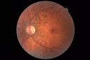
Macular Telangiectasis - Very Mild413 views52 year old female with normal vision and suspicious OCT referred. Multimodal imaging shows very mild mactel (loss of macular pigment, ring on multicolor image and very mild OCT findings)Jan 28, 2019
|
|

Macular Telangiectasis - Very Mild491 views52 year old female with normal vision and suspicious OCT referred. Multimodal imaging shows very mild mactel (loss of macular pigment, ring on multicolor image and very mild OCT findings)Jan 28, 2019
|
|
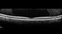
Macular Telangiectasis - Very Mild412 views52 year old female with normal vision and suspicious OCT referred. Multimodal imaging shows very mild mactel (loss of macular pigment, ring on multicolor image and very mild OCT findings)Jan 28, 2019
|
|

Macular Telangiectasis - Very Mild434 views52 year old female with normal vision and suspicious OCT referred. Multimodal imaging shows very mild mactel (loss of macular pigment, ring on multicolor image and very mild OCT findings)Jan 28, 2019
|
|

Macular Telangiectasis - Very Mild426 views52 year old female with normal vision and suspicious OCT referred. Multimodal imaging shows very mild mactel (loss of macular pigment, ring on multicolor image and very mild OCT findings)Jan 28, 2019
|
|

Macular Telangiectasis - Very Mild445 views52 year old female with normal vision and suspicious OCT referred. Multimodal imaging shows very mild mactel (loss of macular pigment, ring on multicolor image and very mild OCT findings)Jan 28, 2019
|
|

Macular Telangiectasis - Very Mild568 views52 year old female with normal vision and suspicious OCT referred. Multimodal imaging shows very mild mactel (loss of macular pigment, ring on multicolor image and very mild OCT findings)Jan 28, 2019
|
|

MacTel - Juxtafoveal Telangiectasis490 views56 year old man with a lot of difficulty adjusting to vision lossOct 16, 2018
|
|

MacTel - Juxtafoveal Telangiectasis425 views56 year old man with a lot of difficulty adjusting to vision lossOct 16, 2018
|
|

MacTel - Juxtafoveal Telangiectasis401 views56 year old man with a lot of difficulty adjusting to vision lossOct 16, 2018
|
|

MacTel - Juxtafoveal Telangiectasis420 views56 year old man with a lot of difficulty adjusting to vision lossOct 16, 2018
|
|

MacTel - Juxtafoveal Telangiectasis439 views56 year old man with a lot of difficulty adjusting to vision lossOct 16, 2018
|
|

MacTel - Juxtafoveal Telangiectasis464 views56 year old man with a lot of difficulty adjusting to vision lossOct 16, 2018
|
|

MacTel - Juxtafoveal Telangiectasis403 views56 year old man with a lot of difficulty adjusting to vision lossOct 16, 2018
|
|

MacTel - Juxtafoveal Telangiectasis447 views56 year old man with a lot of difficulty adjusting to vision lossOct 16, 2018
|
|

MacTel - Juxtafoveal Telangiectasis417 views56 year old man with a lot of difficulty adjusting to vision lossOct 16, 2018
|
|

MacTel - Juxtafoveal Telangiectasis443 views56 year old man with a lot of difficulty adjusting to vision lossOct 16, 2018
|
|

MacTel - Juxtafoveal Telangiectasis429 views56 year old man with a lot of difficulty adjusting to vision lossOct 16, 2018
|
|

MacTel - Juxtafoveal Telangiectasis453 views56 year old man with a lot of difficulty adjusting to vision lossOct 16, 2018
|
|

MacTel - Juxtafoveal Telangiectasis404 views56 year old man with a lot of difficulty adjusting to vision lossOct 16, 2018
|
|

MacTel - Juxtafoveal Telangiectasis460 views56 year old man with a lot of difficulty adjusting to vision lossOct 16, 2018
|
|

Macular Telangiectasis with Symptomatic Blind Spots458 views55 year old man with non-neovascular MacTel. VA 20/20 right eye 20/50 left eye. The color images show gray areas temporally. The FAF shows decreased macular pigment. The OCT shows outer retinal atrophy.Aug 06, 2017
|
|

Macular Telangiectasis with Symptomatic Blind Spots433 views55 year old man with non-neovascular MacTel. VA 20/20 right eye 20/50 left eye. The color images show gray areas temporally. The FAF shows decreased macular pigment. The OCT shows outer retinal atrophy.Aug 06, 2017
|
|

Macular Telangiectasis with Symptomatic Blind Spots448 views55 year old man with non-neovascular MacTel. VA 20/20 right eye 20/50 left eye. The color images show gray areas temporally. The FAF shows decreased macular pigment. The OCT shows outer retinal atrophy.Aug 06, 2017
|
|

Macular Telangiectasis with Symptomatic Blind Spots424 views55 year old man with non-neovascular MacTel. VA 20/20 right eye 20/50 left eye. The color images show gray areas temporally. The FAF shows decreased macular pigment. The OCT shows outer retinal atrophy.Aug 06, 2017
|
|

Macular Telangiectasis with Symptomatic Blind Spots440 views55 year old man with non-neovascular MacTel. VA 20/20 right eye 20/50 left eye. The color images show gray areas temporally. The FAF shows decreased macular pigment. The OCT shows outer retinal atrophy.Aug 06, 2017
|
|

Macular Telangiectasis with Symptomatic Blind Spots448 views55 year old man with non-neovascular MacTel. VA 20/20 right eye 20/50 left eye. The color images show gray areas temporally. The FAF shows decreased macular pigment. The OCT shows outer retinal atrophy.Aug 06, 2017
|
|

Macular Telangiectasis with Symptomatic Blind Spots493 views55 year old man with non-neovascular MacTel. VA 20/20 right eye 20/50 left eye. The color images show gray areas temporally. The FAF shows decreased macular pigment. The OCT shows outer retinal atrophy.Aug 06, 2017
|
|

Macular Telangiectasis with Symptomatic Blind Spots471 views55 year old man with non-neovascular MacTel. VA 20/20 right eye 20/50 left eye. The color images show gray areas temporally. The FAF shows decreased macular pigment. The OCT shows outer retinal atrophy.Aug 06, 2017
|
|

Macular Telangiectasis with Symptomatic Blind Spots495 views55 year old man with non-neovascular MacTel. VA 20/20 right eye 20/50 left eye. The color images show gray areas temporally. The FAF shows decreased macular pigment. The OCT shows outer retinal atrophy.Aug 06, 2017
|
|
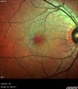
Macular Telangiectasis with Symptomatic Blind Spots582 views55 year old man with non-neovascular MacTel. VA 20/20 right eye 20/50 left eye. The color images show gray areas temporally. The FAF shows decreased macular pigment. The OCT shows outer retinal atrophy.Aug 06, 2017
|
|
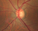
Macular Telangiectasis with Symptomatic Blind Spots423 views55 year old man with non-neovascular MacTel. VA 20/20 right eye 20/50 left eye. The color images show gray areas temporally. The FAF shows decreased macular pigment. The OCT shows outer retinal atrophy.Aug 06, 2017
|
|

Macular Telangiectasis with Symptomatic Blind Spots461 views55 year old man with non-neovascular MacTel. VA 20/20 right eye 20/50 left eye. The color images show gray areas temporally. The FAF shows decreased macular pigment. The OCT shows outer retinal atrophy.Aug 06, 2017
|
|

Macular Telangiectasis with Symptomatic Blind Spots506 views55 year old man with non-neovascular MacTel. VA 20/20 right eye 20/50 left eye. The color images show gray areas temporally. The FAF shows decreased macular pigment. The OCT shows outer retinal atrophy.Aug 06, 2017
|
|

Macular Telangiectasis - visible telangiectasia on color photos471 viewsMulticolor photos too show pathology. This patient had 20/50 vision with pinhole to 20/25Sep 29, 2016
|
|

Macular Telangiectasis - visible telangiectasia on color photos511 viewsMulticolor photos too show pathology. This patient had 20/50 vision with pinhole to 20/25Sep 29, 2016
|
|

Macular Telangiectasis - visible telangiectasia on color photos516 viewsMulticolor photos too show pathology. This patient had 20/50 vision with pinhole to 20/25Sep 29, 2016
|
|

Macular Telangiectasis - visible telangiectasia on color photos528 viewsMulticolor photos too show pathology. This patient had 20/50 vision with pinhole to 20/25Sep 29, 2016
|
|

Macular Telangiectasis - visible telangiectasia on color photos486 viewsMulticolor photos too show pathology. This patient had 20/50 vision with pinhole to 20/25Sep 29, 2016
|
|

Macular Telangiectasis - visible telangiectasia on color photos545 viewsMulticolor photos too show pathology. This patient had 20/50 vision with pinhole to 20/25Sep 29, 2016
|
|
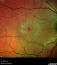
Macular Telangiectasis - visible telangiectasia on color photos471 viewsMulticolor photos too show pathology. This patient had 20/50 vision with pinhole to 20/25Sep 29, 2016
|
|

Macular Telangiectasis - visible telangiectasia on color photos1235 viewsMulticolor photos too show pathology. This patient had 20/50 vision with pinhole to 20/25Sep 29, 2016
|
|

Macular Telangiectasis - visible telangiectasia on color photos543 viewsMulticolor photos too show pathology. This patient had 20/50 vision with pinhole to 20/25Sep 29, 2016
|
|

Macular Telangiectasis - visible telangiectasia on color photos513 viewsMulticolor photos too show pathology. This patient had 20/50 vision with pinhole to 20/25Sep 29, 2016
|
|

Macular Telangiectasis - visible telangiectasia on color photos487 viewsMulticolor photos too show pathology. This patient had 20/50 vision with pinhole to 20/25Sep 29, 2016
|
|

Macular Telangiectasis - visible telangiectasia on color photos511 viewsMulticolor photos too show pathology. This patient had 20/50 vision with pinhole to 20/25Sep 29, 2016
|
|

Macular Telangiectasia and dry AMD - Simulating wet AMD566 viewsThis patient was told different things by different retina specialists. Her case is complex because she has OCT findings of Macular Telangiectasis and dry AMD. This makes it look like she has wet AMD. Her vision is stable.Sep 29, 2016
|
|
| 433 files on 5 page(s) |
2 |  |
|
|


