Last additions - Macular Degeneration - Age Related, Myopic, Choroidal Neovascular Membranes
|

Angioid Streaks - CNVM OS with vision loss469 views48 year old soccer player. Prior trauma OS (with soccer ball). Vision loss for 3 weeks to 20/60 - with Lucentis vision improved to 20/30Dec 18, 2018
|
|

Angioid Streaks - CNVM OS with vision loss395 views48 year old soccer player. Prior trauma OS (with soccer ball). Vision loss for 3 weeks to 20/60 - with Lucentis vision improved to 20/30Dec 18, 2018
|
|

Angioid Streaks - CNVM OS with vision loss412 views48 year old soccer player. Prior trauma OS (with soccer ball). Vision loss for 3 weeks to 20/60 - with Lucentis vision improved to 20/30Dec 18, 2018
|
|
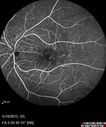
Angioid Streaks - CNVM OS with vision loss404 views48 year old soccer player. Prior trauma OS (with soccer ball). Vision loss for 3 weeks to 20/60 - with Lucentis vision improved to 20/30Dec 18, 2018
|
|

Angioid Streaks - CNVM OS with vision loss459 views48 year old soccer player. Prior trauma OS (with soccer ball). Vision loss for 3 weeks to 20/60 - with Lucentis vision improved to 20/30Dec 18, 2018
|
|
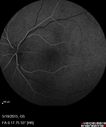
Angioid Streaks - CNVM OS with vision loss413 views48 year old soccer player. Prior trauma OS (with soccer ball). Vision loss for 3 weeks to 20/60 - with Lucentis vision improved to 20/30Dec 18, 2018
|
|
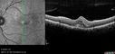
Angioid Streaks - CNVM OS with vision loss442 views48 year old soccer player. Prior trauma OS (with soccer ball). Vision loss for 3 weeks to 20/60 - with Lucentis vision improved to 20/30Dec 18, 2018
|
|

Angioid Streaks - CNVM OS with vision loss414 views48 year old soccer player. Prior trauma OS (with soccer ball). Vision loss for 3 weeks to 20/60 - with Lucentis vision improved to 20/30Dec 18, 2018
|
|

Angioid Streaks - CNVM OS with vision loss474 views48 year old soccer player. Prior trauma OS (with soccer ball). Vision loss for 3 weeks to 20/60 - with Lucentis vision improved to 20/30Dec 18, 2018
|
|

Angioid Streaks - CNVM OS with vision loss460 views48 year old soccer player. Prior trauma OS (with soccer ball). Vision loss for 3 weeks to 20/60 - with Lucentis vision improved to 20/30Dec 18, 2018
|
|

Angioid Streaks - CNVM OS with vision loss406 views48 year old soccer player. Prior trauma OS (with soccer ball). Vision loss for 3 weeks to 20/60 - with Lucentis vision improved to 20/30Dec 18, 2018
|
|

Angioid Streaks - CNVM OS with vision loss405 views48 year old soccer player. Prior trauma OS (with soccer ball). Vision loss for 3 weeks to 20/60 - with Lucentis vision improved to 20/30Dec 18, 2018
|
|

Angioid Streaks - CNVM OS with vision loss447 views48 year old soccer player. Prior trauma OS (with soccer ball). Vision loss for 3 weeks to 20/60 - with Lucentis vision improved to 20/30Dec 18, 2018
|
|
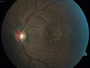
Angioid Streaks - CNVM OS with vision loss482 views48 year old soccer player. Prior trauma OS (with soccer ball). Vision loss for 3 weeks to 20/60 - with Lucentis vision improved to 20/30Dec 18, 2018
|
|

Pattern Dystrophy - Foveal Atrophy419 views73 year old female with vision loss - looks like macular holeNov 05, 2018
|
|

Pattern Dystrophy - Foveal Atrophy430 views73 year old female with vision loss - looks like macular holeNov 05, 2018
|
|

Pattern Dystrophy - Foveal Atrophy475 views73 year old female with vision loss - looks like macular holeNov 05, 2018
|
|

Pattern Dystrophy - Foveal Atrophy507 views73 year old female with vision loss - looks like macular holeNov 05, 2018
|
|

Pattern Dystrophy - Foveal Atrophy478 views73 year old female with vision loss - looks like macular holeNov 05, 2018
|
|

Pattern Dystrophy - Foveal Atrophy430 views73 year old female with vision loss - looks like macular holeNov 05, 2018
|
|

Pattern Dystrophy - Foveal Atrophy431 views73 year old female with vision loss - looks like macular holeNov 05, 2018
|
|

Pattern Dystrophy - Foveal Atrophy448 views73 year old female with vision loss - looks like macular holeNov 05, 2018
|
|

Pattern Dystrophy - Foveal Atrophy500 views73 year old female with vision loss - looks like macular holeNov 05, 2018
|
|

Pattern Dystrophy - Foveal Atrophy506 views73 year old female with vision loss - looks like macular holeNov 05, 2018
|
|

Pattern Dystrophy - Foveal Atrophy473 views73 year old female with vision loss - looks like macular holeNov 05, 2018
|
|

Pattern Dystrophy - Foveal Atrophy478 views73 year old female with vision loss - looks like macular holeNov 05, 2018
|
|

Pattern Dystrophy - Foveal Atrophy640 views73 year old female with vision loss - looks like macular holeNov 05, 2018
|
|

Pattern Dystrophy - Foveal Atrophy493 views73 year old female with vision loss - looks like macular holeNov 05, 2018
|
|

Pattern Dystrophy - Foveal Atrophy503 views73 year old female with vision loss - looks like macular holeNov 05, 2018
|
|

Pattern Dystrophy - Foveal Atrophy488 views73 year old female with vision loss - looks like macular holeNov 05, 2018
|
|

Outer Retinal Tubulation539 views78 year old man chronic wet AMD - 20/200Aug 29, 2018
|
|

Outer Retinal Tubulation400 views78 year old man chronic wet AMD - 20/200Aug 29, 2018
|
|

Outer Retinal Tubulation412 views78 year old man chronic wet AMD - 20/200Aug 29, 2018
|
|

Outer Retinal Tubulation435 views78 year old man chronic wet AMD - 20/200Aug 29, 2018
|
|

Outer Retinal Tubulation429 views78 year old man chronic wet AMD - 20/200Aug 29, 2018
|
|

Outer Retinal Tubulation460 views78 year old man chronic wet AMD - 20/200Aug 29, 2018
|
|

Outer Retinal Tubulation453 views78 year old man chronic wet AMD - 20/200Aug 29, 2018
|
|

Outer Retinal Tubulation431 views78 year old man chronic wet AMD - 20/200Aug 29, 2018
|
|

Outer Retinal Tubulation455 views78 year old man chronic wet AMD - 20/200Aug 29, 2018
|
|
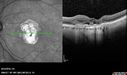
Outer Retinal Tubulation463 views78 year old man chronic wet AMD - 20/200Aug 29, 2018
|
|

Outer Retinal Tubulation445 views78 year old man chronic wet AMD - 20/200Aug 29, 2018
|
|

Outer Retinal Tubulation473 views78 year old man chronic wet AMD - 20/200Aug 29, 2018
|
|

Outer Retinal Tubulation439 views78 year old man chronic wet AMD - 20/200Aug 29, 2018
|
|

Outer Retinal Tubulation482 views78 year old man chronic wet AMD - 20/200Aug 29, 2018
|
|
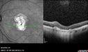
Outer Retinal Tubulation467 views78 year old man chronic wet AMD - 20/200Aug 29, 2018
|
|

Outer Retinal Tubulation419 views78 year old man chronic wet AMD - 20/200Aug 29, 2018
|
|

Outer Retinal Tubulation546 views78 year old man chronic wet AMD - 20/200Aug 29, 2018
|
|

Outer Retinal Tubulation467 views78 year old man chronic wet AMD - 20/200Aug 29, 2018
|
|

Outer Retinal Tubulation480 views78 year old man chronic wet AMD - 20/200Aug 29, 2018
|
|

Outer Retinal Tubulation462 views78 year old man chronic wet AMD - 20/200Aug 29, 2018
|
|

Outer Retinal Tubulation422 views78 year old man chronic wet AMD - 20/200Aug 29, 2018
|
|

Outer Retinal Tubulation436 views78 year old man chronic wet AMD - 20/200Aug 29, 2018
|
|

Outer Retinal Tubulation448 views78 year old man chronic wet AMD - 20/200Aug 29, 2018
|
|

Outer Retinal Tubulation468 views78 year old man chronic wet AMD - 20/200Aug 29, 2018
|
|

Outer Retinal Tubulation418 views78 year old man chronic wet AMD - 20/200Aug 29, 2018
|
|

Outer Retinal Tubulation453 views78 year old man chronic wet AMD - 20/200Aug 29, 2018
|
|

Outer Retinal Tubulation479 views78 year old man chronic wet AMD - 20/200Aug 29, 2018
|
|

Outer Retinal Tubulation490 views78 year old man chronic wet AMD - 20/200Aug 29, 2018
|
|

Outer Retinal Tubulation403 views78 year old man chronic wet AMD - 20/200Aug 29, 2018
|
|

Outer Retinal Tubulation431 views78 year old man chronic wet AMD - 20/200Aug 29, 2018
|
|

Outer Retinal Tubulation442 views78 year old man chronic wet AMD - 20/200Aug 29, 2018
|
|

Outer Retinal Tubulation480 views78 year old man chronic wet AMD - 20/200Aug 29, 2018
|
|

Outer Retinal Tubulation468 views78 year old man chronic wet AMD - 20/200Aug 29, 2018
|
|

Outer Retinal Tubulation456 views78 year old man chronic wet AMD - 20/200Aug 29, 2018
|
|

Outer Retinal Tubulation559 views78 year old man chronic wet AMD - 20/200Aug 29, 2018
|
|

Outer Retinal Tubulation502 views78 year old man chronic wet AMD - 20/200Aug 29, 2018
|
|

Outer Retinal Tubulation567 views78 year old man chronic wet AMD - 20/200Aug 29, 2018
|
|

Juxtapapillary CNVM - Hemorrhage423 views83 year old female VA 20/200 improved to 20/32 within 2 months with LucentisJul 27, 2018
|
|

Juxtapapillary CNVM - Hemorrhage408 views83 year old female VA 20/200 improved to 20/32 within 2 months with LucentisJul 27, 2018
|
|

Juxtapapillary CNVM - Hemorrhage413 views83 year old female VA 20/200 improved to 20/32 within 2 months with LucentisJul 27, 2018
|
|

Juxtapapillary CNVM - Hemorrhage417 views83 year old female VA 20/200 improved to 20/32 within 2 months with LucentisJul 27, 2018
|
|

Juxtapapillary CNVM - Hemorrhage415 views83 year old female VA 20/200 improved to 20/32 within 2 months with LucentisJul 27, 2018
|
|

Juxtapapillary CNVM - Hemorrhage502 views83 year old female VA 20/200 improved to 20/32 within 2 months with LucentisJul 27, 2018
|
|

Juxtapapillary CNVM - Hemorrhage404 views83 year old female VA 20/200 improved to 20/32 within 2 months with LucentisJul 27, 2018
|
|

Juxtapapillary CNVM - Hemorrhage418 views83 year old female VA 20/200 improved to 20/32 within 2 months with LucentisJul 27, 2018
|
|

Juxtapapillary CNVM - Hemorrhage410 views83 year old female VA 20/200 improved to 20/32 within 2 months with LucentisJul 27, 2018
|
|
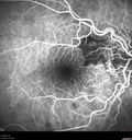
Juxtapapillary CNVM - Hemorrhage415 views83 year old female VA 20/200 improved to 20/32 within 2 months with LucentisJul 27, 2018
|
|

Juxtapapillary CNVM - Hemorrhage425 views83 year old female VA 20/200 improved to 20/32 within 2 months with LucentisJul 27, 2018
|
|

Juxtapapillary CNVM - Hemorrhage395 views83 year old female VA 20/200 improved to 20/32 within 2 months with LucentisJul 27, 2018
|
|

Juxtapapillary CNVM - Hemorrhage494 views83 year old female VA 20/200 improved to 20/32 within 2 months with LucentisJul 27, 2018
|
|

Juxtapapillary CNVM - Hemorrhage384 views83 year old female VA 20/200 improved to 20/32 within 2 months with LucentisJul 27, 2018
|
|

Juxtapapillary CNVM - Hemorrhage432 views83 year old female VA 20/200 improved to 20/32 within 2 months with LucentisJul 27, 2018
|
|

Juxtapapillary CNVM - Hemorrhage413 views83 year old female VA 20/200 improved to 20/32 within 2 months with LucentisJul 27, 2018
|
|

Juxtapapillary CNVM - Hemorrhage570 views83 year old female VA 20/200 improved to 20/32 within 2 months with LucentisJul 27, 2018
|
|

Juxtapapillary CNVM - Hemorrhage556 views83 year old female VA 20/200 improved to 20/32 within 2 months with LucentisJul 27, 2018
|
|

Polypoidal Choroidal Vasculopathy Left Eye455 views74 year old man with vision loss to 20/100 over a few months. He improved to 20/20 with Lucentis. The color and ICG show the polyps.Apr 17, 2018
|
|

Polypoidal Choroidal Vasculopathy Left Eye441 views74 year old man with vision loss to 20/100 over a few months. He improved to 20/20 with Lucentis. The color and ICG show the polyps.Apr 17, 2018
|
|

Polypoidal Choroidal Vasculopathy Left Eye452 views74 year old man with vision loss to 20/100 over a few months. He improved to 20/20 with Lucentis. The color and ICG show the polyps.Apr 17, 2018
|
|

Polypoidal Choroidal Vasculopathy Left Eye434 views74 year old man with vision loss to 20/100 over a few months. He improved to 20/20 with Lucentis. The color and ICG show the polyps.Apr 17, 2018
|
|

Polypoidal Choroidal Vasculopathy Left Eye434 views74 year old man with vision loss to 20/100 over a few months. He improved to 20/20 with Lucentis. The color and ICG show the polyps.Apr 17, 2018
|
|

Polypoidal Choroidal Vasculopathy Left Eye404 views74 year old man with vision loss to 20/100 over a few months. He improved to 20/20 with Lucentis. The color and ICG show the polyps.Apr 17, 2018
|
|

Polypoidal Choroidal Vasculopathy Left Eye446 views74 year old man with vision loss to 20/100 over a few months. He improved to 20/20 with Lucentis. The color and ICG show the polyps.Apr 17, 2018
|
|

Polypoidal Choroidal Vasculopathy Left Eye481 views74 year old man with vision loss to 20/100 over a few months. He improved to 20/20 with Lucentis. The color and ICG show the polyps.Apr 17, 2018
|
|

Polypoidal Choroidal Vasculopathy Left Eye492 views74 year old man with vision loss to 20/100 over a few months. He improved to 20/20 with Lucentis. The color and ICG show the polyps.Apr 17, 2018
|
|

Polypoidal Choroidal Vasculopathy Left Eye436 views74 year old man with vision loss to 20/100 over a few months. He improved to 20/20 with Lucentis. The color and ICG show the polyps.Apr 17, 2018
|
|

Polypoidal Choroidal Vasculopathy Left Eye444 views74 year old man with vision loss to 20/100 over a few months. He improved to 20/20 with Lucentis. The color and ICG show the polyps.Apr 17, 2018
|
|

Polypoidal Choroidal Vasculopathy Left Eye508 views74 year old man with vision loss to 20/100 over a few months. He improved to 20/20 with Lucentis. The color and ICG show the polyps.Apr 17, 2018
|
|

Polypoidal Choroidal Vasculopathy Left Eye534 views74 year old man with vision loss to 20/100 over a few months. He improved to 20/20 with Lucentis. The color and ICG show the polyps.Apr 17, 2018
|
|

Polypoidal Choroidal Vasculopathy Left Eye531 views74 year old man with vision loss to 20/100 over a few months. He improved to 20/20 with Lucentis. The color and ICG show the polyps.Apr 17, 2018
|
|

Polypoidal Choroidal Vasculopathy Left Eye406 views74 year old man with vision loss to 20/100 over a few months. He improved to 20/20 with Lucentis. The color and ICG show the polyps.Apr 17, 2018
|
|
| 2694 files on 27 page(s) |
 |
|
|
|
|
14 | |
|
|
|
|


