Derniers ajouts - Central Retinal Artery Occlusion
|
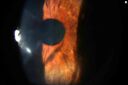
CRAO and NVI (rubeosis)vu 106 fois13 Août 2025
|
|
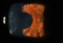
CRAO and NVI (rubeosis)vu 153 fois13 Août 2025
|
|

Central retinal artery occlusion and paracentral acute middle maculopathy and embolivu 359 fois86 year old female with sudden vision loss a week ago and imaging shows PAMM lesions and emboli. Her vision is 20/200 in the left eye01 Décembre 2022
|
|

Central retinal artery occlusion and paracentral acute middle maculopathy and embolivu 365 fois86 year old female with sudden vision loss a week ago and imaging shows PAMM lesions and emboli. Her vision is 20/200 in the left eye01 Décembre 2022
|
|

Central retinal artery occlusion and paracentral acute middle maculopathy and embolivu 356 fois86 year old female with sudden vision loss a week ago and imaging shows PAMM lesions and emboli. Her vision is 20/200 in the left eye01 Décembre 2022
|
|

Central retinal artery occlusion and paracentral acute middle maculopathy and embolivu 304 fois86 year old female with sudden vision loss a week ago and imaging shows PAMM lesions and emboli. Her vision is 20/200 in the left eye01 Décembre 2022
|
|

Central retinal artery occlusion and paracentral acute middle maculopathy and embolivu 306 fois86 year old female with sudden vision loss a week ago and imaging shows PAMM lesions and emboli. Her vision is 20/200 in the left eye01 Décembre 2022
|
|

Central retinal artery occlusion and paracentral acute middle maculopathy and embolivu 355 fois86 year old female with sudden vision loss a week ago and imaging shows PAMM lesions and emboli. Her vision is 20/200 in the left eye01 Décembre 2022
|
|

Central retinal artery occlusion and paracentral acute middle maculopathy and embolivu 328 fois86 year old female with sudden vision loss a week ago and imaging shows PAMM lesions and emboli. Her vision is 20/200 in the left eye01 Décembre 2022
|
|

Central retinal artery occlusion and paracentral acute middle maculopathy and embolivu 318 fois86 year old female with sudden vision loss a week ago and imaging shows PAMM lesions and emboli. Her vision is 20/200 in the left eye01 Décembre 2022
|
|
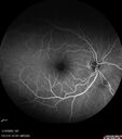
Central retinal artery occlusion and paracentral acute middle maculopathy and embolivu 351 fois86 year old female with sudden vision loss a week ago and imaging shows PAMM lesions and emboli. Her vision is 20/200 in the left eye01 Décembre 2022
|
|

Central retinal artery occlusion and paracentral acute middle maculopathy and embolivu 334 fois86 year old female with sudden vision loss a week ago and imaging shows PAMM lesions and emboli. Her vision is 20/200 in the left eye01 Décembre 2022
|
|

Central retinal artery occlusion and paracentral acute middle maculopathy and embolivu 294 fois86 year old female with sudden vision loss a week ago and imaging shows PAMM lesions and emboli. Her vision is 20/200 in the left eye01 Décembre 2022
|
|

Central retinal artery occlusion and paracentral acute middle maculopathy and embolivu 321 fois86 year old female with sudden vision loss a week ago and imaging shows PAMM lesions and emboli. Her vision is 20/200 in the left eye01 Décembre 2022
|
|

Central retinal artery occlusion and paracentral acute middle maculopathy and embolivu 298 fois86 year old female with sudden vision loss a week ago and imaging shows PAMM lesions and emboli. Her vision is 20/200 in the left eye01 Décembre 2022
|
|

Central retinal artery occlusion and paracentral acute middle maculopathy and embolivu 287 fois86 year old female with sudden vision loss a week ago and imaging shows PAMM lesions and emboli. Her vision is 20/200 in the left eye01 Décembre 2022
|
|

Central retinal artery occlusion and paracentral acute middle maculopathy and embolivu 326 fois86 year old female with sudden vision loss a week ago and imaging shows PAMM lesions and emboli. Her vision is 20/200 in the left eye01 Décembre 2022
|
|

Central retinal artery occlusion and paracentral acute middle maculopathy and embolivu 306 fois86 year old female with sudden vision loss a week ago and imaging shows PAMM lesions and emboli. Her vision is 20/200 in the left eye01 Décembre 2022
|
|

Central retinal artery occlusion and paracentral acute middle maculopathy and embolivu 344 fois86 year old female with sudden vision loss a week ago and imaging shows PAMM lesions and emboli. Her vision is 20/200 in the left eye01 Décembre 2022
|
|

Central retinal artery occlusion and paracentral acute middle maculopathy and embolivu 320 fois86 year old female with sudden vision loss a week ago and imaging shows PAMM lesions and emboli. Her vision is 20/200 in the left eye01 Décembre 2022
|
|

Central retinal artery occlusion and paracentral acute middle maculopathy and embolivu 306 fois86 year old female with sudden vision loss a week ago and imaging shows PAMM lesions and emboli. Her vision is 20/200 in the left eye01 Décembre 2022
|
|

Central Retinal Artery Occlusion - Fluorescein Angiogram Video showing slow fillvu 358 fois63 year old man Sudden severe vision loss in the left eye for 3 days. Six months following macula off retinal detachment repair, Pneumatic then PPV, then PPV buckle. VA was 20/63 on 4/2021 (3 months post RD repair).
PMHx – HTN. Meds: HCT, Meloxicam
VA OD 20/25, OS HM17 Octobre 2021
|
|

Central Retinal Artery Occlusion - Fluorescein Angiogram Videovu 362 fois63 year old man Sudden severe vision loss in the left eye for 3 days. Six months following macula off retinal detachment repair, Pneumatic then PPV, then PPV buckle. VA was 20/63 on 4/2021 (3 months post RD repair).
PMHx – HTN. Meds: HCT, Meloxicam
VA OD 20/25, OS HM17 Octobre 2021
|
|

Central Retinal Artery Occlusion - Fluorescein Angiogram Videovu 400 fois63 year old man Sudden severe vision loss in the left eye for 3 days. Six months following macula off retinal detachment repair, Pneumatic then PPV, then PPV buckle. VA was 20/63 on 4/2021 (3 months post RD repair).
PMHx – HTN. Meds: HCT, Meloxicam
VA OD 20/25, OS HM17 Octobre 2021
|
|

Central Retinal Artery Occlusion - Fluorescein Angiogram Videovu 339 fois63 year old man Sudden severe vision loss in the left eye for 3 days. Six months following macula off retinal detachment repair, Pneumatic then PPV, then PPV buckle. VA was 20/63 on 4/2021 (3 months post RD repair).
PMHx – HTN. Meds: HCT, Meloxicam
VA OD 20/25, OS HM17 Octobre 2021
|
|

Central Retinal Artery Occlusion - Fluorescein Angiogram Videovu 387 fois63 year old man Sudden severe vision loss in the left eye for 3 days. Six months following macula off retinal detachment repair, Pneumatic then PPV, then PPV buckle. VA was 20/63 on 4/2021 (3 months post RD repair).
PMHx – HTN. Meds: HCT, Meloxicam
VA OD 20/25, OS HM17 Octobre 2021
|
|

Central Retinal Artery Occlusion - Fluorescein Angiogram Videovu 351 fois63 year old man Sudden severe vision loss in the left eye for 3 days. Six months following macula off retinal detachment repair, Pneumatic then PPV, then PPV buckle. VA was 20/63 on 4/2021 (3 months post RD repair).
PMHx – HTN. Meds: HCT, Meloxicam
VA OD 20/25, OS HM17 Octobre 2021
|
|

Central Retinal Artery Occlusion - Fluorescein Angiogram Videovu 356 fois63 year old man Sudden severe vision loss in the left eye for 3 days. Six months following macula off retinal detachment repair, Pneumatic then PPV, then PPV buckle. VA was 20/63 on 4/2021 (3 months post RD repair).
PMHx – HTN. Meds: HCT, Meloxicam
VA OD 20/25, OS HM17 Octobre 2021
|
|

Central Retinal Artery Occlusion - Fluorescein Angiogram Videovu 377 fois63 year old man Sudden severe vision loss in the left eye for 3 days. Six months following macula off retinal detachment repair, Pneumatic then PPV, then PPV buckle. VA was 20/63 on 4/2021 (3 months post RD repair).
PMHx – HTN. Meds: HCT, Meloxicam
VA OD 20/25, OS HM17 Octobre 2021
|
|

Central Retinal Artery Occlusion - Fluorescein Angiogram Videovu 387 fois63 year old man Sudden severe vision loss in the left eye for 3 days. Six months following macula off retinal detachment repair, Pneumatic then PPV, then PPV buckle. VA was 20/63 on 4/2021 (3 months post RD repair).
PMHx – HTN. Meds: HCT, Meloxicam
VA OD 20/25, OS HM17 Octobre 2021
|
|

Central Retinal Artery Occlusion - Fluorescein Angiogram Videovu 377 fois63 year old man Sudden severe vision loss in the left eye for 3 days. Six months following macula off retinal detachment repair, Pneumatic then PPV, then PPV buckle. VA was 20/63 on 4/2021 (3 months post RD repair).
PMHx – HTN. Meds: HCT, Meloxicam
VA OD 20/25, OS HM17 Octobre 2021
|
|

Central Retinal Artery Occlusion - Fluorescein Angiogram Videovu 334 fois63 year old man Sudden severe vision loss in the left eye for 3 days. Six months following macula off retinal detachment repair, Pneumatic then PPV, then PPV buckle. VA was 20/63 on 4/2021 (3 months post RD repair).
PMHx – HTN. Meds: HCT, Meloxicam
VA OD 20/25, OS HM17 Octobre 2021
|
|

Central Retinal Artery Occlusion - Fluorescein Angiogram Videovu 353 fois63 year old man Sudden severe vision loss in the left eye for 3 days. Six months following macula off retinal detachment repair, Pneumatic then PPV, then PPV buckle. VA was 20/63 on 4/2021 (3 months post RD repair).
PMHx – HTN. Meds: HCT, Meloxicam
VA OD 20/25, OS HM17 Octobre 2021
|
|

Central Retinal Artery Occlusionvu 390 fois80 Year old man Sudden severe vision loss about 2 weeks ago. Today he can see light for the first time. The right eye is OK. His neuroimaging showed a brain stroke along with the eye stroke. He was stepping in the shower and the vision was suddenly worse. He went, later, to the ER and had a full evaluation. VA 20/25 right eye, LP left eye22 Février 2021
|
|
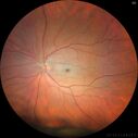
Central Retinal Artery Occlusionvu 481 fois80 Year old man Sudden severe vision loss about 2 weeks ago. Today he can see light for the first time. The right eye is OK. His neuroimaging showed a brain stroke along with the eye stroke. He was stepping in the shower and the vision was suddenly worse. He went, later, to the ER and had a full evaluation. VA 20/25 right eye, LP left eye22 Février 2021
|
|
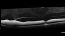
Central Retinal Artery Occlusionvu 378 fois80 Year old man Sudden severe vision loss about 2 weeks ago. Today he can see light for the first time. The right eye is OK. His neuroimaging showed a brain stroke along with the eye stroke. He was stepping in the shower and the vision was suddenly worse. He went, later, to the ER and had a full evaluation. VA 20/25 right eye, LP left eye22 Février 2021
|
|

Central Retinal Artery Occlusionvu 408 fois80 Year old man Sudden severe vision loss about 2 weeks ago. Today he can see light for the first time. The right eye is OK. His neuroimaging showed a brain stroke along with the eye stroke. He was stepping in the shower and the vision was suddenly worse. He went, later, to the ER and had a full evaluation. VA 20/25 right eye, LP left eye22 Février 2021
|
|

Central Retinal Artery Occlusionvu 368 fois80 Year old man Sudden severe vision loss about 2 weeks ago. Today he can see light for the first time. The right eye is OK. His neuroimaging showed a brain stroke along with the eye stroke. He was stepping in the shower and the vision was suddenly worse. He went, later, to the ER and had a full evaluation. VA 20/25 right eye, LP left eye22 Février 2021
|
|

Central Retinal Artery Occlusionvu 410 fois80 Year old man Sudden severe vision loss about 2 weeks ago. Today he can see light for the first time. The right eye is OK. His neuroimaging showed a brain stroke along with the eye stroke. He was stepping in the shower and the vision was suddenly worse. He went, later, to the ER and had a full evaluation. VA 20/25 right eye, LP left eye22 Février 2021
|
|

vu 426 fois23 Avril 2020
|
|

vu 406 fois23 Avril 2020
|
|

vu 396 fois23 Avril 2020
|
|

vu 435 fois23 Avril 2020
|
|

vu 432 fois23 Avril 2020
|
|

vu 442 fois23 Avril 2020
|
|

vu 429 fois23 Avril 2020
|
|

vu 392 fois23 Avril 2020
|
|

vu 412 fois23 Avril 2020
|
|

vu 455 fois23 Avril 2020
|
|

vu 458 fois23 Avril 2020
|
|

vu 414 fois23 Avril 2020
|
|

vu 422 fois23 Avril 2020
|
|

vu 450 fois23 Avril 2020
|
|

vu 406 fois23 Avril 2020
|
|

vu 439 fois23 Avril 2020
|
|

vu 469 fois23 Avril 2020
|
|

vu 435 fois23 Avril 2020
|
|

vu 408 fois23 Avril 2020
|
|

vu 490 fois23 Avril 2020
|
|

vu 432 fois23 Avril 2020
|
|

Acute Central Retinal Artery Occlusionvu 428 fois1 day of vision loss 79 year old man to HM. NVG developed 2 months later.26 Mars 2019
|
|

Acute Central Retinal Artery Occlusionvu 477 fois1 day of vision loss 79 year old man to HM. NVG developed 2 months later.26 Mars 2019
|
|

Acute Central Retinal Artery Occlusionvu 437 fois1 day of vision loss 79 year old man to HM. NVG developed 2 months later.26 Mars 2019
|
|

Acute Central Retinal Artery Occlusionvu 465 fois1 day of vision loss 79 year old man to HM. NVG developed 2 months later.26 Mars 2019
|
|

Acute Central Retinal Artery Occlusionvu 446 fois1 day of vision loss 79 year old man to HM. NVG developed 2 months later.26 Mars 2019
|
|

Acute Central Retinal Artery Occlusionvu 459 fois1 day of vision loss 79 year old man to HM. NVG developed 2 months later.26 Mars 2019
|
|

Acute Central Retinal Artery Occlusionvu 440 fois1 day of vision loss 79 year old man to HM. NVG developed 2 months later.26 Mars 2019
|
|

Acute Central Retinal Artery Occlusionvu 403 fois1 day of vision loss 79 year old man to HM. NVG developed 2 months later.26 Mars 2019
|
|

Acute Central Retinal Artery Occlusionvu 462 fois1 day of vision loss 79 year old man to HM. NVG developed 2 months later.26 Mars 2019
|
|

Acute Central Retinal Artery Occlusionvu 493 fois1 day of vision loss 79 year old man to HM. NVG developed 2 months later.26 Mars 2019
|
|

Acute Central Retinal Artery Occlusionvu 445 fois1 day of vision loss 79 year old man to HM. NVG developed 2 months later.26 Mars 2019
|
|

Acute Central Retinal Artery Occlusionvu 506 fois1 day of vision loss 79 year old man to HM. NVG developed 2 months later.26 Mars 2019
|
|

Central Retinal Artery Occlusion - Diabetic Retinopathy - NO cherry red spotvu 446 fois58 year old man with sudden vision loss in the right eye. The vision had been 20/40 and dropped to 20/200 the day prior to presentation.
VA 20/200 OD, 20/20 OS
FA shows CRAO. No cherry red spot.27 Février 2019
|
|

Central Retinal Artery Occlusion - Diabetic Retinopathy - NO cherry red spotvu 532 fois58 year old man with sudden vision loss in the right eye. The vision had been 20/40 and dropped to 20/200 the day prior to presentation.
VA 20/200 OD, 20/20 OS
FA shows CRAO. No cherry red spot.27 Février 2019
|
|

Central Retinal Artery Occlusion - Diabetic Retinopathy - NO cherry red spotvu 471 fois58 year old man with sudden vision loss in the right eye. The vision had been 20/40 and dropped to 20/200 the day prior to presentation.
VA 20/200 OD, 20/20 OS
FA shows CRAO. No cherry red spot.27 Février 2019
|
|

Central Retinal Artery Occlusion - Diabetic Retinopathy - NO cherry red spotvu 527 fois58 year old man with sudden vision loss in the right eye. The vision had been 20/40 and dropped to 20/200 the day prior to presentation.
VA 20/200 OD, 20/20 OS
FA shows CRAO. No cherry red spot.27 Février 2019
|
|
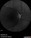
Central Retinal Artery Occlusion - Diabetic Retinopathy - NO cherry red spotvu 491 fois58 year old man with sudden vision loss in the right eye. The vision had been 20/40 and dropped to 20/200 the day prior to presentation.
VA 20/200 OD, 20/20 OS
FA shows CRAO. No cherry red spot.27 Février 2019
|
|

Central Retinal Artery Occlusion - Diabetic Retinopathy - NO cherry red spotvu 547 fois58 year old man with sudden vision loss in the right eye. The vision had been 20/40 and dropped to 20/200 the day prior to presentation.
VA 20/200 OD, 20/20 OS
FA shows CRAO. No cherry red spot.27 Février 2019
|
|

Central Retinal Artery Occlusion - Diabetic Retinopathy - NO cherry red spotvu 478 fois58 year old man with sudden vision loss in the right eye. The vision had been 20/40 and dropped to 20/200 the day prior to presentation.
VA 20/200 OD, 20/20 OS
FA shows CRAO. No cherry red spot.27 Février 2019
|
|

Central Retinal Artery Occlusion - Diabetic Retinopathy - NO cherry red spotvu 520 fois58 year old man with sudden vision loss in the right eye. The vision had been 20/40 and dropped to 20/200 the day prior to presentation.
VA 20/200 OD, 20/20 OS
FA shows CRAO. No cherry red spot.27 Février 2019
|
|

Central Retinal Artery Occlusion - Diabetic Retinopathy - NO cherry red spotvu 439 fois58 year old man with sudden vision loss in the right eye. The vision had been 20/40 and dropped to 20/200 the day prior to presentation.
VA 20/200 OD, 20/20 OS
FA shows CRAO. No cherry red spot.27 Février 2019
|
|

Central Retinal Artery Occlusion - Diabetic Retinopathy - NO cherry red spotvu 556 fois58 year old man with sudden vision loss in the right eye. The vision had been 20/40 and dropped to 20/200 the day prior to presentation.
VA 20/200 OD, 20/20 OS
FA shows CRAO. No cherry red spot.27 Février 2019
|
|

Central Retinal Artery Occlusion - Diabetic Retinopathy - NO cherry red spotvu 570 fois58 year old man with sudden vision loss in the right eye. The vision had been 20/40 and dropped to 20/200 the day prior to presentation.
VA 20/200 OD, 20/20 OS
FA shows CRAO. No cherry red spot.27 Février 2019
|
|

Central Retinal Artery Occlusion - Diabetic Retinopathy - NO cherry red spotvu 617 fois58 year old man with sudden vision loss in the right eye. The vision had been 20/40 and dropped to 20/200 the day prior to presentation.
VA 20/200 OD, 20/20 OS
FA shows CRAO. No cherry red spot.27 Février 2019
|
|

Central Retinal Artery Occlusion then Neovascular Glaucomavu 495 fois81 year old male with sudden vision loss. 2 months later developed NVG27 Juillet 2018
|
|

Central Retinal Artery Occlusion then Neovascular Glaucomavu 565 fois81 year old male with sudden vision loss. 2 months later developed NVG27 Juillet 2018
|
|

Central Retinal Artery Occlusion then Neovascular Glaucomavu 463 fois81 year old male with sudden vision loss. 2 months later developed NVG27 Juillet 2018
|
|

Central Retinal Artery Occlusion then Neovascular Glaucomavu 424 fois81 year old male with sudden vision loss. 2 months later developed NVG27 Juillet 2018
|
|

Central Retinal Artery Occlusion then Neovascular Glaucomavu 412 fois81 year old male with sudden vision loss. 2 months later developed NVG27 Juillet 2018
|
|

Central Retinal Artery Occlusion then Neovascular Glaucomavu 508 fois81 year old male with sudden vision loss. 2 months later developed NVG27 Juillet 2018
|
|

Central Retinal Artery Occlusion then Neovascular Glaucomavu 543 fois81 year old male with sudden vision loss. 2 months later developed NVG27 Juillet 2018
|
|

Central Retinal Artery Occlusion then Neovascular Glaucomavu 435 fois81 year old male with sudden vision loss. 2 months later developed NVG27 Juillet 2018
|
|

Central Retinal Artery Occlusion then Neovascular Glaucomavu 496 fois81 year old male with sudden vision loss. 2 months later developed NVG27 Juillet 2018
|
|

Central Retinal Artery Occlusion then Neovascular Glaucomavu 490 fois81 year old male with sudden vision loss. 2 months later developed NVG27 Juillet 2018
|
|

Central Retinal Artery Occlusion - MAPvu 755 foisProgression from acute to atrophy - decreasing retinal thickenss28 Novembre 2016
|
|

Retinal Artery Occlusionvu 1182 foisPatient comes in for eval on artery occlusions in both eyes. VA is 20/400, right eye and NLP, left eye. Fundus photos show paniretinal scars in the right eye with arterial narrowing and the left eye has arterial narrowing as well.23 Septembre 2014
|
|
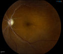
Retinal Artery Occlusionvu 1688 foisPatient comes in for eval on artery occlusions in both eyes. VA is 20/400, right eye and NLP, left eye. Fundus photos show paniretinal scars in the right eye with arterial narrowing and the left eye has arterial narrowing as well.23 Septembre 2014
|
|

Retinal Artery Occlusionvu 1023 foisPatient comes in for eval on artery occlusions in both eyes. VA is 20/400, right eye and NLP, left eye. Fundus photos show paniretinal scars in the right eye with arterial narrowing and the left eye has arterial narrowing as well.23 Septembre 2014
|
|

Retinal Artery Occlusionvu 589 foisPatient comes in for eval on artery occlusions in both eyes. VA is 20/400, right eye and NLP, left eye. Fundus photos show paniretinal scars in the right eye with arterial narrowing and the left eye has arterial narrowing as well.23 Septembre 2014
|
|

Central Retinal Artery Occlusionvu 1097 foisCentral Retinal Artery Occlusion with cilio artery perfusion. 15 Mai 2014
|
|
| 192 photos sur 2 page(s) |
1 |
|


