Last additions - Diabetic Retinopathy
|

Proliferative Diabetic Retinopathy - NVD regressed with PRP laser497 views76 year old diabetic man - Presented one year ago with NVD in the left eye. This regressed with laser but then a year later worsened. Additional PRP was done and the NVD regressed again.Jun 02, 2017
|
|

Proliferative Diabetic Retinopathy - NVD regressed with PRP laser562 views76 year old diabetic man - Presented one year ago with NVD in the left eye. This regressed with laser but then a year later worsened. Additional PRP was done and the NVD regressed again.Jun 02, 2017
|
|

Proliferative Diabetic Retinopathy - NVD regressed with PRP laser522 views76 year old diabetic man - Presented one year ago with NVD in the left eye. This regressed with laser but then a year later worsened. Additional PRP was done and the NVD regressed again.Jun 02, 2017
|
|

Proliferative Diabetic Retinopathy - NVD regressed with PRP laser454 views76 year old diabetic man - Presented one year ago with NVD in the left eye. This regressed with laser but then a year later worsened. Additional PRP was done and the NVD regressed again.Jun 02, 2017
|
|

Proliferative Diabetic Retinopathy - NVD regressed with PRP laser432 views76 year old diabetic man - Presented one year ago with NVD in the left eye. This regressed with laser but then a year later worsened. Additional PRP was done and the NVD regressed again.Jun 02, 2017
|
|

Proliferative Diabetic Retinopathy - NVD regressed with PRP laser424 views76 year old diabetic man - Presented one year ago with NVD in the left eye. This regressed with laser but then a year later worsened. Additional PRP was done and the NVD regressed again.Jun 02, 2017
|
|

Proliferative Diabetic Retinopathy - NVD regressed with PRP laser563 views76 year old diabetic man - Presented one year ago with NVD in the left eye. This regressed with laser but then a year later worsened. Additional PRP was done and the NVD regressed again.Jun 02, 2017
|
|

Proliferative Diabetic Retinopathy - NVD regressed with PRP laser489 views76 year old diabetic man - Presented one year ago with NVD in the left eye. This regressed with laser but then a year later worsened. Additional PRP was done and the NVD regressed again.Jun 02, 2017
|
|

Proliferative Diabetic Retinopathy - NVD regressed with PRP laser494 views76 year old diabetic man - Presented one year ago with NVD in the left eye. This regressed with laser but then a year later worsened. Additional PRP was done and the NVD regressed again.Jun 02, 2017
|
|

Proliferative Diabetic Retinopathy - NVD regressed with PRP laser503 views76 year old diabetic man - Presented one year ago with NVD in the left eye. This regressed with laser but then a year later worsened. Additional PRP was done and the NVD regressed again.Jun 02, 2017
|
|

Proliferative Diabetic Retinopathy - NVD regressed with PRP laser411 views76 year old diabetic man - Presented one year ago with NVD in the left eye. This regressed with laser but then a year later worsened. Additional PRP was done and the NVD regressed again.Jun 02, 2017
|
|

Proliferative Diabetic Retinopathy - NVD regressed with PRP laser437 views76 year old diabetic man - Presented one year ago with NVD in the left eye. This regressed with laser but then a year later worsened. Additional PRP was done and the NVD regressed again.Jun 02, 2017
|
|

Proliferative Diabetic Retinopathy - NVD regressed with PRP laser453 views76 year old diabetic man - Presented one year ago with NVD in the left eye. This regressed with laser but then a year later worsened. Additional PRP was done and the NVD regressed again.Jun 02, 2017
|
|
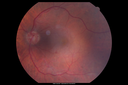
Proliferative Diabetic Retinopathy - NVD regressed with PRP laser524 views76 year old diabetic man - Presented one year ago with NVD in the left eye. This regressed with laser but then a year later worsened. Additional PRP was done and the NVD regressed again.Jun 02, 2017
|
|

Proliferative Diabetic Retinopathy - NVD regressed with PRP laser525 views76 year old diabetic man - Presented one year ago with NVD in the left eye. This regressed with laser but then a year later worsened. Additional PRP was done and the NVD regressed again.Jun 02, 2017
|
|

Proliferative Diabetic Retinopathy - NVD regressed with PRP laser684 views76 year old diabetic man - Presented one year ago with NVD in the left eye. This regressed with laser but then a year later worsened. Additional PRP was done and the NVD regressed again.Jun 02, 2017
|
|

PDR NVD left eye and NVE right eye632 views41 year old diabetic woman with PDR in both eyes. High risk in the left eye. She had PRP, Avastin, and vitrectomy x 2 in the left eye with final vision of 20/25. The right eye had eventually PRP laserApr 07, 2017
|
|
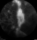
PDR NVD left eye and NVE right eye457 views41 year old diabetic woman with PDR in both eyes. High risk in the left eye. She had PRP, Avastin, and vitrectomy x 2 in the left eye with final vision of 20/25. The right eye had eventually PRP laserApr 07, 2017
|
|

PDR NVD left eye and NVE right eye460 views41 year old diabetic woman with PDR in both eyes. High risk in the left eye. She had PRP, Avastin, and vitrectomy x 2 in the left eye with final vision of 20/25. The right eye had eventually PRP laserApr 07, 2017
|
|

PDR NVD left eye and NVE right eye500 views41 year old diabetic woman with PDR in both eyes. High risk in the left eye. She had PRP, Avastin, and vitrectomy x 2 in the left eye with final vision of 20/25. The right eye had eventually PRP laserApr 07, 2017
|
|

PDR NVD left eye and NVE right eye519 views41 year old diabetic woman with PDR in both eyes. High risk in the left eye. She had PRP, Avastin, and vitrectomy x 2 in the left eye with final vision of 20/25. The right eye had eventually PRP laserApr 07, 2017
|
|

PDR NVD left eye and NVE right eye531 views41 year old diabetic woman with PDR in both eyes. High risk in the left eye. She had PRP, Avastin, and vitrectomy x 2 in the left eye with final vision of 20/25. The right eye had eventually PRP laserApr 07, 2017
|
|

PDR NVD left eye and NVE right eye529 views41 year old diabetic woman with PDR in both eyes. High risk in the left eye. She had PRP, Avastin, and vitrectomy x 2 in the left eye with final vision of 20/25. The right eye had eventually PRP laserApr 07, 2017
|
|

PDR NVD left eye and NVE right eye448 views41 year old diabetic woman with PDR in both eyes. High risk in the left eye. She had PRP, Avastin, and vitrectomy x 2 in the left eye with final vision of 20/25. The right eye had eventually PRP laserApr 07, 2017
|
|

PDR NVD left eye and NVE right eye477 views41 year old diabetic woman with PDR in both eyes. High risk in the left eye. She had PRP, Avastin, and vitrectomy x 2 in the left eye with final vision of 20/25. The right eye had eventually PRP laserApr 07, 2017
|
|

PDR NVD left eye and NVE right eye658 views41 year old diabetic woman with PDR in both eyes. High risk in the left eye. She had PRP, Avastin, and vitrectomy x 2 in the left eye with final vision of 20/25. The right eye had eventually PRP laserApr 07, 2017
|
|

PDR NVD left eye and NVE right eye515 views41 year old diabetic woman with PDR in both eyes. High risk in the left eye. She had PRP, Avastin, and vitrectomy x 2 in the left eye with final vision of 20/25. The right eye had eventually PRP laserApr 07, 2017
|
|

PDR NVD left eye and NVE right eye481 views41 year old diabetic woman with PDR in both eyes. High risk in the left eye. She had PRP, Avastin, and vitrectomy x 2 in the left eye with final vision of 20/25. The right eye had eventually PRP laserApr 07, 2017
|
|

PDR NVD left eye and NVE right eye434 views41 year old diabetic woman with PDR in both eyes. High risk in the left eye. She had PRP, Avastin, and vitrectomy x 2 in the left eye with final vision of 20/25. The right eye had eventually PRP laserApr 07, 2017
|
|

PDR NVD left eye and NVE right eye473 views41 year old diabetic woman with PDR in both eyes. High risk in the left eye. She had PRP, Avastin, and vitrectomy x 2 in the left eye with final vision of 20/25. The right eye had eventually PRP laserApr 07, 2017
|
|

PDR NVD left eye and NVE right eye469 views41 year old diabetic woman with PDR in both eyes. High risk in the left eye. She had PRP, Avastin, and vitrectomy x 2 in the left eye with final vision of 20/25. The right eye had eventually PRP laserApr 07, 2017
|
|

PDR NVD left eye and NVE right eye459 views41 year old diabetic woman with PDR in both eyes. High risk in the left eye. She had PRP, Avastin, and vitrectomy x 2 in the left eye with final vision of 20/25. The right eye had eventually PRP laserApr 07, 2017
|
|

PDR NVD left eye and NVE right eye521 views41 year old diabetic woman with PDR in both eyes. High risk in the left eye. She had PRP, Avastin, and vitrectomy x 2 in the left eye with final vision of 20/25. The right eye had eventually PRP laserApr 07, 2017
|
|

PDR NVD left eye and NVE right eye462 views41 year old diabetic woman with PDR in both eyes. High risk in the left eye. She had PRP, Avastin, and vitrectomy x 2 in the left eye with final vision of 20/25. The right eye had eventually PRP laserApr 07, 2017
|
|

PDR NVD left eye and NVE right eye504 views41 year old diabetic woman with PDR in both eyes. High risk in the left eye. She had PRP, Avastin, and vitrectomy x 2 in the left eye with final vision of 20/25. The right eye had eventually PRP laserApr 07, 2017
|
|

PDR NVD left eye and NVE right eye446 views41 year old diabetic woman with PDR in both eyes. High risk in the left eye. She had PRP, Avastin, and vitrectomy x 2 in the left eye with final vision of 20/25. The right eye had eventually PRP laserApr 07, 2017
|
|

PDR NVD left eye and NVE right eye464 views41 year old diabetic woman with PDR in both eyes. High risk in the left eye. She had PRP, Avastin, and vitrectomy x 2 in the left eye with final vision of 20/25. The right eye had eventually PRP laserApr 07, 2017
|
|

PDR NVD left eye and NVE right eye502 views41 year old diabetic woman with PDR in both eyes. High risk in the left eye. She had PRP, Avastin, and vitrectomy x 2 in the left eye with final vision of 20/25. The right eye had eventually PRP laserApr 07, 2017
|
|

PDR NVD left eye and NVE right eye505 views41 year old diabetic woman with PDR in both eyes. High risk in the left eye. She had PRP, Avastin, and vitrectomy x 2 in the left eye with final vision of 20/25. The right eye had eventually PRP laserApr 07, 2017
|
|

PDR NVD left eye and NVE right eye505 views41 year old diabetic woman with PDR in both eyes. High risk in the left eye. She had PRP, Avastin, and vitrectomy x 2 in the left eye with final vision of 20/25. The right eye had eventually PRP laserApr 07, 2017
|
|

PDR NVD left eye and NVE right eye754 views41 year old diabetic woman with PDR in both eyes. High risk in the left eye. She had PRP, Avastin, and vitrectomy x 2 in the left eye with final vision of 20/25. The right eye had eventually PRP laserApr 07, 2017
|
|

PDR NVD left eye and NVE right eye555 views41 year old diabetic woman with PDR in both eyes. High risk in the left eye. She had PRP, Avastin, and vitrectomy x 2 in the left eye with final vision of 20/25. The right eye had eventually PRP laserApr 07, 2017
|
|

PDR NVD left eye and NVE right eye516 views41 year old diabetic woman with PDR in both eyes. High risk in the left eye. She had PRP, Avastin, and vitrectomy x 2 in the left eye with final vision of 20/25. The right eye had eventually PRP laserApr 07, 2017
|
|

PDR NVD left eye and NVE right eye580 views41 year old diabetic woman with PDR in both eyes. High risk in the left eye. She had PRP, Avastin, and vitrectomy x 2 in the left eye with final vision of 20/25. The right eye had eventually PRP laserApr 07, 2017
|
|

PDR NVD left eye and NVE right eye476 views41 year old diabetic woman with PDR in both eyes. High risk in the left eye. She had PRP, Avastin, and vitrectomy x 2 in the left eye with final vision of 20/25. The right eye had eventually PRP laserApr 07, 2017
|
|

PDR NVD left eye and NVE right eye555 views41 year old diabetic woman with PDR in both eyes. High risk in the left eye. She had PRP, Avastin, and vitrectomy x 2 in the left eye with final vision of 20/25. The right eye had eventually PRP laserApr 07, 2017
|
|

PDR NVD left eye and NVE right eye542 views41 year old diabetic woman with PDR in both eyes. High risk in the left eye. She had PRP, Avastin, and vitrectomy x 2 in the left eye with final vision of 20/25. The right eye had eventually PRP laserApr 07, 2017
|
|
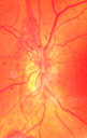
PDR NVD left eye and NVE right eye656 views41 year old diabetic woman with PDR in both eyes. High risk in the left eye. She had PRP, Avastin, and vitrectomy x 2 in the left eye with final vision of 20/25. The right eye had eventually PRP laserApr 07, 2017
|
|

PDR NVD left eye and NVE right eye598 views41 year old diabetic woman with PDR in both eyes. High risk in the left eye. She had PRP, Avastin, and vitrectomy x 2 in the left eye with final vision of 20/25. The right eye had eventually PRP laserApr 07, 2017
|
|

PDR NVD left eye and NVE right eye565 views41 year old diabetic woman with PDR in both eyes. High risk in the left eye. She had PRP, Avastin, and vitrectomy x 2 in the left eye with final vision of 20/25. The right eye had eventually PRP laserApr 07, 2017
|
|

PDR NVD left eye and NVE right eye687 views41 year old diabetic woman with PDR in both eyes. High risk in the left eye. She had PRP, Avastin, and vitrectomy x 2 in the left eye with final vision of 20/25. The right eye had eventually PRP laserApr 07, 2017
|
|

Diabetic Retimopathy with Peripherial Ischemia610 views30 year old woman with decreased vision right eyeJul 08, 2015
|
|

Fluorescence Angiography of BDR418 viewsPatient is here for eval on diabetic retinopathy. FA photos show significant diabetic retinopathy with hemorrhage inferior temporal to the optic nerve. Sep 23, 2014
|
|

Fluorescence Angiography of BDR556 viewsPatient is here for eval on diabetic retinopathy. FA photos show significant diabetic retinopathy with hemorrhage inferior temporal to the optic nerve. Sep 23, 2014
|
|

Fluorescence Angiography of BDR505 viewsPatient is here for eval on diabetic retinopathy. FA photos show significant diabetic retinopathy with hemorrhage inferior temporal to the optic nerve. Sep 23, 2014
|
|

Vitreo-Traction due to Diabetic Retinopathy656 viewsPatient comes in for follow up on her diabetes. VA is 20/30 in the right eye. Fundus photos shows thick traction nasally to the optic nerve forming a ring around the macula. Will follow up in 6-months.Sep 16, 2014
|
|

Vitreo-Traction due to Diabetic Retinopathy588 viewsPatient comes in for follow up on her diabetes. VA is 20/30 in the right eye. Fundus photos shows thick traction nasally to the optic nerve forming a ring around the macula. Will follow up in 6-months.Sep 16, 2014
|
|

Vitreo-Traction due to Diabetic Retinopathy1311 viewsPatient comes in for follow up on her diabetes. VA is 20/30 in the right eye. Fundus photos shows thick traction nasally to the optic nerve forming a ring around the macula. Will follow up in 6-months.Sep 16, 2014
|
|

NVD896 viewsDiabetic patient with NVD and NVEAug 05, 2014
|
|

Diabetic Retinopathy with Scattered Hemorrhages. 868 viewsDiabetic patient with uncontrolled A1C comes in with blurred vision in the right eye. Fundus photo reveals scattered hemorrhages with cotton wool spots and some edema in the macula. Patient was treated with Avastin for edema.Jun 03, 2014
|
|

Diabetic Retinopathy727 viewsAnnual diabetic exam. VA 20/30, right eye and 20/50, left eye. Fundus photos shows hemorrhages with cotton wool spots. Exudates are visible around the macula in the right eye. Scattered hemorrhages in the left eye.Mar 28, 2014
|
|

Diabetic Retinopathy1468 viewsAnnual diabetic exam. VA 20/30, right eye and 20/50, left eye. Fundus photos shows hemorrhages with cotton wool spots. Exudates are visible around the macula in the right eye. Scattered hemorrhages in the left eye.Mar 28, 2014
|
|

Diabetic Retinopathy816 viewsAnnual diabetic exam. VA 20/30, right eye and 20/50, left eye. Fundus photos shows hemorrhages with cotton wool spots. Exudates are visible around the macula in the right eye. Scattered hemorrhages in the left eye.Mar 28, 2014
|
|

Diabetic Retinopathy959 viewsAnnual diabetic exam. VA 20/30, right eye and 20/50, left eye. Fundus photos shows hemorrhages with cotton wool spots. Exudates are visible around the macula in the right eye. Scattered hemorrhages in the left eye.Mar 28, 2014
|
|

Proliferative Diabetic Retinopathy945 viewsFundus photography shows severe fibrosis and arterial narrowing. Peripheral laser scars in both eyes. VA is 20/40, right eye and 20/50, left eye. Jan 23, 2014
|
|

Proliferative Diabetic Retinopathy1346 viewsFundus photography shows severe fibrosis and arterial narrowing. Peripheral laser scars in both eyes. VA is 20/40, right eye and 20/50, left eye. Jan 23, 2014
|
|

Diabetic Retinopathy with CME620 viewsSecond opinion for diabetic retinopathy. VA is 20/30, right eye, 20/50, left eye. Patient has mild swelling in the macula of the right eye. No evidence of CME but does have a moderate cataract in the left eye. Dec 19, 2013
|
|

Diabetic Retinopathy with CME801 viewsSecond opinion for diabetic retinopathy. VA is 20/30, right eye, 20/50, left eye. Patient has mild swelling in the macula of the right eye. No evidence of CME but does have a moderate cataract in the left eye. Dec 19, 2013
|
|

Diabetic Retinopathy with CME1270 viewsSecond opinion for diabetic retinopathy. VA is 20/30, right eye, 20/50, left eye. Patient has mild swelling in the macula of the right eye. No evidence of CME but does have a moderate cataract in the left eye. Dec 19, 2013
|
|

Diabetic Retinopathy with CME586 viewsSecond opinion for diabetic retinopathy. VA is 20/30, right eye, 20/50, left eye. Patient has mild swelling in the macula of the right eye. No evidence of CME but does have a moderate cataract in the left eye. Dec 19, 2013
|
|

Proliferative Diabetic Retinopathy - Moderate - mild NVD with NVE 689 viewsFA shows leakage from neovascularization and dark areas in periphery from non-perfusionDec 06, 2013
|
|

Proliferative Diabetic Retinopathy - Moderate - mild NVD with NVE 538 viewsFA shows leakage from neovascularization and dark areas in periphery from non-perfusionDec 06, 2013
|
|

Proliferative Diabetic Retinopathy - Moderate - mild NVD with NVE 597 viewsFA shows leakage from neovascularization and dark areas in periphery from non-perfusionDec 06, 2013
|
|

Proliferative Diabetic Retinopathy - Moderate - mild NVD with NVE 764 viewsFA shows leakage from neovascularization and dark areas in periphery from non-perfusionDec 06, 2013
|
|

Proliferative Diabetic Retinopathy - Moderate - mild NVD with NVE 594 viewsFA shows leakage from neovascularization and dark areas in periphery from non-perfusionDec 06, 2013
|
|

Proliferative Diabetic Retinopathy - Moderate - mild NVD with NVE 551 viewsFA shows leakage from neovascularization and dark areas in periphery from non-perfusionDec 06, 2013
|
|

Proliferative Diabetic Retinopathy - Moderate - mild NVD with NVE 931 viewsFA shows leakage from neovascularization and dark areas in periphery from non-perfusionDec 06, 2013
|
|

Proliferative Diabetic Retinopathy - Moderate - mild NVD with NVE 553 viewsFA shows leakage from neovascularization and dark areas in periphery from non-perfusionDec 06, 2013
|
|

Proliferative Diabetic Retinopathy - Moderate - mild NVD with NVE 525 viewsFA shows leakage from neovascularization and dark areas in periphery from non-perfusionDec 06, 2013
|
|

Proliferative Diabetic Retinopathy - Moderate - mild NVD with NVE 576 viewsFA shows leakage from neovascularization and dark areas in periphery from non-perfusionDec 06, 2013
|
|

Proliferative Diabetic Retinopathy - Moderate - mild NVD with NVE 504 viewsFA shows leakage from neovascularization and dark areas in periphery from non-perfusionDec 06, 2013
|
|

Proliferative Diabetic Retinopathy - Moderate - mild NVD with NVE 544 viewsFA shows leakage from neovascularization and dark areas in periphery from non-perfusionDec 06, 2013
|
|

Proliferative Diabetic Retinopathy - Moderate - mild NVD with NVE 999 viewsFA shows leakage from neovascularization and dark areas in periphery from non-perfusionDec 06, 2013
|
|

Proliferative Diabetic Retinopathy - Moderate - mild NVD with NVE 560 viewsFA shows leakage from neovascularization and dark areas in periphery from non-perfusionDec 06, 2013
|
|

Proliferative Diabetic Retinopathy - Moderate - mild NVD with NVE 557 viewsFA shows leakage from neovascularization and dark areas in periphery from non-perfusionDec 06, 2013
|
|

Proliferative Diabetic Retinopathy - Moderate - mild NVD with NVE 484 viewsFA shows leakage from neovascularization and dark areas in periphery from non-perfusionDec 06, 2013
|
|

Proliferative Diabetic Retinopathy - Moderate - mild NVD with NVE 533 viewsFA shows leakage from neovascularization and dark areas in periphery from non-perfusionDec 06, 2013
|
|

Proliferative Diabetic Retinopathy - Moderate - mild NVD with NVE 468 viewsFA shows leakage from neovascularization and dark areas in periphery from non-perfusionDec 06, 2013
|
|

Proliferative Diabetic Retinopathy - Moderate - mild NVD with NVE 591 viewsFA shows leakage from neovascularization and dark areas in periphery from non-perfusionDec 06, 2013
|
|

Proliferative Diabetic Retinopathy - Moderate - mild NVD with NVE 612 viewsFA shows leakage from neovascularization and dark areas in periphery from non-perfusionDec 06, 2013
|
|

Proliferative Diabetic Retinopathy - Moderate - mild NVD with NVE 528 viewsFAFDec 06, 2013
|
|

Proliferative Diabetic Retinopathy - Moderate - mild NVD with NVE 628 viewsFAFDec 06, 2013
|
|

Proliferative Diabetic Retinopathy - Moderate - mild NVD with NVE 553 viewsIR reflectanceDec 06, 2013
|
|

Proliferative Diabetic Retinopathy - Moderate - mild NVD with NVE 531 viewsIR reflectanceDec 06, 2013
|
|

Proliferative Diabetic Retinopathy - Moderate - mild NVD with NVE 457 viewsIR reflectanceDec 06, 2013
|
|

Proliferative Diabetic Retinopathy - Moderate - mild NVD with NVE 470 viewsIR reflectanceDec 06, 2013
|
|

Proliferative Diabetic Retinopathy - Moderate - mild NVD with NVE 472 viewsSD OCT shows no significant macular edemaDec 06, 2013
|
|

Proliferative Diabetic Retinopathy - Moderate - mild NVD with NVE 529 viewsSD OCT shows no significant macular edemaDec 06, 2013
|
|

Proliferative Diabetic Retinopathy - Moderate - mild NVD with NVE 525 viewsSD OCT shows no significant macular edemaDec 06, 2013
|
|

Proliferative Diabetic Retinopathy - Moderate - mild NVD with NVE 512 viewsSD OCT shows no significant macular edemaDec 06, 2013
|
|
| 1432 files on 15 page(s) |
 |
|
|
|
|
8 | |
|
|
|
|


