Last additions - Proliferative Diabetic Retinopathy
|

Pre-proliferative Diabetic Retinopathy - Peripheral non-perfusion visible on FA630 viewsApr 25, 2013
|
|
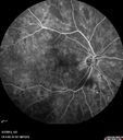
Pre-proliferative Diabetic Retinopathy - Peripheral non-perfusion visible on FA560 viewsApr 25, 2013
|
|

Pre-proliferative Diabetic Retinopathy - Peripheral non-perfusion visible on FA597 viewsApr 25, 2013
|
|

PDR - IR547 viewsApr 25, 2013
|
|

PDR - FAF664 viewsApr 25, 2013
|
|

BDR FAF593 viewsApr 25, 2013
|
|

548 viewsApr 25, 2013
|
|

590 viewsApr 25, 2013
|
|

Neovascularization elsewhere (NVE) Left Eye with Vitreous Hemorrhage933 viewsApr 25, 2013
|
|

Neovascularization elsewhere (NVE) Left Eye with Vitreous Hemorrhage624 viewsApr 25, 2013
|
|

Neovascularization elsewhere (NVE) Left Eye with Vitreous Hemorrhage819 viewsApr 25, 2013
|
|

Neovascularization elsewhere (NVE) Left Eye with Vitreous Hemorrhage760 viewsApr 25, 2013
|
|

Neovascularization elsewhere (NVE) Left Eye with Vitreous Hemorrhage631 viewsApr 25, 2013
|
|

BDR OD with Macular Edema600 viewsApr 25, 2013
|
|

BDR OD with Macular Edema643 viewsApr 25, 2013
|
|

Post-Op Vitrectomy with Membrane Stripping and Laser487 viewsPatient had surgery to help clear up some vision in the left eye. Pre-op VA was count fingers at 1-ft. Post op VA was 20/200 in the left eye. Patient will return in 3-months for follow-up.Apr 08, 2013
|
|

Post-Op Vitrectomy with Membrane Stripping and Laser477 viewsPatient had surgery to help clear up some vision in the left eye. Pre-op VA was count fingers at 1-ft. Post op VA was 20/200 in the left eye. Patient will return in 3-months for follow-up.Apr 08, 2013
|
|

Post-Op Vitrectomy with Membrane Stripping and Laser508 viewsPatient had surgery to help clear up some vision in the left eye. Pre-op VA was count fingers at 1-ft. Post op VA was 20/200 in the left eye. Patient will return in 3-months for follow-up.Apr 08, 2013
|
|

Post-Op Vitrectomy with Membrane Stripping and Laser447 viewsPatient had surgery to help clear up some vision in the left eye. Pre-op VA was count fingers at 1-ft. Post op VA was 20/200 in the left eye. Patient will return in 3-months for follow-up.Apr 08, 2013
|
|

Post-Op Vitrectomy with Membrane Stripping and Laser533 viewsPatient had surgery to help clear up some vision in the left eye. Pre-op VA was count fingers at 1-ft. Post op VA was 20/200 in the left eye. Patient will return in 3-months for follow-up.Apr 08, 2013
|
|

Post-Op Vitrectomy with Membrane Stripping and Laser543 viewsPatient had surgery to help clear up some vision in the left eye. Pre-op VA was count fingers at 1-ft. Post op VA was 20/200 in the left eye. Patient will return in 3-months for follow-up.Apr 08, 2013
|
|

Post-Op Vitrectomy with Membrane Stripping and Laser451 viewsPatient had surgery to help clear up some vision in the left eye. Pre-op VA was count fingers at 1-ft. Post op VA was 20/200 in the left eye. Patient will return in 3-months for follow-up.Apr 08, 2013
|
|

Post-Op Vitrectomy with Membrane Stripping and Laser570 viewsPatient had surgery to help clear up some vision in the left eye. Pre-op VA was count fingers at 1-ft. Post op VA was 20/200 in the left eye. Patient will return in 3-months for follow-up.Apr 08, 2013
|
|

Post-Op Vitrectomy with Membrane Stripping and Laser973 viewsPatient had surgery to help clear up some vision in the left eye. Pre-op VA was count fingers at 1-ft. Post op VA was 20/200 in the left eye. Patient will return in 3-months for follow-up.Apr 08, 2013
|
|

Proliferative Diabetic Retinopathy - Fresh and Old Scatter (Panretinal) Photocoagulation (Laser) - Neovascularization is regressing853 views1 month after initial laserApr 03, 2013
|
|

Proliferative Diabetic Retinopathy - Fresh and Old Scatter (Panretinal) Photocoagulation (Laser) - Neovascularization is regressing673 views1 month after initial laserApr 03, 2013
|
|

Proliferative Diabetic Retinopathy - Fresh and Old Scatter (Panretinal) Photocoagulation (Laser) - Neovascularization is regressing682 views1 month after initial laserApr 03, 2013
|
|

Proliferative Diabetic Retinopathy - Fresh and Old Scatter (Panretinal) Photocoagulation (Laser) - Neovascularization is regressing705 views1 month after initial laserApr 03, 2013
|
|

Proliferative Diabetic Retinopathy - Fresh and Old Scatter (Panretinal) Photocoagulation (Laser) - Neovascularization is regressing619 views1 month after initial laserApr 03, 2013
|
|

Proliferative Diabetic Retinopathy - Fresh and Old Scatter (Panretinal) Photocoagulation (Laser) - Neovascularization is regressing594 views1 month after initial laserApr 03, 2013
|
|

Proliferative Diabetic Retinopathy - Fresh and Old Scatter (Panretinal) Photocoagulation (Laser) - Neovascularization is regressing615 views1 month after initial laserApr 03, 2013
|
|

Panretinal (Scatter) Photocoagulation (Fresh)730 viewsMar 01, 2013
|
|

Panretinal (Scatter) Photocoagulation (Fresh)548 viewsMar 01, 2013
|
|

Panretinal (Scatter) Photocoagulation (Fresh)503 viewsMar 01, 2013
|
|

Panretinal (Scatter) Photocoagulation (Fresh)540 viewsMar 01, 2013
|
|

Neovascularization of the Disc - Heidelberg false color 530 viewsMar 01, 2013
|
|

FA showing NVE, NVD, non- perfusion left eye inferonasally with neovascularization at the margin of the non-perfusion700 viewsMar 01, 2013
|
|

FA showing NVE, NVD, non- perfusion left eye inferonasally with neovascularization at the margin of the non-perfusion618 viewsMar 01, 2013
|
|

FA showing NVE, NVD, non- perfusion left eye inferonasally with neovascularization at the margin of the non-perfusion578 viewsMar 01, 2013
|
|

FA showing NVE, NVD, non- perfusion left eye inferonasally with neovascularization at the margin of the non-perfusion (heavy leakage from NV)711 viewsMar 01, 2013
|
|

FA showing NVE, NVD, non- perfusion left eye inferonasally with neovascularization at the margin of the non-perfusion581 viewsMar 01, 2013
|
|

FA showing NVE, NVD, non- perfusion left eye inferonasally with neovascularization at the margin of the non-perfusion537 viewsMar 01, 2013
|
|

FA showing NVE, NVD, non- perfusion left eye inferonasally with neovascularization at the margin of the non-perfusion619 viewsMar 01, 2013
|
|

FA showing NVE, NVD, non- perfusion left eye inferonasally with neovascularization at the margin of the non-perfusion528 viewsMar 01, 2013
|
|

FA showing NVE, NVD, non- perfusion left eye inferonasally with neovascularization at the margin of the non-perfusion778 viewsMar 01, 2013
|
|

FA showing NVE, NVD, non- perfusion left eye inferonasally with neovascularization at the margin of the non-perfusion574 viewsMar 01, 2013
|
|

FA showing NVE, NVD, non- perfusion left eye inferonasally with neovascularization at the margin of the non-perfusion567 viewsMar 01, 2013
|
|

FA showing NVE, NVD, non- perfusion left eye inferonasally with neovascularization at the margin of the non-perfusion588 viewsMar 01, 2013
|
|

FA showing NVE, NVD, non- perfusion left eye inferonasally with neovascularization at the margin of the non-perfusion587 viewsMar 01, 2013
|
|

FA showing NVE, NVD, non- perfusion left eye inferonasally with neovascularization at the margin of the non-perfusion510 viewsMar 01, 2013
|
|

FA showing NVE, NVD, non- perfusion left eye inferonasally with neovascularization at the margin of the non-perfusion577 viewsMar 01, 2013
|
|

BDR right eye - previous laser564 viewsMar 01, 2013
|
|

BDR right eye - previous laser540 viewsMar 01, 2013
|
|

No significant macular edema510 viewsMar 01, 2013
|
|

No significant macular edema546 viewsMar 01, 2013
|
|

FAF598 viewsMar 01, 2013
|
|
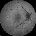
FAF583 viewsMar 01, 2013
|
|

NVD - proliferative diabetic retinopathy650 viewsMar 01, 2013
|
|

NVD - proliferative diabetic retinopathy Heidelberg False Color710 viewsMar 01, 2013
|
|

NVD - proliferative diabetic retinopathy Heidelberg False Color647 viewsMar 01, 2013
|
|

NVD - proliferative diabetic retinopathy Heidelberg False Color693 viewsMar 01, 2013
|
|

Heidelberg False Color607 viewsMar 01, 2013
|
|

Lacy Neovascularization of the Disk - Color Photo737 viewsMar 01, 2013
|
|

PDR572 viewsMar 01, 2013
|
|

PDR637 viewsMar 01, 2013
|
|

BDR571 viewsMar 01, 2013
|
|

bdr612 viewsMar 01, 2013
|
|

bdr760 viewsMar 01, 2013
|
|

BDR656 viewsMar 01, 2013
|
|

NVE frond629 viewsFeb 05, 2013
|
|

PDR, NVE - interesting shape536 views24 yr. old with uncontrolled blood sugar. Decreased vision from 20/40 O.U. in August of 2012, to 20/200: OD and 20/CF: OS in January 2013.Jan 30, 2013
|
|
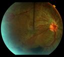
PDR w/ERM and severe CSDME745 views24 year old woman with uncontrolled blood sugar; progressively worsening vision OU - from level of 20/40 in August 2012 to 20/200:OD and 20/CF:OS in January 2013.Jan 30, 2013
|
|

PDR w/TRD464 viewsPDR w/heme and traction retinal detachmentJan 30, 2013
|
|

NVE OD517 viewsJan 30, 2013
|
|

PDR483 viewsWorst florid PDR to date 1/30/2013Jan 30, 2013
|
|

PDR and subhyloid hem678 viewsSep 05, 2012
|
|

Angiogram of PDR and subhyloid hem548 viewsSep 05, 2012
|
|
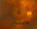
PDR with subhyloid hem1297 viewsPDR with subhyloid hemSep 05, 2012
|
|

Angiogram of PDR1331 viewsSep 05, 2012
|
|

Subhyloid Hem with PDR1638 viewsSep 05, 2012
|
|

Proliferative Diabetic Retinopathy with NVD, NVE, and Pre Retinal Hemorrhage620 viewsJuvenile Onset Diabetic with NVD, NVE and Pre Retinal Hemorrhage.Aug 03, 2012
|
|

Juvenile Onset Diabetic - Proliferative Diabetic retinopathy with NVE and NVD4772 viewsProliferative Diabetic Retinopathy with Neovascularization of the Disc and Neovascularization Elsewhere.
James L. Perron, C.R.A.Jul 02, 2012
|
|

Severe Hemorrhage from NVD and NVE Due To PDR421 views60 year old female with Diabetic Retinopathy. VA 20/40 OD HM OS. History of Dot and Blot hemorrhages and cotton-wool spots associated with Diabetes. Severe Hemorrhaging due to NVD and NVE. Pt stated sight decreased very rapidly.May 25, 2012
|
|

Severe Hemorrhage from NVD and NVE Due To PDR506 views60 year old female with Diabetic Retinopathy. VA 20/40 OD HM OS. History of Dot and Blot hemorrhages and cotton-wool spots associated with Diabetes. Severe Hemorrhaging due to NVD and NVE. Pt stated sight decreased very rapidly.May 25, 2012
|
|

Capillary Drop Out of the FAZ697 viewsHigh magnification image of a damaged foveal avascular zone secondary to proliferative diabetic retinopathy. Seen here are multiple laser scars and capillary drop out in FAZ.May 08, 2012
|
|

Proliferative Diabetic Retinopathy - Preretinal Hemorrhage1815 viewsPreretinal Hemorrhage secondary to Proliferative Diabetic Retinopathy (PDR)
James L. Perron, C.R.A.May 04, 2012
|
|

Regressed Proliferative Diabetic Retinopathy with Recurrent Vitreous Hemorrhage from Vitreoretinal Traction over a Major Retinal Vessel (Vein)659 views56-year-old woman has had recurrent vitreous hemorrhage in the left eye from vitreoretinal traction. Diabetes Mellitus for 20 years and excellent response to vitrectomy in the right eye about 1 year ago for recurrent vitreous hemorrhage from traction over a major retinal vessel.
VISUAL ACUITY: Vision OS is 20/200Feb 07, 2012
|
|

Regressed Proliferative Diabetic Retinopathy with Recurrent Vitreous Hemorrhage from Vitreoretinal Traction over a Major Retinal Vessel (Vein)884 views56-year-old woman has had recurrent vitreous hemorrhage in the left eye from vitreoretinal traction. Diabetes Mellitus for 20 years and excellent response to vitrectomy in the right eye about 1 year ago for recurrent vitreous hemorrhage from traction over a major retinal vessel.
VISUAL ACUITY: Vision OS is 20/200Feb 07, 2012
|
|

Regressed Proliferative Diabetic Retinopathy with Recurrent Vitreous Hemorrhage from Vitreoretinal Traction over a Major Retinal Vessel (Vein)1264 views56-year-old woman has had recurrent vitreous hemorrhage in the left eye from vitreoretinal traction. Diabetes Mellitus for 20 years and excellent response to vitrectomy in the right eye about 1 year ago for recurrent vitreous hemorrhage from traction over a major retinal vessel.
VISUAL ACUITY: Vision OS is 20/200Feb 07, 2012
|
|

Regressed Proliferative Diabetic Retinopathy with Recurrent Vitreous Hemorrhage from Vitreoretinal Traction over a Major Retinal Vessel (Vein)1195 views56-year-old woman has had recurrent vitreous hemorrhage in the left eye from vitreoretinal traction. Diabetes Mellitus for 20 years and excellent response to vitrectomy in the right eye about 1 year ago for recurrent vitreous hemorrhage from traction over a major retinal vessel.
VISUAL ACUITY: Vision OS is 20/200Feb 07, 2012
|
|
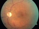
Proliferative Diabetic Retinopathy and Mild Vitreous Hemorrhage850 views58-year-old woman has diabetic retinopathy in both eyes with neovascularization of the optic nerve, worse in the right eye than the left eye. Vision OD is 20/25, OS is 20/20Jul 07, 2011
|
|
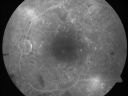
Proliferative Diabetic Retinopathy and Mild Vitreous Hemorrhage526 views58-year-old woman has diabetic retinopathy in both eyes with neovascularization of the optic nerve, worse in the right eye than the left eye. Vision OD is 20/25, OS is 20/20Jul 07, 2011
|
|

Proliferative Diabetic Retinopathy and Mild Vitreous Hemorrhage473 views58-year-old woman has diabetic retinopathy in both eyes with neovascularization of the optic nerve, worse in the right eye than the left eye. Vision OD is 20/25, OS is 20/20Jul 07, 2011
|
|

Proliferative Diabetic Retinopathy and Mild Vitreous Hemorrhage501 views58-year-old woman has diabetic retinopathy in both eyes with neovascularization of the optic nerve, worse in the right eye than the left eye. Vision OD is 20/25, OS is 20/20Jul 07, 2011
|
|
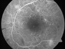
Proliferative Diabetic Retinopathy and Mild Vitreous Hemorrhage538 views58-year-old woman has diabetic retinopathy in both eyes with neovascularization of the optic nerve, worse in the right eye than the left eye. Vision OD is 20/25, OS is 20/20Jul 07, 2011
|
|
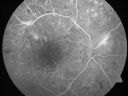
Proliferative Diabetic Retinopathy and Mild Vitreous Hemorrhage499 views58-year-old woman has diabetic retinopathy in both eyes with neovascularization of the optic nerve, worse in the right eye than the left eye. Vision OD is 20/25, OS is 20/20Jul 07, 2011
|
|
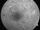
Proliferative Diabetic Retinopathy and Mild Vitreous Hemorrhage449 views58-year-old woman has diabetic retinopathy in both eyes with neovascularization of the optic nerve, worse in the right eye than the left eye. Vision OD is 20/25, OS is 20/20Jul 07, 2011
|
|

Proliferative Diabetic Retinopathy and Mild Vitreous Hemorrhage465 views58-year-old woman has diabetic retinopathy in both eyes with neovascularization of the optic nerve, worse in the right eye than the left eye. Vision OD is 20/25, OS is 20/20Jul 07, 2011
|
|
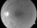
Proliferative Diabetic Retinopathy and Mild Vitreous Hemorrhage476 views58-year-old woman has diabetic retinopathy in both eyes with neovascularization of the optic nerve, worse in the right eye than the left eye. Vision OD is 20/25, OS is 20/20Jul 07, 2011
|
|

Proliferative Diabetic Retinopathy and Mild Vitreous Hemorrhage511 views58-year-old woman has diabetic retinopathy in both eyes with neovascularization of the optic nerve, worse in the right eye than the left eye. Vision OD is 20/25, OS is 20/20Jul 07, 2011
|
|
|
|


