Last additions - Anterior Segment
|

Cataract Morgagnian336 viewsMar 09, 2023
|
|

Iris Arteriovenous Malformation320 viewsDec 12, 2022
|
|

Iris Arteriovenous Malformation371 viewsDec 12, 2022
|
|

Iris Arteriovenous Malformation373 viewsDec 12, 2022
|
|

Idiopathic Iris Cyst372 views85 year old man History of wet AMD OD x 7 years under treatment with Lucentis. VA 20/40 OD, 20/32 OS
PMHx High cholesterol, GERD, COPD, A-Fib,
MEDS: coumadin and ASA and othersOct 28, 2021
|
|

Idiopathic Iris Cyst388 views85 year old man History of wet AMD OD x 7 years under treatment with Lucentis. VA 20/40 OD, 20/32 OS
PMHx High cholesterol, GERD, COPD, A-Fib,
MEDS: coumadin and ASA and othersOct 28, 2021
|
|

Idiopathic Iris Cyst443 views85 year old man History of wet AMD OD x 7 years under treatment with Lucentis. VA 20/40 OD, 20/32 OS
PMHx High cholesterol, GERD, COPD, A-Fib,
MEDS: coumadin and ASA and othersOct 28, 2021
|
|

Idiopathic Iris Cyst394 views85 year old man History of wet AMD OD x 7 years under treatment with Lucentis. VA 20/40 OD, 20/32 OS
PMHx High cholesterol, GERD, COPD, A-Fib,
MEDS: coumadin and ASA and othersOct 28, 2021
|
|

Idiopathic Iris Cyst383 views85 year old man History of wet AMD OD x 7 years under treatment with Lucentis. VA 20/40 OD, 20/32 OS
PMHx High cholesterol, GERD, COPD, A-Fib,
MEDS: coumadin and ASA and othersOct 28, 2021
|
|

Idiopathic Iris Cyst475 views85 year old man History of wet AMD OD x 7 years under treatment with Lucentis. VA 20/40 OD, 20/32 OS
PMHx High cholesterol, GERD, COPD, A-Fib,
MEDS: coumadin and ASA and othersOct 28, 2021
|
|
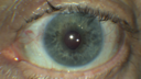
Fuch's Heterochromic Iridocyclitis - Iris Photos549 views71 year old man with inflammation in the left eye for over 50 years. The left eye got a tube shunt 15 years ago. Vision is 20/32 OD, 20/40 OSApr 27, 2020
|
|

Fuch's Heterochromic Iridocyclitis - Iris Photos501 views71 year old man with inflammation in the left eye for over 50 years. The left eye got a tube shunt 15 years ago. Vision is 20/32 OD, 20/40 OSApr 27, 2020
|
|

Fuch's Heterochromic Iridocyclitis - Iris Photos522 views71 year old man with inflammation in the left eye for over 50 years. The left eye got a tube shunt 15 years ago. Vision is 20/32 OD, 20/40 OSApr 27, 2020
|
|

Anterior Uveitis Vemurafenib - Zelboraf518 views60 year old man on 8 years of vemurafenib with 4 days of pain and floaters - images show 360 degree synechia. There is an iris nodule not visibleMar 02, 2018
|
|

Anterior Uveitis Vemurafenib - Zelboraf469 views60 year old man on 8 years of vemurafenib with 4 days of pain and floaters - images show 360 degree synechia. There is an iris nodule not visibleMar 02, 2018
|
|

Anterior Uveitis Vemurafenib - Zelboraf482 views60 year old man on 8 years of vemurafenib with 4 days of pain and floaters - images show 360 degree synechia. There is an iris nodule not visibleMar 02, 2018
|
|

Fuchs Heterochromic Iridocyclitis - Left eye has inflammation1647 viewsThe right eye is darker than the left eye which has chronic iritisNov 06, 2017
|
|

Scleromalacia and Uveitis656 viewsPoor vision from uveitis and cataract. She was treated with Remicade and the uveitis abated and she had the right eye cataract done. The initial vision was 20/400 OD, 20/200 OS Final vision 20/60 OD, 20/32 OSMay 05, 2017
|
|

Scleromalacia and Uveitis537 viewsPoor vision from uveitis and cataract. She was treated with Remicade and the uveitis abated and she had the right eye cataract done. The initial vision was 20/400 OD, 20/200 OS Final vision 20/60 OD, 20/32 OSMay 05, 2017
|
|

Scleromalacia and Uveitis523 viewsPoor vision from uveitis and cataract. She was treated with Remicade and the uveitis abated and she had the right eye cataract done. The initial vision was 20/400 OD, 20/200 OS Final vision 20/60 OD, 20/32 OSMay 05, 2017
|
|

Scleromalacia and Uveitis562 viewsPoor vision from uveitis and cataract. She was treated with Remicade and the uveitis abated and she had the right eye cataract done. The initial vision was 20/400 OD, 20/200 OS Final vision 20/60 OD, 20/32 OSMay 05, 2017
|
|

Scleromalacia and Uveitis590 viewsPoor vision from uveitis and cataract. She was treated with Remicade and the uveitis abated and she had the right eye cataract done. The initial vision was 20/400 OD, 20/200 OS Final vision 20/60 OD, 20/32 OSMay 05, 2017
|
|

Scleromalacia and Uveitis562 viewsPoor vision from uveitis and cataract. She was treated with Remicade and the uveitis abated and she had the right eye cataract done. The initial vision was 20/400 OD, 20/200 OS Final vision 20/60 OD, 20/32 OSMay 05, 2017
|
|

Scleromalacia and Uveitis578 viewsPoor vision from uveitis and cataract. She was treated with Remicade and the uveitis abated and she had the right eye cataract done. The initial vision was 20/400 OD, 20/200 OS Final vision 20/60 OD, 20/32 OSMay 05, 2017
|
|

Scleromalacia and Uveitis562 viewsPoor vision from uveitis and cataract. She was treated with Remicade and the uveitis abated and she had the right eye cataract done. The initial vision was 20/400 OD, 20/200 OS Final vision 20/60 OD, 20/32 OSMay 05, 2017
|
|

Scleromalacia and Uveitis523 viewsPoor vision from uveitis and cataract. She was treated with Remicade and the uveitis abated and she had the right eye cataract done. The initial vision was 20/400 OD, 20/200 OS Final vision 20/60 OD, 20/32 OSMay 05, 2017
|
|

Dislocated IOL - Astigmastism - Corrected with new prescription718 views82 year old woman with prior vitrectomy for macular hole - vision dropped from 20/32 to 20/200. Corrected with new rx: 11/2016 old RX OD +0.5 -0.75 x 63; new rx -0.50 -3.00 x 65Jan 08, 2017
|
|

Dislocated IOL - Astigmastism - Corrected with new prescription716 views82 year old woman with prior vitrectomy for macular hole - vision dropped from 20/32 to 20/200. Corrected with new rx: 11/2016 old RX OD +0.5 -0.75 x 63; new rx -0.50 -3.00 x 65Jan 08, 2017
|
|

Leucoma vascolarizzato845 views67 female failed PKP outcomeNov 12, 2014
|
|

Free Floating Dislocated Lens in Vitreous488 viewsPatient comes in aphakic with dislocated lens floating to the back of the eye when laying down. Lens is laying up against the endothelium of the cornea when patient is right side up..Sep 23, 2014
|
|

Free Floating Dislocated Lens in Vitreous423 viewsPatient comes in aphakic with dislocated lens floating to the back of the eye when laying down. Lens is laying up against the endothelium of the cornea when patient is right side up..Sep 23, 2014
|
|

Free Floating Dislocated Lens in Vitreous460 viewsPatient comes in aphakic with dislocated lens floating to the back of the eye when laying down. Lens is laying up against the endothelium of the cornea when patient is right side up..Sep 23, 2014
|
|

Free Floating Dislocated Lens in Vitreous406 viewsPatient comes in aphakic with dislocated lens floating to the back of the eye when laying down. Lens is laying up against the endothelium of the cornea when patient is right side up..Sep 23, 2014
|
|

Free Floating Dislocated Lens in Vitreous462 viewsPatient comes in aphakic with dislocated lens floating to the back of the eye when laying down. Lens is laying up against the endothelium of the cornea when patient is right side up..Sep 23, 2014
|
|

Free Floating Dislocated Lens in Vitreous868 viewsPatient comes in aphakic with dislocated lens floating to the back of the eye when laying down. Lens is laying up against the endothelium of the cornea when patient is right side up..Sep 23, 2014
|
|

Free Floating Dislocated Lens in Vitreous550 viewsPatient comes in aphakic with dislocated lens floating to the back of the eye when laying down. Lens is laying up against the endothelium of the cornea when patient is right side up..Sep 23, 2014
|
|

Free Floating Dislocated Lens in Vitreous1133 viewsPatient comes in aphakic with dislocated lens floating to the back of the eye when laying down. Lens is laying up against the endothelium of the cornea when patient is right side up..Sep 23, 2014
|
|

Free Floating Dislocated Lens in Vitreous496 viewsPatient comes in aphakic with dislocated lens floating to the back of the eye when laying down. Lens is laying up against the endothelium of the cornea when patient is right side up..Sep 23, 2014
|
|
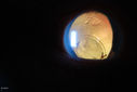
Free Floating Dislocated Lens in Vitreous561 viewsPatient comes in aphakic with dislocated lens floating to the back of the eye when laying down. Lens is laying up against the endothelium of the cornea when patient is right side up..Sep 23, 2014
|
|

Flower Cataract987 viewsPatient with ahmed tube and flower cataractSep 23, 2014
|
|

Flower Cataract1524 viewsPatient with ahmed tube and flower cataractSep 23, 2014
|
|
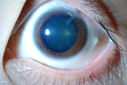
Flower Cataract1219 viewsPatient with ahmed tube and flower cataractSep 23, 2014
|
|

415 viewsSep 12, 2014
|
|

418 viewsSep 11, 2014
|
|

Gonioscopy, Pigment Dispersion Syndrome1242 viewsPatient comes in with elevated pressures. Goinoscopy shows heavy pigment dusted off the iris into the trabecular meshwork. Laser procedure was done to break up the pigment for more aqueous flow.Sep 08, 2014
|
|

Gonioscopy, Pigment Dispersion Syndrome1123 viewsPatient comes in with elevated pressures. Goinoscopy shows heavy pigment dusted off the iris into the trabecular meshwork. Laser procedure was done to break up the pigment for more aqueous flow.Sep 08, 2014
|
|

Gonioscopy, Pigment Dispersion Syndrome1473 viewsPatient comes in with elevated pressures. Goinoscopy shows heavy pigment dusted off the iris into the trabecular meshwork. Laser procedure was done to break up the pigment for more aqueous flow.Sep 08, 2014
|
|

Gonioscopy, Pigment Dispersion Syndrome1086 viewsPatient comes in with elevated pressures. Goinoscopy shows heavy pigment dusted off the iris into the trabecular meshwork. Laser procedure was done to break up the pigment for more aqueous flow.Sep 08, 2014
|
|

Corneal Foreign Body687 viewsPatient comes in to the ER with foreign body sensation in his left eye. Patient was referred to Ophthalmology. Slit lamp shows metallic foreign body with rust ring nasally to the limbus in the left eye. Foreign body was removed with no complications..Aug 29, 2014
|
|
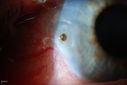
Corneal Foreign Body520 viewsPatient comes in to the ER with foreign body sensation in his left eye. Patient was referred to Ophthalmology. Slit lamp shows metallic foreign body with rust ring nasally to the limbus in the left eye. Foreign body was removed with no complications..Aug 29, 2014
|
|

Corneal Foreign Body669 viewsPatient comes in to the ER with foreign body sensation in his left eye. Patient was referred to Ophthalmology. Slit lamp shows metallic foreign body with rust ring nasally to the limbus in the left eye. Foreign body was removed with no complications..Aug 29, 2014
|
|

Corneal Foreign Body974 viewsPatient comes in to the ER with foreign body sensation in his left eye. Patient was referred to Ophthalmology. Slit lamp shows metallic foreign body with rust ring nasally to the limbus in the left eye. Foreign body was removed with no complications..Aug 29, 2014
|
|

Dislocated IOL with Tube Shunt1184 viewsPatient comes in with a dislocated lens in the right eye. Decreased vision in the right eye. Slit lamp photos shows lens dislocated halfway inferior. Ahmed Tube shut is visible at 11-o'clock superiorly in the right eye. Aug 25, 2014
|
|

Dislocated IOL with Tube Shunt575 viewsPatient comes in with a dislocated lens in the right eye. Decreased vision in the right eye. Slit lamp photos shows lens dislocated halfway inferior. Ahmed Tube shut is visible at 11-o'clock superiorly in the right eye. Aug 25, 2014
|
|

Dislocated IOL with Tube Shunt541 viewsPatient comes in with a dislocated lens in the right eye. Decreased vision in the right eye. Slit lamp photos shows lens dislocated halfway inferior. Ahmed Tube shut is visible at 11-o'clock superiorly in the right eye. Aug 25, 2014
|
|
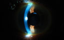
Dislocated IOL with Tube Shunt545 viewsPatient comes in with a dislocated lens in the right eye. Decreased vision in the right eye. Slit lamp photos shows lens dislocated halfway inferior. Ahmed Tube shut is visible at 11-o'clock superiorly in the right eye. Aug 25, 2014
|
|

Gonioscopy, Mass in the Angle of Anterior Chamber469 viewsSlit Lamp and goinoscopy photos show a mass at 7-o'clock i the right eye. The mass extends beneath the iris behind the lens. Aug 25, 2014
|
|
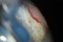
Gonioscopy, Mass in the Angle of Anterior Chamber456 viewsSlit Lamp and goinoscopy photos show a mass at 7-o'clock i the right eye. The mass extends beneath the iris behind the lens. Aug 25, 2014
|
|

Gonioscopy, Mass in the Angle of Anterior Chamber1154 viewsSlit Lamp and goinoscopy photos show a mass at 7-o'clock i the right eye. The mass extends beneath the iris behind the lens. Aug 25, 2014
|
|

Gonioscopy, Mass in the Angle of Anterior Chamber430 viewsSlit Lamp and goinoscopy photos show a mass at 7-o'clock i the right eye. The mass extends beneath the iris behind the lens. Aug 25, 2014
|
|

Gonioscopy, Mass in the Angle of Anterior Chamber483 viewsSlit Lamp and goinoscopy photos show a mass at 7-o'clock i the right eye. The mass extends beneath the iris behind the lens. Aug 25, 2014
|
|

Gonioscopy, Mass in the Angle of Anterior Chamber476 viewsSlit Lamp and goinoscopy photos show a mass at 7-o'clock i the right eye. The mass extends beneath the iris behind the lens. Aug 25, 2014
|
|

Gonioscopy, Mass in the Angle of Anterior Chamber470 viewsSlit Lamp and goinoscopy photos show a mass at 7-o'clock i the right eye. The mass extends beneath the iris behind the lens. Aug 25, 2014
|
|

Gonioscopy, Mass in the Angle of Anterior Chamber486 viewsSlit Lamp and goinoscopy photos show a mass at 7-o'clock i the right eye. The mass extends beneath the iris behind the lens. Aug 25, 2014
|
|

Ghost Cell Glaucoma883 viewsPost cataract surgery, patient presents with ghost cell in the anterior chamber after hemorrhage cleared. A small hyphema still is visible in the anterior chamber with a whitish ball appearance. Aug 25, 2014
|
|

Ghost Cell Glaucoma918 viewsPost cataract surgery, patient presents with ghost cell in the anterior chamber after hemorrhage cleared. A small hyphema still is visible in the anterior chamber with a whitish ball appearance. Aug 25, 2014
|
|

Ghost Cell Glaucoma1090 viewsPost cataract surgery, patient presents with ghost cell in the anterior chamber after hemorrhage cleared. A small hyphema still is visible in the anterior chamber with a whitish ball appearance. Aug 25, 2014
|
|

Ghost Cell Glaucoma901 viewsPost cataract surgery, patient presents with ghost cell in the anterior chamber after hemorrhage cleared. A small hyphema still is visible in the anterior chamber with a whitish ball appearance. Aug 25, 2014
|
|

Ghost Cell Glaucoma1211 viewsPost cataract surgery, patient presents with ghost cell in the anterior chamber after hemorrhage cleared. A small hyphema still is visible in the anterior chamber with a whitish ball appearance. Aug 25, 2014
|
|

Ghost Cell Glaucoma894 viewsPost cataract surgery, patient presents with ghost cell in the anterior chamber after hemorrhage cleared. A small hyphema still is visible in the anterior chamber with a whitish ball appearance. Aug 25, 2014
|
|

Ghost Cell Glaucoma1172 viewsPost cataract surgery, patient presents with ghost cell in the anterior chamber after hemorrhage cleared. A small hyphema still is visible in the anterior chamber with a whitish ball appearance. Aug 25, 2014
|
|

Ghost Cell Glaucoma1051 viewsPost cataract surgery, patient presents with ghost cell in the anterior chamber after hemorrhage cleared. A small hyphema still is visible in the anterior chamber with a whitish ball appearance. Aug 25, 2014
|
|

Ghost Cell Glaucoma782 viewsPost cataract surgery, patient presents with ghost cell in the anterior chamber after hemorrhage cleared. A small hyphema still is visible in the anterior chamber with a whitish ball appearance. Aug 25, 2014
|
|

Ghost Cell Glaucoma1176 viewsPost cataract surgery, patient presents with ghost cell in the anterior chamber after hemorrhage cleared. A small hyphema still is visible in the anterior chamber with a whitish ball appearance. Aug 25, 2014
|
|

Ghost Cell Glaucoma1123 viewsPost cataract surgery, patient presents with ghost cell in the anterior chamber after hemorrhage cleared. A small hyphema still is visible in the anterior chamber with a whitish ball appearance. Aug 25, 2014
|
|

Trans-Illumination Defect with Fuch's Dystrophy.398 viewsPatient comes in for eval on post cataract surgery. VA is 20/30 both eyes. Slit lamp exam shows Fuch's Dystrophy on the cornea on the right eye. IOL haptics are chaffing the iris therefore producing trans illumination defect in the right eye. Aug 07, 2014
|
|

Trans-Illumination Defect with Fuch's Dystrophy.456 viewsPatient comes in for eval on post cataract surgery. VA is 20/30 both eyes. Slit lamp exam shows Fuch's Dystrophy on the cornea on the right eye. IOL haptics are chaffing the iris therefore producing trans illumination defect in the right eye. Aug 07, 2014
|
|

Trans-Illumination Defect with Fuch's Dystrophy.419 viewsPatient comes in for eval on post cataract surgery. VA is 20/30 both eyes. Slit lamp exam shows Fuch's Dystrophy on the cornea on the right eye. IOL haptics are chaffing the iris therefore producing trans illumination defect in the right eye. Aug 07, 2014
|
|

Trans-Illumination Defect with Fuch's Dystrophy.356 viewsPatient comes in for eval on post cataract surgery. VA is 20/30 both eyes. Slit lamp exam shows Fuch's Dystrophy on the cornea on the right eye. IOL haptics are chaffing the iris therefore producing trans illumination defect in the right eye. Aug 07, 2014
|
|

Trans-Illumination Defect with Fuch's Dystrophy.441 viewsPatient comes in for eval on post cataract surgery. VA is 20/30 both eyes. Slit lamp exam shows Fuch's Dystrophy on the cornea on the right eye. IOL haptics are chaffing the iris therefore producing trans illumination defect in the right eye. Aug 07, 2014
|
|

Trans-Illumination Defect with Fuch's Dystrophy.498 viewsPatient comes in for eval on post cataract surgery. VA is 20/30 both eyes. Slit lamp exam shows Fuch's Dystrophy on the cornea on the right eye. IOL haptics are chaffing the iris therefore producing trans illumination defect in the right eye. Aug 07, 2014
|
|
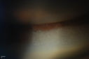
Silicone Oil Bubbles in the Angle732 viewsPatient is evaluated for glaucoma associated with ocular inflammation. VA is CF in the right eye and 20/50, left eye. Gonioscopy photos shows little silicone oil bubbles in the inferior angle of the right eye. Patient to consider removal of silicone oil. Jul 24, 2014
|
|

Silicone Oil Bubbles in the Angle1367 viewsPatient is evaluated for glaucoma associated with ocular inflammation. VA is CF in the right eye and 20/50, left eye. Gonioscopy photos shows little silicone oil bubbles in the inferior angle of the right eye. Patient to consider removal of silicone oil. Jul 24, 2014
|
|

Silicone Oil Bubbles in the Angle678 viewsPatient is evaluated for glaucoma associated with ocular inflammation. VA is CF in the right eye and 20/50, left eye. Gonioscopy photos shows little silicone oil bubbles in the inferior angle of the right eye. Patient to consider removal of silicone oil. Jul 24, 2014
|
|

Silicone Oil Bubbles in the Angle608 viewsPatient is evaluated for glaucoma associated with ocular inflammation. VA is CF in the right eye and 20/50, left eye. Gonioscopy photos shows little silicone oil bubbles in the inferior angle of the right eye. Patient to consider removal of silicone oil. Jul 24, 2014
|
|

Iris Nevus722 viewsPatient came in for cataract evaluation. VA 20/200, right eye, 20/60, left eye. Slit lamp photography shows iris nevus 3-4 mm located nasally at 9-o'clock in the left eye. Jul 18, 2014
|
|

Iris Nevus586 viewsPatient came in for cataract evaluation. VA 20/200, right eye, 20/60, left eye. Slit lamp photography shows iris nevus 3-4 mm located nasally at 9-o'clock in the left eye. Jul 18, 2014
|
|
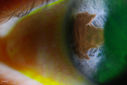
Iris Nevus541 viewsPatient came in for cataract evaluation. VA 20/200, right eye, 20/60, left eye. Slit lamp photography shows iris nevus 3-4 mm located nasally at 9-o'clock in the left eye. Jul 18, 2014
|
|

Iris Nevus561 viewsPatient came in for cataract evaluation. VA 20/200, right eye, 20/60, left eye. Slit lamp photography shows iris nevus 3-4 mm located nasally at 9-o'clock in the left eye. Jul 18, 2014
|
|

Peripheral Iridotomy667 viewsSlit lamp photo of peripheral iridotomy for narrow angles. A hole was place at 10-o'clock superior temporally. Jul 12, 2014
|
|

Peripheral Iridotomy1055 viewsSlit lamp photo of peripheral iridotomy for narrow angles. A hole was place at 10-o'clock superior temporally. Jul 12, 2014
|
|
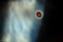
Corneal Foreign Body597 viewsPatient was cutting tile and debris flew in his eyes. Patient complained of discomfort and redness in the right eye. VA is 20/20 in the right eye. Slit lamp photography shows foreign body inferior nasal in the right eye. Removed foreign body and patched with antibiotic and will be re examined the next day. Jul 12, 2014
|
|

Corneal Foreign Body469 viewsPatient was cutting tile and debris flew in his eyes. Patient complained of discomfort and redness in the right eye. VA is 20/20 in the right eye. Slit lamp photography shows foreign body inferior nasal in the right eye. Removed foreign body and patched with antibiotic and will be re examined the next day. Jul 12, 2014
|
|

Corneal Foreign Body494 viewsPatient was cutting tile and debris flew in his eyes. Patient complained of discomfort and redness in the right eye. VA is 20/20 in the right eye. Slit lamp photography shows foreign body inferior nasal in the right eye. Removed foreign body and patched with antibiotic and will be re examined the next day. Jul 12, 2014
|
|

Corneal Foreign Body500 viewsPatient was cutting tile and debris flew in his eyes. Patient complained of discomfort and redness in the right eye. VA is 20/20 in the right eye. Slit lamp photography shows foreign body inferior nasal in the right eye. Removed foreign body and patched with antibiotic and will be re examined the next day. Jul 12, 2014
|
|
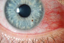
Corneal Foreign Body601 viewsPatient was cutting tile and debris flew in his eyes. Patient complained of discomfort and redness in the right eye. VA is 20/20 in the right eye. Slit lamp photography shows foreign body inferior nasal in the right eye. Removed foreign body and patched with antibiotic and will be re examined the next day. Jul 12, 2014
|
|
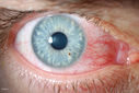
Corneal Foreign Body577 viewsPatient was cutting tile and debris flew in his eyes. Patient complained of discomfort and redness in the right eye. VA is 20/20 in the right eye. Slit lamp photography shows foreign body inferior nasal in the right eye. Removed foreign body and patched with antibiotic and will be re examined the next day. Jul 12, 2014
|
|
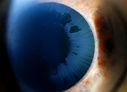
Severe Pseudoexfoliation Ring, Left Eye378 viewsPatient comes in for blurred vision. VA is 20/30 both eyes. Mostly at near which was corrected with refraction. Slit lamp photos show severe pseudoexfoliation in the left eye. Patient will be followed up for glaucoma eval in a few weeks. Jun 25, 2014
|
|

Severe Pseudoexfoliation Ring, Left Eye387 viewsPatient comes in for blurred vision. VA is 20/30 both eyes. Mostly at near which was corrected with refraction. Slit lamp photos show severe pseudoexfoliation in the left eye. Patient will be followed up for glaucoma eval in a few weeks. Jun 25, 2014
|
|

Severe Pseudoexfoliation Ring, Left Eye1001 viewsPatient comes in for blurred vision. VA is 20/30 both eyes. Mostly at near which was corrected with refraction. Slit lamp photos show severe pseudoexfoliation in the left eye. Patient will be followed up for glaucoma eval in a few weeks. Jun 25, 2014
|
|
| 381 files on 4 page(s) |
1 |
 |
|


