Last additions - Anterior Segment
|

Conjunctival Melanoma1300 viewsRight conjunctival melanoma extending into anterior orbit, right eye. Temporally and nasally with pigmented masses/nodules. VA was 20/30 without correction in the right eye. Follow up to proceed with proton beam therapy.Nov 29, 2013
|
|

Correctopia - post laser iridoplasty - 20/20 vision2037 views1 month after the iridoplasty, her vision was 20/20. She began to improve on the drive home from the treatment. The laser setting were : green argon 500 microns, 500 mw, 500 msec.Nov 01, 2013
|
|

Correctopia - post laser iridoplasty - 20/20 vision2641 views1 month after the iridoplasty, her vision was 20/20. She began to improve on the drive home from the treatment. The laser setting were : green argon 500 microns, 500 mw, 500 msec.Nov 01, 2013
|
|

Correctopia - before laser iridoplasty - 20/60 vision1496 viewsIris is occluding visual axisNov 01, 2013
|
|

Correctopia - before laser iridoplasty - 20/60 vision1548 viewsIris is occluding visual axisNov 01, 2013
|
|

Giant Papillary Conjunctivitis, Left Upper Eyelid817 viewsContact lens wearer, comes in for exam. Has rough feeling underneath both eyelids. Patient thought it was through SCL wear. Patient VA was 20/20. right eye, 20/30, left eye. Underneath the left upper eyelid, you can see papillary inflammation and redness. Jul 22, 2013
|
|

Giant Papillary Conjunctivitis, Left Upper Eyelid1289 viewsContact lens wearer, comes in for exam. Has rough feeling underneath both eyelids. Patient thought it was through SCL wear. Patient VA was 20/20. right eye, 20/30, left eye. Underneath the left upper eyelid, you can see papillary inflammation and redness. Jul 22, 2013
|
|

Giant Papillary Conjunctivitis, Left Upper Eyelid827 viewsContact lens wearer, comes in for exam. Has rough feeling underneath both eyelids. Patient thought it was through SCL wear. Patient VA was 20/20. right eye, 20/30, left eye. Underneath the left upper eyelid, you can see papillary inflammation and redness. Jul 22, 2013
|
|

Gonioscopy of Blood in the Angle514 viewsYoung female who has a history of uveitis, glaucoma and juvenile rheumatoid arthritis. VA is hand motion in the left eye. Pigment changes with some blood in the anterior chamber seen here on Gonioscopy. Jul 12, 2013
|
|

Gonioscopy of Blood in the Angle479 viewsYoung female who has a history of uveitis, glaucoma and juvenile rheumatoid arthritis. VA is hand motion in the left eye. Pigment changes with some blood in the anterior chamber seen here on Gonioscopy. Jul 12, 2013
|
|

Gonioscopy of Blood in the Angle541 viewsYoung female who has a history of uveitis, glaucoma and juvenile rheumatoid arthritis. VA is hand motion in the left eye. Pigment changes with some blood in the anterior chamber seen here on Gonioscopy. Jul 12, 2013
|
|

Gonioscopy of Blood in the Angle505 viewsYoung female who has a history of uveitis, glaucoma and juvenile rheumatoid arthritis. VA is hand motion in the left eye. Pigment changes with some blood in the anterior chamber seen here on Gonioscopy. Jul 12, 2013
|
|
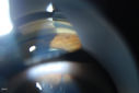
Gonioscopy of Blood in the Angle538 viewsYoung female who has a history of uveitis, glaucoma and juvenile rheumatoid arthritis. VA is hand motion in the left eye. Pigment changes with some blood in the anterior chamber seen here on Gonioscopy. Jul 12, 2013
|
|

Gonioscopy of Blood in the Angle612 viewsYoung female who has a history of uveitis, glaucoma and juvenile rheumatoid arthritis. VA is hand motion in the left eye. Pigment changes with some blood in the anterior chamber seen here on Gonioscopy. Jul 12, 2013
|
|

Gonioscopy of Blood in the Angle585 viewsYoung female who has a history of uveitis, glaucoma and juvenile rheumatoid arthritis. VA is hand motion in the left eye. Pigment changes with some blood in the anterior chamber seen here on Gonioscopy. Jul 12, 2013
|
|

Gonioscopy of Blood in the Angle1246 viewsYoung female who has a history of uveitis, glaucoma and juvenile rheumatoid arthritis. VA is hand motion in the left eye. Pigment changes with some blood in the anterior chamber seen here on Gonioscopy. Jul 12, 2013
|
|

Gonioscopy of Blood in the Angle601 viewsYoung female who has a history of uveitis, glaucoma and juvenile rheumatoid arthritis. VA is hand motion in the left eye. Pigment changes with some blood in the anterior chamber seen here on Gonioscopy. Jul 12, 2013
|
|

Gonioscopy of Blood in the Angle630 viewsYoung female who has a history of uveitis, glaucoma and juvenile rheumatoid arthritis. VA is hand motion in the left eye. Pigment changes with some blood in the anterior chamber seen here on Gonioscopy. Jul 12, 2013
|
|

Gonioscopy of Blood in the Angle712 viewsYoung female who has a history of uveitis, glaucoma and juvenile rheumatoid arthritis. VA is hand motion in the left eye. Pigment changes with some blood in the anterior chamber seen here on Gonioscopy. Jul 12, 2013
|
|

Pseudoexfoliation with Partial Subluxation416 views81-year old female who woke up with loss of vision in the left eye. Patient was hand motion in the left eye. Slit lamp examination shows dislocated lens with pseudoexfoliation material around the iris rim. Proceeded with cataract surgery. Jul 12, 2013
|
|
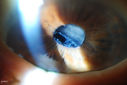
Pseudoexfoliation with Partial Subluxation437 views81-year old female who woke up with loss of vision in the left eye. Patient was hand motion in the left eye. Slit lamp examination shows dislocated lens with pseudoexfoliation material around the iris rim. Proceeded with cataract surgery.Jul 12, 2013
|
|
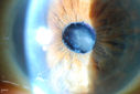
Pseudoexfoliation with Partial Subluxation438 views81-year old female who woke up with loss of vision in the left eye. Patient was hand motion in the left eye. Slit lamp examination shows dislocated lens with pseudoexfoliation material around the iris rim. Proceeded with cataract surgery.Jul 12, 2013
|
|

Polychromatic Cataract (Christmas Tree Cataract)507 viewsPatient comes in with cataract evaluation. Blurred vision in the left eye. Patient will think about surgery and call back to schedule.Jun 21, 2013
|
|
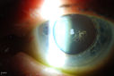
Polychromatic Cataract (Christmas Tree Cataract)474 viewsPatient comes in with cataract evaluation. Blurred vision in the left eye. Patient will think about surgery and call back to schedule.Jun 21, 2013
|
|
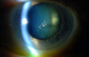
Polychromatic Cataract (Christmas Tree Cataract)522 viewsPatient comes in with cataract evaluation. Blurred vision in the left eye. Patient will think about surgery and call back to schedule.Jun 21, 2013
|
|

Polychromatic Cataract (Christmas Tree Cataract)890 viewsPatient comes in with cataract evaluation. Blurred vision in the left eye. Patient will think about surgery and call back to schedule.Jun 21, 2013
|
|
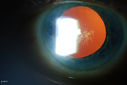
Polychromatic Cataract (Christmas Tree Cataract)557 viewsPatient comes in with cataract evaluation. Blurred vision in the left eye. Patient will think about surgery and call back to schedule.Jun 21, 2013
|
|
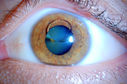
Silicone Oil in the Anterior Chamber1256 viewsPatient comes in for second opinion for retinal detachment in the left eye. VA is hand motion in the left eye. Poor view to look into the retina. Small silicone bubble at 12 o'clock superior in the anterior chamber. Surgery was not recommended due to the silicone oil in the left eye. Jun 21, 2013
|
|

Silicone Oil in the Anterior Chamber1256 viewsPatient comes in for second opinion for retinal detachment in the left eye. VA is hand motion in the left eye. Poor view to look into the retina. Small silicone bubble at 12 o'clock superior in the anterior chamber. Surgery was not recommended due to the silicone oil in the left eye. Jun 21, 2013
|
|

Silicone Oil in the Anterior Chamber548 viewsPatient comes in for second opinion for retinal detachment in the left eye. VA is hand motion in the left eye. Poor view to look into the retina. Small silicone bubble at 12 o'clock superior in the anterior chamber. Surgery was not recommended due to the silicone oil in the left eye. Jun 21, 2013
|
|

Silicone Oil in the Anterior Chamber854 viewsPatient comes in for second opinion for retinal detachment in the left eye. VA is hand motion in the left eye. Poor view to look into the retina. Small silicone bubble at 12 o'clock superior in the anterior chamber. Surgery was not recommended due to the silicone oil in the left eye. Jun 21, 2013
|
|

TID (Trans Illumination Defect) Christmas Tree Effect1016 viewsPatient who is followed up for glaucoma. Retro Illumination reveals the Christmas Tree Effect. Jun 03, 2013
|
|
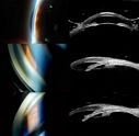
Angle Closure/ UBM & Gonioscopy1755 viewsPatient comes in a day after dilation with severe headache and nausea. Patient's IOP was 64 in the right eye. Patient ended up having angle closure attack. Promoted severe nausea and eye pain. Peripheral Iridotomy was performed and IOP dropped slowly. Patient was followed up the next morning and will be seen 2-days after that. Jun 03, 2013
|
|

360-Degree's Pseudoexfoliation1641 viewsFollow-up on glaucoma. Patient has sever pseudoexfoliation in the right eye. VA is 20/25 with IOP at 27. Patient will return in 6-months for re-evaluation. May 30, 2013
|
|
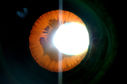
360-Degree's Pseudoexfoliation1183 viewsFollow-up on glaucoma. Patient has sever pseudoexfoliation in the right eye. VA is 20/25 with IOP at 27. Patient will return in 6-months for re-evaluation. May 30, 2013
|
|
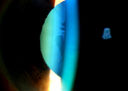
360-Degree's Pseudoexfoliation1372 viewsFollow-up on glaucoma. Patient has sever pseudoexfoliation in the right eye. VA is 20/25 with IOP at 27. Patient will return in 6-months for re-evaluation. May 30, 2013
|
|

Ozurdex Implant in AC1737 views79 yr old male presents with Blurry vison with slight discomfort x 1 in OD. Pt is S/P Ozurdex Implant x 2 mos which has now surfaced in the Anterior Chamber. Mar 22, 2013
|
|
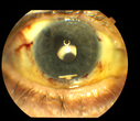
Ozurdex Implant in AC1290 views79yr old male presents Blurry vision with slight discomfort x 1 week in OD . Pt is S/P Ozurdex Implant x 2mos which is now surfaced in the Anterior Chamber.Mar 22, 2013
|
|
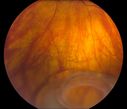
Total Dislocation of Intra Ocular Lens into the Vitreous514 views84-year old female comes in with blurred vision in the left eye. VA was 20/30, right eye and Count Fingers in the left eye. Fundus examination reveals dislocation of the IOL into the vitreous inferiorily @ 6-o'clock. Suggest surgery to fix the problem. Mar 18, 2013
|
|

Total Dislocation of Intra Ocular Lens into the Vitreous432 views84-year old female comes in with blurred vision in the left eye. VA was 20/30, right eye and Count Fingers in the left eye. Fundus examination reveals dislocation of the IOL into the vitreous inferiorily @ 6-o'clock. Suggest surgery to fix the problem. Mar 18, 2013
|
|

Total Dislocation of Intra Ocular Lens into the Vitreous441 views84-year old female comes in with blurred vision in the left eye. VA was 20/30, right eye and Count Fingers in the left eye. Fundus examination reveals dislocation of the IOL into the vitreous inferiorily @ 6-o'clock. Suggest surgery to fix the problem. Mar 18, 2013
|
|
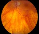
Total Dislocation of Intra Ocular Lens into the Vitreous442 views84-year old female comes in with blurred vision in the left eye. VA was 20/30, right eye and Count Fingers in the left eye. Fundus examination reveals dislocation of the IOL into the vitreous inferiorily @ 6-o'clock. Suggest surgery to fix the problem. Mar 18, 2013
|
|

Total Dislocation of Intra Ocular Lens into the Vitreous429 views84-year old female comes in with blurred vision in the left eye. VA was 20/30, right eye and Count Fingers in the left eye. Fundus examination reveals dislocation of the IOL into the vitreous inferiorily @ 6-o'clock. Suggest surgery to fix the problem. Mar 18, 2013
|
|

Total Dislocation of Intra Ocular Lens into the Vitreous465 views84-year old female comes in with blurred vision in the left eye. VA was 20/30, right eye and Count Fingers in the left eye. Fundus examination reveals dislocation of the IOL into the vitreous inferiorily @ 6-o'clock. Suggest surgery to fix the problem. Mar 18, 2013
|
|

Total Dislocation of Intra Ocular Lens into the Vitreous447 views84-year old female comes in with blurred vision in the left eye. VA was 20/30, right eye and Count Fingers in the left eye. Fundus examination reveals dislocation of the IOL into the vitreous inferiorily @ 6-o'clock. Suggest surgery to fix the problem. Mar 18, 2013
|
|

Total Dislocation of Intra Ocular Lens into the Vitreous451 views84-year old female comes in with blurred vision in the left eye. VA was 20/30, right eye and Count Fingers in the left eye. Fundus examination reveals dislocation of the IOL into the vitreous inferiorily @ 6-o'clock. Suggest surgery to fix the problem. Mar 18, 2013
|
|

Total Dislocation of Intra Ocular Lens into the Vitreous509 views84-year old female comes in with blurred vision in the left eye. VA was 20/30, right eye and Count Fingers in the left eye. Fundus examination reveals dislocation of the IOL into the vitreous inferiorily @ 6-o'clock. Suggest surgery to fix the problem. Mar 18, 2013
|
|

Total Dislocation of Intra Ocular Lens into the Vitreous421 views84-year old female comes in with blurred vision in the left eye. VA was 20/30, right eye and Count Fingers in the left eye. Fundus examination reveals dislocation of the IOL into the vitreous inferiorily @ 6-o'clock. Suggest surgery to fix the problem. Mar 18, 2013
|
|

Total Dislocation of Intra Ocular Lens into the Vitreous409 views84-year old female comes in with blurred vision in the left eye. VA was 20/30, right eye and Count Fingers in the left eye. Fundus examination reveals dislocation of the IOL into the vitreous inferiorily @ 6-o'clock. Suggest surgery to fix the problem. Mar 18, 2013
|
|
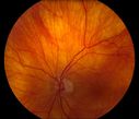
Total Dislocation of Intra Ocular Lens into the Vitreous405 views84-year old female comes in with blurred vision in the left eye. VA was 20/30, right eye and Count Fingers in the left eye. Fundus examination reveals dislocation of the IOL into the vitreous inferiorily @ 6-o'clock. Suggest surgery to fix the problem. Mar 18, 2013
|
|

Total Dislocation of Intra Ocular Lens into the Vitreous413 views84-year old female comes in with blurred vision in the left eye. VA was 20/30, right eye and Count Fingers in the left eye. Fundus examination reveals dislocation of the IOL into the vitreous inferiorily @ 6-o'clock. Suggest surgery to fix the problem. Mar 18, 2013
|
|

Total Dislocation of Intra Ocular Lens into the Vitreous509 views84-year old female comes in with blurred vision in the left eye. VA was 20/30, right eye and Count Fingers in the left eye. Fundus examination reveals dislocation of the IOL into the vitreous inferiorily @ 6-o'clock. Suggest surgery to fix the problem. Mar 18, 2013
|
|

Total Dislocation of Intra Ocular Lens into the Vitreous459 views84-year old female comes in with blurred vision in the left eye. VA was 20/30, right eye and Count Fingers in the left eye. Fundus examination reveals dislocation of the IOL into the vitreous inferiorily @ 6-o'clock. Suggest surgery to fix the problem. Mar 18, 2013
|
|

Total Dislocation of Intra Ocular Lens into the Vitreous519 views84-year old female comes in with blurred vision in the left eye. VA was 20/30, right eye and Count Fingers in the left eye. Fundus examination reveals dislocation of the IOL into the vitreous inferiorily @ 6-o'clock. Suggest surgery to fix the problem. Mar 18, 2013
|
|

Total Dislocation of Intra Ocular Lens into the Vitreous715 views84-year old female comes in with blurred vision in the left eye. VA was 20/30, right eye and Count Fingers in the left eye. Fundus examination reveals dislocation of the IOL into the vitreous inferiorily @ 6-o'clock. Suggest surgery to fix the problem. Mar 18, 2013
|
|

Rubeosis Iridis534 viewsPatient presents with Rubeosis Iridis in the right eye due to neovascular glaucoma. VA is 20/40 in the right eye. Will follow up in 3-months.Mar 13, 2013
|
|

Rubeosis Iridis1487 viewsPatient presents with Rubeosis Iridis in the right eye due to neovascular glaucoma. VA is 20/40 in the right eye. Will follow up in 3-months.Mar 13, 2013
|
|

Rubeosis Iridis468 viewsPatient presents with Rubeosis Iridis in the right eye due to neovascular glaucoma. VA is 20/40 in the right eye. Will follow up in 3-months.Mar 13, 2013
|
|

Rubeosis Iridis511 viewsPatient presents with Rubeosis Iridis in the right eye due to neovascular glaucoma. VA is 20/40 in the right eye. Will follow up in 3-months.Mar 13, 2013
|
|

Rubeosis Iridis511 viewsPatient presents with Rubeosis Iridis in the right eye due to neovascular glaucoma. VA is 20/40 in the right eye. Will follow up in 3-months.Mar 13, 2013
|
|

Rubeosis Iridis466 viewsPatient presents with Rubeosis Iridis in the right eye due to neovascular glaucoma. VA is 20/40 in the right eye. Will follow up in 3-months.Mar 13, 2013
|
|

Rubeosis Iridis377 viewsPatient presents with Rubeosis Iridis in the right eye due to neovascular glaucoma. VA is 20/40 in the right eye. Will follow up in 3-months.Mar 13, 2013
|
|

Rubeosis Iridis503 viewsPatient presents with Rubeosis Iridis in the right eye due to neovascular glaucoma. VA is 20/40 in the right eye. Will follow up in 3-months.Mar 13, 2013
|
|

Rubeosis Iridis511 viewsPatient presents with Rubeosis Iridis in the right eye due to neovascular glaucoma. VA is 20/40 in the right eye. Will follow up in 3-months.Mar 13, 2013
|
|

Rubeosis Iridis482 viewsPatient presents with Rubeosis Iridis in the right eye due to neovascular glaucoma. VA is 20/40 in the right eye. Will follow up in 3-months.Mar 13, 2013
|
|

Rubeosis Iridis437 viewsPatient presents with Rubeosis Iridis in the right eye due to neovascular glaucoma. VA is 20/40 in the right eye. Will follow up in 3-months.Mar 13, 2013
|
|
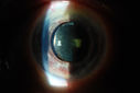
Rubeosis Iridis590 viewsPatient presents with Rubeosis Iridis in the right eye due to neovascular glaucoma. VA is 20/40 in the right eye. Will follow up in 3-months.Mar 13, 2013
|
|

Rubeosis Iridis666 viewsPatient presents with Rubeosis Iridis in the right eye due to neovascular glaucoma. VA is 20/40 in the right eye. Will follow up in 3-months.Mar 13, 2013
|
|
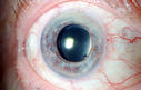
Rubeosis Iridis818 viewsPatient presents with Rubeosis Iridis in the right eye due to neovascular glaucoma. VA is 20/40 in the right eye. Will follow up in 3-months.Mar 13, 2013
|
|

Rubeosis Iridis704 viewsPatient presents with Rubeosis Iridis in the right eye due to neovascular glaucoma. VA is 20/40 in the right eye. Will follow up in 3-months.Mar 13, 2013
|
|

Gonioscopy; Scattered Peripheral Anterior Synechiae1602 viewsPatient comes in for evaluation for glaucoma. Patient also has a history of Uveitis. Last flare up was back in 1990. Patient's VA was 20/30, Right eye and 20/40-1, Left eye. Slit Lamp Gonioscopy reveals iris bow with scattered PAS around the angles of the anterior chamber. You can also see pigmentation in the trabecular meshwork. Patient will follow up in 3-months. Mar 12, 2013
|
|

Gonioscopy; Scattered Peripheral Anterior Synechiae1792 viewsPatient comes in for evaluation for glaucoma. Patient also has a history of Uveitis. Last flare up was back in 1990. Patient's VA was 20/30, Right eye and 20/40-1, Left eye. Slit Lamp Gonioscopy reveals iris bow with scattered PAS around the angles of the anterior chamber. You can also see pigmentation in the trabecular meshwork. Patient will follow up in 3-months. Mar 12, 2013
|
|

Gonioscopy; Scattered Peripheral Anterior Synechiae1347 viewsPatient comes in for evaluation for glaucoma. Patient also has a history of Uveitis. Last flare up was back in 1990. Patient's VA was 20/30, Right eye and 20/40-1, Left eye. Slit Lamp Gonioscopy reveals iris bow with scattered PAS around the angles of the anterior chamber. You can also see pigmentation in the trabecular meshwork. Patient will follow up in 3-months. Mar 12, 2013
|
|

Gonioscopy; Scattered Peripheral Anterior Synechiae1226 viewsPatient comes in for evaluation for glaucoma. Patient also has a history of Uveitis. Last flare up was back in 1990. Patient's VA was 20/30, Right eye and 20/40-1, Left eye. Slit Lamp Gonioscopy reveals iris bow with scattered PAS around the angles of the anterior chamber. You can also see pigmentation in the trabecular meshwork. Patient will follow up in 3-months. Mar 12, 2013
|
|

Gonioscopy; Scattered Peripheral Anterior Synechiae1422 viewsPatient comes in for evaluation for glaucoma. Patient also has a history of Uveitis. Last flare up was back in 1990. Patient's VA was 20/30, Right eye and 20/40-1, Left eye. Slit Lamp Gonioscopy reveals iris bow with scattered PAS around the angles of the anterior chamber. You can also see pigmentation in the trabecular meshwork. Patient will follow up in 3-months. Mar 12, 2013
|
|

Gonioscopy; Scattered Peripheral Anterior Synechiae1686 viewsPatient comes in for evaluation for glaucoma. Patient also has a history of Uveitis. Last flare up was back in 1990. Patient's VA was 20/30, Right eye and 20/40-1, Left eye. Slit Lamp Gonioscopy reveals iris bow with scattered PAS around the angles of the anterior chamber. You can also see pigmentation in the trabecular meshwork. Patient will follow up in 3-months. Mar 12, 2013
|
|

Gonioscopy; Scattered Peripheral Anterior Synechiae1432 viewsPatient comes in for evaluation for glaucoma. Patient also has a history of Uveitis. Last flare up was back in 1990. Patient's VA was 20/30, Right eye and 20/40-1, Left eye. Slit Lamp Gonioscopy reveals iris bow with scattered PAS around the angles of the anterior chamber. You can also see pigmentation in the trabecular meshwork. Patient will follow up in 3-months. Mar 12, 2013
|
|

Gonioscopy; Scattered Peripheral Anterior Synechiae1141 viewsPatient comes in for evaluation for glaucoma. Patient also has a history of Uveitis. Last flare up was back in 1990. Patient's VA was 20/30, Right eye and 20/40-1, Left eye. Slit Lamp Gonioscopy reveals iris bow with scattered PAS around the angles of the anterior chamber. You can also see pigmentation in the trabecular meshwork. Patient will follow up in 3-months. Mar 12, 2013
|
|

Gonioscopy; Scattered Peripheral Anterior Synechiae1073 viewsPatient comes in for evaluation for glaucoma. Patient also has a history of Uveitis. Last flare up was back in 1990. Patient's VA was 20/30, Right eye and 20/40-1, Left eye. Slit Lamp Gonioscopy reveals iris bow with scattered PAS around the angles of the anterior chamber. You can also see pigmentation in the trabecular meshwork. Patient will follow up in 3-months. Mar 12, 2013
|
|
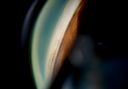
Gonioscopy; Scattered Peripheral Anterior Synechiae1053 viewsPatient comes in for evaluation for glaucoma. Patient also has a history of Uveitis. Last flare up was back in 1990. Patient's VA was 20/30, Right eye and 20/40-1, Left eye. Slit Lamp Gonioscopy reveals iris bow with scattered PAS around the angles of the anterior chamber. You can also see pigmentation in the trabecular meshwork. Patient will follow up in 3-months. Mar 12, 2013
|
|

Gonioscopy; Scattered Peripheral Anterior Synechiae1161 viewsPatient comes in for evaluation for glaucoma. Patient also has a history of Uveitis. Last flare up was back in 1990. Patient's VA was 20/30, Right eye and 20/40-1, Left eye. Slit Lamp Gonioscopy reveals iris bow with scattered PAS around the angles of the anterior chamber. You can also see pigmentation in the trabecular meshwork. Patient will follow up in 3-months. Mar 12, 2013
|
|

Gonioscopy; Scattered Peripheral Anterior Synechiae1431 viewsPatient comes in for evaluation for glaucoma. Patient also has a history of Uveitis. Last flare up was back in 1990. Patient's VA was 20/30, Right eye and 20/40-1, Left eye. Slit Lamp Gonioscopy reveals iris bow with scattered PAS around the angles of the anterior chamber. You can also see pigmentation in the trabecular meshwork. Patient will follow up in 3-months. Mar 12, 2013
|
|

Gonioscopy; Scattered Peripheral Anterior Synechiae1297 viewsPatient comes in for evaluation for glaucoma. Patient also has a history of Uveitis. Last flare up was back in 1990. Patient's VA was 20/30, Right eye and 20/40-1, Left eye. Slit Lamp Gonioscopy reveals iris bow with scattered PAS around the angles of the anterior chamber. You can also see pigmentation in the trabecular meshwork. Patient will follow up in 3-months. Mar 12, 2013
|
|

Lid Verruca Vulgaris764 viewsMar 07, 2013
|
|

Lid Verruca Vulgaris776 viewsMar 07, 2013
|
|

Lid Verruca Vulgaris889 viewsMar 07, 2013
|
|

Accordioning Crystalline Lens with loss of Posterior Capsule683 views71-year old male complains of blurred vision in the left eye. VA 20/40, right eye and 20/400, left eye without correction. Slit lamp exam shows Crystalline lens, both eyes. Right eye IOL is aligned and centered. Left eye shows an accordion of the crystalline lens. Retro Illumination shows the IOL bent inward in the left eye. There was a 6-diopter difference of astigmatism between the right and left eye. Patient will have surgery to correct the issue. Mar 06, 2013
|
|

Accordioning Crystalline Lens with loss of Posterior Capsule1265 views71-year old male complains of blurred vision in the left eye. VA 20/40, right eye and 20/400, left eye without correction. Slit lamp exam shows Crystalline lens, both eyes. Right eye IOL is aligned and centered. Left eye shows an accordion of the crystalline lens. Retro Illumination shows the IOL bent inward in the left eye. There was a 6-diopter difference of astigmatism between the right and left eye. Patient will have surgery to correct the issue. Mar 06, 2013
|
|
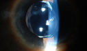
Accordioning Crystalline Lens with loss of Posterior Capsule536 views71-year old male complains of blurred vision in the left eye. VA 20/40, right eye and 20/400, left eye without correction. Slit lamp exam shows Crystalline lens, both eyes. Right eye IOL is aligned and centered. Left eye shows an accordion of the crystalline lens. Retro Illumination shows the IOL bent inward in the left eye. There was a 6-diopter difference of astigmatism between the right and left eye. Patient will have surgery to correct the issue. Mar 06, 2013
|
|

Accordioning Crystalline Lens with loss of Posterior Capsule749 views71-year old male complains of blurred vision in the left eye. VA 20/40, right eye and 20/400, left eye without correction. Slit lamp exam shows Crystalline lens, both eyes. Right eye IOL is aligned and centered. Left eye shows an accordion of the crystalline lens. Retro Illumination shows the IOL bent inward in the left eye. There was a 6-diopter difference of astigmatism between the right and left eye. Patient will have surgery to correct the issue. Mar 06, 2013
|
|

Accordioning Crystalline Lens with loss of Posterior Capsule776 views71-year old male complains of blurred vision in the left eye. VA 20/40, right eye and 20/400, left eye without correction. Slit lamp exam shows Crystalline lens, both eyes. Right eye IOL is aligned and centered. Left eye shows an accordion of the crystalline lens. Retro Illumination shows the IOL bent inward in the left eye. There was a 6-diopter difference of astigmatism between the right and left eye. Patient will have surgery to correct the issue. Mar 06, 2013
|
|

Accordioning Crystalline Lens with loss of Posterior Capsule875 views71-year old male complains of blurred vision in the left eye. VA 20/40, right eye and 20/400, left eye without correction. Slit lamp exam shows Crystalline lens, both eyes. Right eye IOL is aligned and centered. Left eye shows an accordion of the crystalline lens. Retro Illumination shows the IOL bent inward in the left eye. There was a 6-diopter difference of astigmatism between the right and left eye. Patient will have surgery to correct the issue. Mar 06, 2013
|
|

Accordioning Crystalline Lens with loss of Posterior Capsule907 views71-year old male complains of blurred vision in the left eye. VA 20/40, right eye and 20/400, left eye without correction. Slit lamp exam shows Crystalline lens, both eyes. Right eye IOL is aligned and centered. Left eye shows an accordion of the crystalline lens. Retro Illumination shows the IOL bent inward in the left eye. There was a 6-diopter difference of astigmatism between the right and left eye. Patient will have surgery to correct the issue. Mar 06, 2013
|
|

Corneal Abrasion with Foreign Body Present 1143 viewsER patient comes in with corneal abrasion in the left eye which was getting worst. VA 20/25. Slit lamp exam showed corneal abrasion superiorly at 2-o'clock. Flipped eyelid and foreign body appeared on the upper tarsal plate. Foreign body was removed. Feb 26, 2013
|
|
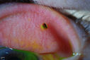
Corneal Abrasion with Foreign Body Present 2632 viewsER patient comes in with corneal abrasion in the left eye which was getting worst. VA 20/25. Slit lamp exam showed corneal abrasion superiorly at 2-o'clock. Flipped eyelid and foreign body appeared on the upper tarsal plate. Foreign body was removed. Feb 26, 2013
|
|

Corneal Abrasion with Foreign Body Present 1339 viewsER patient comes in with corneal abrasion in the left eye which was getting worst. VA 20/25. Slit lamp exam showed corneal abrasion superiorly at 2-o'clock. Flipped eyelid and foreign body appeared on the upper tarsal plate. Foreign body was removed. Feb 26, 2013
|
|

Corneal Abrasion with Foreign Body Present 2436 viewsER patient comes in with corneal abrasion in the left eye which was getting worst. VA 20/25. Slit lamp exam showed corneal abrasion superiorly at 2-o'clock. Flipped eyelid and foreign body appeared on the upper tarsal plate. Foreign body was removed. Feb 26, 2013
|
|
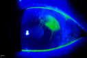
Corneal Abrasion with Foreign Body Present 664 viewsER patient comes in with corneal abrasion in the left eye which was getting worst. VA 20/25. Slit lamp exam showed corneal abrasion superiorly at 2-o'clock. Flipped eyelid and foreign body appeared on the upper tarsal plate. Foreign body was removed. Feb 26, 2013
|
|

Corneal Abrasion with Foreign Body Present 631 viewsER patient comes in with corneal abrasion in the left eye which was getting worst. VA 20/25. Slit lamp exam showed corneal abrasion superiorly at 2-o'clock. Flipped eyelid and foreign body appeared on the upper tarsal plate. Foreign body was removed. Feb 26, 2013
|
|

Corneal Abrasion with Foreign Body Present 982 viewsER patient comes in with corneal abrasion in the left eye which was getting worst. VA 20/25. Slit lamp exam showed corneal abrasion superiorly at 2-o'clock. Flipped eyelid and foreign body appeared on the upper tarsal plate. Foreign body was removed. Feb 26, 2013
|
|
| 398 files on 4 page(s) |
 |
3 |
|


