neueste Dateien - Anterior Segment
|
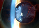
Subluxated Crystalline IOL with Pseudoexfoliation735 x angesehen82-year old male comes in with blurriness on the bottom half of his vision. Patient's VA was 20/25. Slit lamp examination shows subluxated crystalline lens inferiorily. Pseudoexfoliation debris superiorly at 10 o'clock with no vitreous present in the anterior chamber. Patient scheduled for surgery for CE IOL in the right eye. 04. Februar 2013
|
|

Subluxated Crystalline IOL with Pseudoexfoliation810 x angesehen82-year old male comes in with blurriness on the bottom half of his vision. Patient's VA was 20/25. Slit lamp examination shows subluxated crystalline lens inferiorily. Pseudoexfoliation debris superiorly at 10 o'clock with no vitreous present in the anterior chamber. Patient scheduled for surgery for CE IOL in the right eye. 04. Februar 2013
|
|

Subluxated Crystalline IOL with Pseudoexfoliation771 x angesehen82-year old male comes in with blurriness on the bottom half of his vision. Patient's VA was 20/25. Slit lamp examination shows subluxated crystalline lens inferiorily. Pseudoexfoliation debris superiorly at 10 o'clock with no vitreous present in the anterior chamber. Patient scheduled for surgery for CE IOL in the right eye. 04. Februar 2013
|
|

Subluxated Crystalline IOL with Pseudoexfoliation772 x angesehen82-year old male comes in with blurriness on the bottom half of his vision. Patient's VA was 20/25. Slit lamp examination shows subluxated crystalline lens inferiorily. Pseudoexfoliation debris superiorly at 10 o'clock with no vitreous present in the anterior chamber. Patient scheduled for surgery for CE IOL in the right eye. 04. Februar 2013
|
|
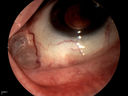
Displaced Scleral Buckle765 x angesehenDisplaced Scleral Buckle in OD of a 75 yr old male08. Januar 2013
|
|

Displaced Scleral Buckle1356 x angesehenDisplaced Scleral Buckle in OD of a 75 yr old male08. Januar 2013
|
|

Phacoantigenic Reaction1289 x angesehenPicture shows a Left eye enucleation with H&E staining.
Anterior capsule of lens is broken with evidence of granulomatous inflammation. 03. Dezember 2012
|
|

Conjunctival Vascular Anomaly841 x angesehenPatient presents a vascular pattern of blood vessels @ 10 o'clock of the superior-temporal quadrant of the right eye. Patient has had flare ups with anterior scleritis in the past which is controlled by Methotrexate. Patient's VA is currently 20/40 with PH 20/30, right eye. 05. November 2012
|
|

Conjunctival Vascular Anomaly882 x angesehenPatient presents a vascular pattern of blood vessels @ 10 o'clock of the superior-temporal quadrant of the right eye. Patient has had flare ups with anterior scleritis in the past which is controlled by Methotrexate. Patient's VA is currently 20/40 with PH 20/30, right eye. 05. November 2012
|
|

Conjunctival Vascular Anomaly938 x angesehenPatient presents a vascular pattern of blood vessels @ 10 o'clock of the superior-temporal quadrant of the right eye. Patient has had flare ups with anterior scleritis in the past which is controlled by Methotrexate. Patient's VA is currently 20/40 with PH 20/30, right eye. 05. November 2012
|
|

Pigmentary Glaucoma, Zonules visible Inferiorly...446 x angesehenYoung gentleman with pigmentary glaucoma. Heavy pigment lining trabecular meshwork with Goinoscopy. Crisp image of the zonules. 15. Oktober 2012
|
|

Gonioscopy- Pigmentary Glaucoma Suspect1307 x angesehenPatient with no family history of glaucoma, comes in with elevated IOP. During Gonioscopy exam. brown pigment overlying the trabecular meshwork. Also, trans-illumination defects on the iris.29. August 2012
|
|

Gonioscopy- Pigmentary Glaucoma Suspect1356 x angesehenPatient with no family history of glaucoma, comes in with elevated IOP. During Gonioscopy exam. brown pigment overlying the trabecular meshwork. Also, trans-illumination defects on the iris.29. August 2012
|
|
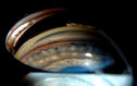
Gonioscopy- Pigmentary Glaucoma Suspect1717 x angesehenPatient with no family history of glaucoma, comes in with elevated IOP. During Gonioscopy exam. brown pigment overlying the trabecular meshwork. Also, trans-illumination defects on the iris.29. August 2012
|
|

Gonioscopy- Pigmentary Glaucoma Suspect1435 x angesehenPatient with no family history of glaucoma, comes in with elevated IOP. During Gonioscopy exam. brown pigment overlying the trabecular meshwork. Also, trans-illumination defects on the iris.29. August 2012
|
|

Gonioscopy- Pigmentary Glaucoma Suspect1224 x angesehenPatient with no family history of glaucoma, comes in with elevated IOP. During Gonioscopy exam. brown pigment overlying the trabecular meshwork. Also, trans-illumination defects on the iris.29. August 2012
|
|
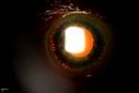
Trans-Illumination Defects626 x angesehen74-year old patient who VA 20/70 OD, 20/50 OS. Complaints of blurred vision. Iris atrophy, both eyes. ERM, right eye, Patient to have cataract surgery to improve distance vision. 28. August 2012
|
|
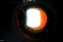
Trans-Illumination Defects657 x angesehen74-year old patient who VA 20/70 OD, 20/50 OS. Complaints of blurred vision. Iris atrophy, both eyes. ERM, right eye, Patient to have cataract surgery to improve distance vision. 28. August 2012
|
|
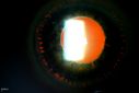
Trans-Illumination Defects613 x angesehen74-year old patient who VA 20/70 OD, 20/50 OS. Complaints of blurred vision. Iris atrophy, both eyes. ERM, right eye, Patient to have cataract surgery to improve distance vision. 28. August 2012
|
|
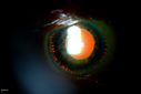
Trans-Illumination Defects749 x angesehen74-year old patient who VA 20/70 OD, 20/50 OS. Complaints of blurred vision. Iris atrophy, both eyes. ERM, right eye, Patient to have cataract surgery to improve distance vision. 28. August 2012
|
|
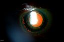
Trans-Illumination Defects767 x angesehen74-year old patient who VA 20/70 OD, 20/50 OS. Complaints of blurred vision. Iris atrophy, both eyes. ERM, right eye, Patient to have cataract surgery to improve distance vision. 28. August 2012
|
|
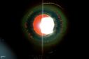
Trans-Illumination Defects737 x angesehen74-year old patient who VA 20/70 OD, 20/50 OS. Complaints of blurred vision. Iris atrophy, both eyes. ERM, right eye, Patient to have cataract surgery to improve distance vision. 28. August 2012
|
|

Pseudoexfoliation Ring564 x angesehenPatient's VA 20/50 with pinhole no improvement. Patient will have cataract surgery in the future. Notice Pseudoexfoliation ring centrally.28. August 2012
|
|
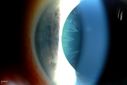
Pseudoexfoliation Ring464 x angesehenPatient's VA 20/50 with pinhole no improvement. Patient will have cataract surgery in the future. Notice Pseudoexfoliation ring centrally.28. August 2012
|
|

Pseudoexfoliation Ring488 x angesehenPatient's VA 20/50 with pinhole no improvement. Patient will have cataract surgery in the future. Notice Pseudoexfoliation ring centrally.28. August 2012
|
|

Pseudoexfoliation Ring496 x angesehenPatient's VA 20/50 with pinhole no improvement. Patient will have cataract surgery in the future. Notice Pseudoexfoliation ring centrally.28. August 2012
|
|

Pseudoexfoliation Ring518 x angesehenPatient's VA 20/50 with pinhole no improvement. Patient will have cataract surgery in the future. Notice Pseudoexfoliation ring centrally.28. August 2012
|
|
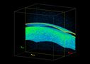
3-D HD OCT Image of Soft Contact Lens over Cornea756 x angesehen11. Juli 2012
|
|

Blood Strand through Anterior Chamber821 x angesehen11. Juli 2012
|
|

Blood Strand through Anterior Chamber798 x angesehen11. Juli 2012
|
|
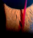
Blood strand into the anterior chamber759 x angesehen10. Juli 2012
|
|

Endophthalmitis961 x angesehen10. Juli 2012
|
|

Endophthalmitis with Hypopion1333 x angesehen10. Juli 2012
|
|

Endophthalmitis with Hypopion1376 x angesehen10. Juli 2012
|
|

Juvenile Xanthogranuloma758 x angesehenPresented with hyphaema after minor trauma05. Juli 2012
|
|

Juvenile Xanthogranuloma1421 x angesehenPresented with hyphaema after minor trauma05. Juli 2012
|
|
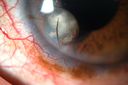
Cilia and Cement Intraocular Foreign Body938 x angesehenConstruction worker jack-hammering 3 months prior to pictures. Asymptomatic. 20/20 OU. Corneal wound seen superiorly (not in pictures)05. Juni 2012
|
|

Cilia and Cement Intraocular Foreign Body1187 x angesehenConstruction worker jack-hammering 3 months prior to pictures. Asymptomatic. 20/20 OU. Corneal wound seen superiorly (not in pictures)05. Juni 2012
|
|
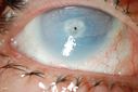
Corneal Ulcer. 609 x angesehenA hole in the cornea. Aqueous Humor leak16. Mai 2012
|
|

Corneal Ulcer. 546 x angesehenA hole in the cornea. Aqueous Humor leak16. Mai 2012
|
|

Corneal Ulcer. 493 x angesehenA hole in the cornea. Aqueous Humor leak16. Mai 2012
|
|
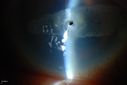
Corneal Ulcer. 606 x angesehenA hole in the cornea. Aqueous Humor leak16. Mai 2012
|
|
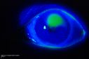
Cornea Ulcer/Abrasion589 x angesehen16. Mai 2012
|
|

Cornea Ulcer/Abrasion1377 x angesehen16. Mai 2012
|
|

Cornea Ulcer/Abrasion639 x angesehen16. Mai 2012
|
|

Cornea Ulcer/Abrasion531 x angesehen16. Mai 2012
|
|

Cornea Ulcer/Abrasion545 x angesehen16. Mai 2012
|
|
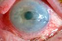
Cornea Ulcer/Abrasion645 x angesehen16. Mai 2012
|
|

Cornea Ulcer/Abrasion614 x angesehen16. Mai 2012
|
|

Cornea Ulcer544 x angesehen16. Mai 2012
|
|

4-up of Cornea Ulcer with Iris Prolaps843 x angesehen16. Mai 2012
|
|

Cornea Ulcer with Iris Prolaps1058 x angesehen16. Mai 2012
|
|

Cornea Ulcer654 x angesehen16. Mai 2012
|
|

Cornea Ulcer677 x angesehen16. Mai 2012
|
|

Cornea Ulcer683 x angesehen16. Mai 2012
|
|

Cornea Ulcer692 x angesehen16. Mai 2012
|
|

Cornea Ulcer scar871 x angesehen16. Mai 2012
|
|

426 x angesehen08. Mai 2012
|
|
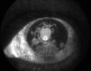
Iris Neovascularization1293 x angesehenDiabetic patient with advanced iris neovascularization08. Mai 2012
|
|

Tick on Lower Lid565 x angesehenPt. came in for cataract surgery and a tick was found on the lower lid. Pt. had no idea the tick was attached and did not know how long it had been there. Tick was photographed and then removed by physician.25. April 2012
|
|
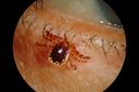
Tick on Lower Lid757 x angesehenPt. came in for cataract surgery and a tick was found on the lower lid. Pt. had no idea the tick was attached and did not know how long it had been there. Tick was photographed and then removed by physician.25. April 2012
|
|

Tick on Lower Lid575 x angesehenPt. came in for cataract surgery and a tick was found on the lower lid. Pt. had no idea the tick was attached and did not know how long it had been there. Tick was photographed and then removed by physician.25. April 2012
|
|

Tick on Lower Lid843 x angesehenPt. came in for cataract surgery and a tick was found on the lower lid. Pt. had no idea the tick was attached and did not know how long it had been there. Tick was photographed and then removed by physician.25. April 2012
|
|

Fuch's heterochromic iridocyclitis - inflammed blue eye1116 x angesehen46-year-old man has had blurred vision in the left eye for the last two years. He had cataract surgery in that eye eight years ago. He has also had a lot of floaters in the left eye for several years. His left eye is blue and his right eye is brown. OD 20/16, OS 20/50. 02. Januar 2012
|
|

Fuch's heterochromic iridocyclitis - uninvolved brown eye1264 x angesehen46-year-old man has had blurred vision in the left eye for the last two years. He had cataract surgery in that eye eight years ago. He has also had a lot of floaters in the left eye for several years. His left eye is blue and his right eye is brown. OD 20/16, OS 20/50. 02. Januar 2012
|
|
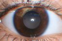
Sectoral Heterochromia Iridum - 3 year old boy916 x angesehen3-year-old child was seen in the office on September 14, 2011. Since he was an infant a white spot was noticed on the iris in the right eye, it seems to be growing and he comes in because of that. 15. Dezember 2011
|
|

Sectoral Heterochromia Iridum - 3 year old boy998 x angesehen3-year-old child was seen in the office on September 14, 2011. Since he was an infant a white spot was noticed on the iris in the right eye, it seems to be growing and he comes in because of that. 15. Dezember 2011
|
|
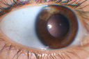
Sectoral Heterochromia Iridum - 3 year old boy912 x angesehen3-year-old child was seen in the office on September 14, 2011. Since he was an infant a white spot was noticed on the iris in the right eye, it seems to be growing and he comes in because of that. 15. Dezember 2011
|
|
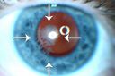
Post Lasik Epithelial Ingrowth735 x angesehen58-year-old man had a vitrectomy for a macular pucker in the right eye almost two years ago. His vision was good after his cornea surgeon removed the cells from his flap but then recently the cells have regrown and his vision has declined again.
OD is 20/100.13. Dezember 2011
|
|
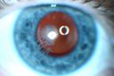
Post Lasik Epithelial Ingrowth710 x angesehen58-year-old man had a vitrectomy for a macular pucker in the right eye almost two years ago. His vision was good after his cornea surgeon removed the cells from his flap but then recently the cells have regrown and his vision has declined again.
OD is 20/100.13. Dezember 2011
|
|
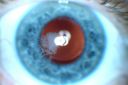
Post Lasik Epithelial Ingrowth922 x angesehen58-year-old man had a vitrectomy for a macular pucker in the right eye almost two years ago. His vision was good after his cornea surgeon removed the cells from his flap but then recently the cells have regrown and his vision has declined again.
OD is 20/100.13. Dezember 2011
|
|
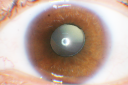
Ectopia Lentis and Cataract - Marfan's Syndrome1050 x angesehen40-year-old woman has had dislocated lenses since she was a child. The right eye is seeing well with an aphakic correction. The left eye unfortunately has declining vision for 4 months. OD 20/32, OS 20/125. Pinhole 20/5011. Dezember 2011
|
|
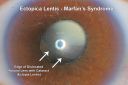
Ectopia Lentis and Cataract - Marfan's Syndrome1454 x angesehen40-year-old woman has had dislocated lenses since she was a child. The right eye is seeing well with an aphakic correction. The left eye unfortunately has declining vision for 4 months. OD 20/32, OS 20/125. Pinhole 20/5011. Dezember 2011
|
|

Uveitis - Iris Nodule - Koeppe Nodule1325 x angesehen29-year-old woman has had problems with uveitis in the right eye for about the last two years. Her symptoms are usually floaters. Occasionally the eye gets red, it has not been very uncomfortable, and it has always been in the right eye. Has had Iris Nodule like one in photo in left eye and Right eye. They clear with or without steroids after about a month.08. November 2011
|
|

Uveitis - Iris Nodule - Koeppe Nodule2123 x angesehen29-year-old woman has had problems with uveitis in the right eye for about the last two years. Her symptoms are usually floaters. Occasionally the eye gets red, it has not been very uncomfortable, and it has always been in the right eye. Has had Iris Nodule like one in photo in left eye and Right eye. They clear with or without steroids after about a month.08. November 2011
|
|

Uveitis - Iris Nodule - Koeppe Nodule1290 x angesehen29-year-old woman has had problems with uveitis in the right eye for about the last two years. Her symptoms are usually floaters. Occasionally the eye gets red, it has not been very uncomfortable, and it has always been in the right eye. Has had Iris Nodule like one in photo in left eye and Right eye. They clear with or without steroids after about a month.08. November 2011
|
|

Uveitis - Iris Nodule - Koeppe Nodule1446 x angesehen29-year-old woman has had problems with uveitis in the right eye for about the last two years. Her symptoms are usually floaters. Occasionally the eye gets red, it has not been very uncomfortable, and it has always been in the right eye. Has had Iris Nodule like one in photo in left eye and Right eye. They clear with or without steroids after about a month.08. November 2011
|
|
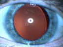
Ectopia Lentis - Marfan's Syndrome - Intermittent Pupillary Block Glaucoma Right Eye1210 x angesehen41-year-old woman decreased vision right eye and intermittent pupillary block glaucoma for 6 months from a dislocated natural lens in the eye, probably associated with Marfan syndrome.Vision OD is 20/160, PH 20/40; OS is 20/30, PH 20/2527. März 2011
|
|

Ectopia Lentis - Marfan's Syndrome - Intermittent Pupillary Block Glaucoma Right Eye1495 x angesehen41-year-old woman decreased vision right eye and intermittent pupillary block glaucoma for 6 months from a dislocated natural lens in the eye, probably associated with Marfan syndrome.Vision OD is 20/160, PH 20/40; OS is 20/30, PH 20/2527. März 2011
|
|

Ectopia Lentis - Marfan's Syndrome - Intermittent Pupillary Block Glaucoma Right Eye1660 x angesehen41-year-old woman decreased vision right eye and intermittent pupillary block glaucoma for 6 months from a dislocated natural lens in the eye, probably associated with Marfan syndrome.Vision OD is 20/160, PH 20/40; OS is 20/30, PH 20/2527. März 2011
|
|

Ectopia Lentis - Marfan's Syndrome - Intermittent Pupillary Block Glaucoma Right Eye1972 x angesehen41-year-old woman decreased vision right eye and intermittent pupillary block glaucoma for 6 months from a dislocated natural lens in the eye, probably associated with Marfan syndrome.Vision OD is 20/160, PH 20/40; OS is 20/30, PH 20/2527. März 2011
|
|
|
|
|
| 381 Dateien auf 4 Seite(n) |
 |
4 |
|


