Last additions - Congenital Anomalies
|

Congenital Hypetrophy of the Retinal Pigment Epithelium (CHRPE)440 views63 year old female with normal vision and CHRPE lesion in the right eye.Apr 27, 2020
|
|

Congenital Hypetrophy of the Retinal Pigment Epithelium (CHRPE)440 views63 year old female with normal vision and CHRPE lesion in the right eye.Apr 27, 2020
|
|
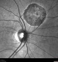
Congenital Hypetrophy of the Retinal Pigment Epithelium (CHRPE)728 views63 year old female with normal vision and CHRPE lesion in the right eye.Apr 27, 2020
|
|

Congenital Hypetrophy of the Retinal Pigment Epithelium (CHRPE)447 views63 year old female with normal vision and CHRPE lesion in the right eye.Apr 27, 2020
|
|

Congenital Hypetrophy of the Retinal Pigment Epithelium (CHRPE)439 views63 year old female with normal vision and CHRPE lesion in the right eye.Apr 27, 2020
|
|

Congenital Hypetrophy of the Retinal Pigment Epithelium (CHRPE)477 views63 year old female with normal vision and CHRPE lesion in the right eye.Apr 27, 2020
|
|

Congenital Hypetrophy of the Retinal Pigment Epithelium (CHRPE)451 views63 year old female with normal vision and CHRPE lesion in the right eye.Apr 27, 2020
|
|

Congenital Hypetrophy of the Retinal Pigment Epithelium (CHRPE)536 views63 year old female with normal vision and CHRPE lesion in the right eye.Apr 27, 2020
|
|

Coats Disease - Parafoveal Telangiectasis Type I - Right Eye510 views67 year old man with long history of vision loss in the right eye. Previously treated with laser, avastin. VA 20/200.Feb 11, 2020
|
|

Coats Disease - Parafoveal Telangiectasis Type I - Right Eye480 views67 year old man with long history of vision loss in the right eye. Previously treated with laser, avastin. VA 20/200.Feb 11, 2020
|
|

Coats Disease - Parafoveal Telangiectasis Type I - Right Eye472 views67 year old man with long history of vision loss in the right eye. Previously treated with laser, avastin. VA 20/200.Feb 11, 2020
|
|

Coats Disease - Parafoveal Telangiectasis Type I - Right Eye506 views67 year old man with long history of vision loss in the right eye. Previously treated with laser, avastin. VA 20/200.Feb 11, 2020
|
|

Coats Disease - Parafoveal Telangiectasis Type I - Right Eye436 views67 year old man with long history of vision loss in the right eye. Previously treated with laser, avastin. VA 20/200.Feb 11, 2020
|
|

Coats Disease - Parafoveal Telangiectasis Type I - Right Eye476 views67 year old man with long history of vision loss in the right eye. Previously treated with laser, avastin. VA 20/200.Feb 11, 2020
|
|

Coats Disease - Parafoveal Telangiectasis Type I - Right Eye479 views67 year old man with long history of vision loss in the right eye. Previously treated with laser, avastin. VA 20/200.Feb 11, 2020
|
|

Coats Disease - Parafoveal Telangiectasis Type I - Right Eye472 views67 year old man with long history of vision loss in the right eye. Previously treated with laser, avastin. VA 20/200.Feb 11, 2020
|
|

Coats Disease - Parafoveal Telangiectasis Type I - Right Eye498 views67 year old man with long history of vision loss in the right eye. Previously treated with laser, avastin. VA 20/200.Feb 11, 2020
|
|

Coats Disease - Parafoveal Telangiectasis Type I - Right Eye459 views67 year old man with long history of vision loss in the right eye. Previously treated with laser, avastin. VA 20/200.Feb 11, 2020
|
|

Coats Disease - Parafoveal Telangiectasis Type I - Right Eye446 views67 year old man with long history of vision loss in the right eye. Previously treated with laser, avastin. VA 20/200.Feb 11, 2020
|
|

Coats Disease - Parafoveal Telangiectasis Type I - Right Eye447 views67 year old man with long history of vision loss in the right eye. Previously treated with laser, avastin. VA 20/200.Feb 11, 2020
|
|
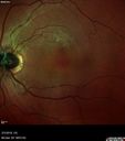
Coats Disease - Parafoveal Telangiectasis Type I - Right Eye476 views67 year old man with long history of vision loss in the right eye. Previously treated with laser, avastin. VA 20/200.Feb 11, 2020
|
|

Coats Disease - Parafoveal Telangiectasis Type I - Right Eye512 views67 year old man with long history of vision loss in the right eye. Previously treated with laser, avastin. VA 20/200.Feb 11, 2020
|
|

Coats Disease - Parafoveal Telangiectasis Type I - Right Eye480 views67 year old man with long history of vision loss in the right eye. Previously treated with laser, avastin. VA 20/200.Feb 11, 2020
|
|

Coats Disease - Parafoveal Telangiectasis Type I - Right Eye453 views67 year old man with long history of vision loss in the right eye. Previously treated with laser, avastin. VA 20/200.Feb 11, 2020
|
|

Coats Disease - Parafoveal Telangiectasis Type I - Right Eye478 views67 year old man with long history of vision loss in the right eye. Previously treated with laser, avastin. VA 20/200.Feb 11, 2020
|
|

Torpedo Maculopathy - Left Eye549 views25 year old man with normal vision. Left macula has torpedo lesionNov 17, 2019
|
|

Torpedo Maculopathy - Left Eye486 views25 year old man with normal vision. Left macula has torpedo lesionNov 17, 2019
|
|

Torpedo Maculopathy - Left Eye481 views25 year old man with normal vision. Left macula has torpedo lesionNov 17, 2019
|
|

Torpedo Maculopathy - Left Eye606 views25 year old man with normal vision. Left macula has torpedo lesionNov 17, 2019
|
|

Torpedo Maculopathy - Left Eye514 views25 year old man with normal vision. Left macula has torpedo lesionNov 17, 2019
|
|

Torpedo Maculopathy - Left Eye445 views25 year old man with normal vision. Left macula has torpedo lesionNov 17, 2019
|
|

Torpedo Maculopathy - Left Eye489 views25 year old man with normal vision. Left macula has torpedo lesionNov 17, 2019
|
|

Torpedo Maculopathy - Left Eye447 views25 year old man with normal vision. Left macula has torpedo lesionNov 17, 2019
|
|

Torpedo Maculopathy - Left Eye498 views25 year old man with normal vision. Left macula has torpedo lesionNov 17, 2019
|
|

Torpedo Maculopathy - Left Eye528 views25 year old man with normal vision. Left macula has torpedo lesionNov 17, 2019
|
|

Torpedo Maculopathy - Left Eye506 views25 year old man with normal vision. Left macula has torpedo lesionNov 17, 2019
|
|

Congenital hypertrophy of the retinal pigment epithelium CHRPE773 viewsFAF, Optos Photo and FAJun 11, 2019
|
|

Congenital hypertrophy of the retinal pigment epithelium CHRPE500 viewsJun 11, 2019
|
|

Congenital hypertrophy of the retinal pigment epithelium CHRPE536 viewsFA was done for scleritis in the fellow eyeJun 11, 2019
|
|

Nanophthalmous with Choroidal Folds458 views71 year old female with 20/20 vision and worsening cataracts. There is also a CHRPE lesion in the peripheryApr 06, 2019
|
|

Nanophthalmous with Choroidal Folds414 views71 year old female with 20/20 vision and worsening cataracts. There is also a CHRPE lesion in the peripheryApr 06, 2019
|
|

Nanophthalmous with Choroidal Folds457 views71 year old female with 20/20 vision and worsening cataracts. There is also a CHRPE lesion in the peripheryApr 06, 2019
|
|

Nanophthalmous with Choroidal Folds446 views71 year old female with 20/20 vision and worsening cataracts. There is also a CHRPE lesion in the peripheryApr 06, 2019
|
|

Nanophthalmous with Choroidal Folds411 views71 year old female with 20/20 vision and worsening cataracts. There is also a CHRPE lesion in the peripheryApr 06, 2019
|
|

Nanophthalmous with Choroidal Folds425 views71 year old female with 20/20 vision and worsening cataracts. There is also a CHRPE lesion in the peripheryApr 06, 2019
|
|

Nanophthalmous with Choroidal Folds484 views71 year old female with 20/20 vision and worsening cataracts. There is also a CHRPE lesion in the peripheryApr 06, 2019
|
|

Nanophthalmous with Choroidal Folds450 views71 year old female with 20/20 vision and worsening cataracts. There is also a CHRPE lesion in the peripheryApr 06, 2019
|
|

Nanophthalmous with Choroidal Folds434 views71 year old female with 20/20 vision and worsening cataracts. There is also a CHRPE lesion in the peripheryApr 06, 2019
|
|

Nanophthalmous with Choroidal Folds461 views71 year old female with 20/20 vision and worsening cataracts. There is also a CHRPE lesion in the peripheryApr 06, 2019
|
|

Nanophthalmous with Choroidal Folds495 views71 year old female with 20/20 vision and worsening cataracts. There is also a CHRPE lesion in the peripheryApr 06, 2019
|
|

Nanophthalmous with Choroidal Folds476 views71 year old female with 20/20 vision and worsening cataracts. There is also a CHRPE lesion in the peripheryApr 06, 2019
|
|

Nanophthalmous with Choroidal Folds454 views71 year old female with 20/20 vision and worsening cataracts. There is also a CHRPE lesion in the peripheryApr 06, 2019
|
|

Nanophthalmous with Choroidal Folds449 views71 year old female with 20/20 vision and worsening cataracts. There is also a CHRPE lesion in the peripheryApr 06, 2019
|
|
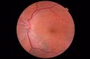
Nanophthalmous with Choroidal Folds465 views71 year old female with 20/20 vision and worsening cataracts. There is also a CHRPE lesion in the peripheryApr 06, 2019
|
|

Nanophthalmous with Choroidal Folds515 views71 year old female with 20/20 vision and worsening cataracts. There is also a CHRPE lesion in the peripheryApr 06, 2019
|
|

Nanophthalmous with Choroidal Folds435 views71 year old female with 20/20 vision and worsening cataracts. There is also a CHRPE lesion in the peripheryApr 06, 2019
|
|

Macular Macroaneurysm with Coats Disease473 views61 year old man with increased leakage from macular macroaneurysm right eyeApr 06, 2019
|
|
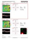
Macular Macroaneurysm with Coats Disease456 views61 year old man with increased leakage from macular macroaneurysm right eyeApr 06, 2019
|
|

Macular Macroaneurysm with Coats Disease454 views61 year old man with increased leakage from macular macroaneurysm right eyeApr 06, 2019
|
|

Macular Macroaneurysm with Coats Disease469 views61 year old man with increased leakage from macular macroaneurysm right eyeApr 06, 2019
|
|

Macular Macroaneurysm with Coats Disease561 views61 year old man with increased leakage from macular macroaneurysm right eyeApr 06, 2019
|
|

Macular Macroaneurysm with Coats Disease467 views61 year old man with increased leakage from macular macroaneurysm right eyeApr 06, 2019
|
|

Macular Macroaneurysm with Coats Disease496 views61 year old man with increased leakage from macular macroaneurysm right eyeApr 06, 2019
|
|

Macular Macroaneurysm with Coats Disease541 views61 year old man with increased leakage from macular macroaneurysm right eyeApr 06, 2019
|
|

Familial Exudative Vitreoretinopathy - FEVR - Stage 4b OD452 views10 year old child with poor vision OD from birth. The left eye had vascular remodelling in the temporal periphery with preretinal abnormalitlies seen on OCT. The patient never returned for a fluorescein angiogram. Left eye is either stage 1 or stage 2. no family historyJan 01, 2019
|
|

Familial Exudative Vitreoretinopathy - FEVR - Stage 4b OD669 views10 year old child with poor vision OD from birth. The left eye had vascular remodelling in the temporal periphery with preretinal abnormalitlies seen on OCT. The patient never returned for a fluorescein angiogram. Left eye is either stage 1 or stage 2. no family historyJan 01, 2019
|
|

Familial Exudative Vitreoretinopathy - FEVR - Stage 4b OD529 views10 year old child with poor vision OD from birth. The left eye had vascular remodelling in the temporal periphery with preretinal abnormalitlies seen on OCT. The patient never returned for a fluorescein angiogram. Left eye is either stage 1 or stage 2. no family historyJan 01, 2019
|
|

Familial Exudative Vitreoretinopathy - FEVR - Stage 4b OD476 views10 year old child with poor vision OD from birth. The left eye had vascular remodelling in the temporal periphery with preretinal abnormalitlies seen on OCT. The patient never returned for a fluorescein angiogram. Left eye is either stage 1 or stage 2. no family historyJan 01, 2019
|
|

Familial Exudative Vitreoretinopathy - FEVR - Stage 4b OD467 views10 year old child with poor vision OD from birth. The left eye had vascular remodelling in the temporal periphery with preretinal abnormalitlies seen on OCT. The patient never returned for a fluorescein angiogram. Left eye is either stage 1 or stage 2. no family historyJan 01, 2019
|
|

Familial Exudative Vitreoretinopathy - FEVR - Stage 4b OD463 views10 year old child with poor vision OD from birth. The left eye had vascular remodelling in the temporal periphery with preretinal abnormalitlies seen on OCT. The patient never returned for a fluorescein angiogram. Left eye is either stage 1 or stage 2. no family historyJan 01, 2019
|
|

Familial Exudative Vitreoretinopathy - FEVR - Stage 4b OD515 views10 year old child with poor vision OD from birth. The left eye had vascular remodelling in the temporal periphery with preretinal abnormalitlies seen on OCT. The patient never returned for a fluorescein angiogram. Left eye is either stage 1 or stage 2. no family historyJan 01, 2019
|
|

Familial Exudative Vitreoretinopathy - FEVR - Stage 4b OD492 views10 year old child with poor vision OD from birth. The left eye had vascular remodelling in the temporal periphery with preretinal abnormalitlies seen on OCT. The patient never returned for a fluorescein angiogram. Left eye is either stage 1 or stage 2. no family historyJan 01, 2019
|
|
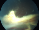
Familial Exudative Vitreoretinopathy - FEVR - Stage 4b OD544 views10 year old child with poor vision OD from birth. The left eye had vascular remodelling in the temporal periphery with preretinal abnormalitlies seen on OCT. The patient never returned for a fluorescein angiogram. Left eye is either stage 1 or stage 2. no family historyJan 01, 2019
|
|

Familial Exudative Vitreoretinopathy - FEVR - Stage 4b OD618 views10 year old child with poor vision OD from birth. The left eye had vascular remodelling in the temporal periphery with preretinal abnormalitlies seen on OCT. The patient never returned for a fluorescein angiogram. Left eye is either stage 1 or stage 2. no family historyJan 01, 2019
|
|

Familial Exudative Vitreoretinopathy - FEVR - Stage 4b OD623 views10 year old child with poor vision OD from birth. The left eye had vascular remodelling in the temporal periphery with preretinal abnormalitlies seen on OCT. The patient never returned for a fluorescein angiogram. Left eye is either stage 1 or stage 2. no family historyJan 01, 2019
|
|

Familial Exudative Vitreoretinopathy - FEVR - Stage 4b OD560 views10 year old child with poor vision OD from birth. The left eye had vascular remodelling in the temporal periphery with preretinal abnormalitlies seen on OCT. The patient never returned for a fluorescein angiogram. Left eye is either stage 1 or stage 2. no family historyJan 01, 2019
|
|

Familial Exudative Vitreoretinopathy - FEVR - Stage 4b OD737 views10 year old child with poor vision OD from birth. The left eye had vascular remodelling in the temporal periphery with preretinal abnormalitlies seen on OCT. The patient never returned for a fluorescein angiogram. Left eye is either stage 1 or stage 2. no family historyJan 01, 2019
|
|
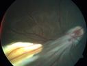
Familial Exudative Vitreoretinopathy - FEVR - Stage 4b OD822 views10 year old child with poor vision OD from birth. The left eye had vascular remodelling in the temporal periphery with preretinal abnormalitlies seen on OCT. The patient never returned for a fluorescein angiogram. Left eye is either stage 1 or stage 2. no family historyJan 01, 2019
|
|

Foveal hypoplasia - aplasia573 viewsSD OCT shows persistent plexiform layers in the center of the macula
EXTENDED HPI: He is having some trouble seeing his gadgets (like his watch). He was diagnosed with morning glory syndrome at about 3 yeras old. This last visit you saw fluid under the retina and asked him to come here. The vision is a little worse.
VA OD: Dcc20/40+2 NccJ2-2
VA OS: Dcc20/25-2 NccJ1-2Dec 20, 2018
|
|

Foveal hypoplasia - aplasia591 viewsSD OCT shows persistent plexiform layers in the center of the macula
EXTENDED HPI: He is having some trouble seeing his gadgets (like his watch). He was diagnosed with morning glory syndrome at about 3 yeras old. This last visit you saw fluid under the retina and asked him to come here. The vision is a little worse.
VA OD: Dcc20/40+2 NccJ2-2
VA OS: Dcc20/25-2 NccJ1-2Dec 20, 2018
|
|

Foveal hypoplasia - aplasia668 viewsSD OCT shows persistent plexiform layers in the center of the macula
EXTENDED HPI: He is having some trouble seeing his gadgets (like his watch). He was diagnosed with morning glory syndrome at about 3 yeras old. This last visit you saw fluid under the retina and asked him to come here. The vision is a little worse.
VA OD: Dcc20/40+2 NccJ2-2
VA OS: Dcc20/25-2 NccJ1-2Dec 20, 2018
|
|

Foveal hypoplasia - aplasia774 viewsSD OCT shows persistent plexiform layers in the center of the macula
EXTENDED HPI: He is having some trouble seeing his gadgets (like his watch). He was diagnosed with morning glory syndrome at about 3 yeras old. This last visit you saw fluid under the retina and asked him to come here. The vision is a little worse.
VA OD: Dcc20/40+2 NccJ2-2
VA OS: Dcc20/25-2 NccJ1-2Dec 20, 2018
|
|

Foveal hypoplasia - aplasia1056 viewsSD OCT shows persistent plexiform layers in the center of the macula
EXTENDED HPI: He is having some trouble seeing his gadgets (like his watch). He was diagnosed with morning glory syndrome at about 3 yeras old. This last visit you saw fluid under the retina and asked him to come here. The vision is a little worse.
VA OD: Dcc20/40+2 NccJ2-2
VA OS: Dcc20/25-2 NccJ1-2Dec 20, 2018
|
|

Foveal hypoplasia - aplasia581 viewsSD OCT shows persistent plexiform layers in the center of the macula
EXTENDED HPI: He is having some trouble seeing his gadgets (like his watch). He was diagnosed with morning glory syndrome at about 3 yeras old. This last visit you saw fluid under the retina and asked him to come here. The vision is a little worse.
VA OD: Dcc20/40+2 NccJ2-2
VA OS: Dcc20/25-2 NccJ1-2Dec 20, 2018
|
|

Foveal hypoplasia - aplasia511 viewsSD OCT shows persistent plexiform layers in the center of the macula
EXTENDED HPI: He is having some trouble seeing his gadgets (like his watch). He was diagnosed with morning glory syndrome at about 3 yeras old. This last visit you saw fluid under the retina and asked him to come here. The vision is a little worse.
VA OD: Dcc20/40+2 NccJ2-2
VA OS: Dcc20/25-2 NccJ1-2Dec 20, 2018
|
|

Foveal hypoplasia - aplasia529 viewsSD OCT shows persistent plexiform layers in the center of the macula
EXTENDED HPI: He is having some trouble seeing his gadgets (like his watch). He was diagnosed with morning glory syndrome at about 3 yeras old. This last visit you saw fluid under the retina and asked him to come here. The vision is a little worse.
VA OD: Dcc20/40+2 NccJ2-2
VA OS: Dcc20/25-2 NccJ1-2Dec 20, 2018
|
|

Coats' Disease -51 year old asymptomatic male469 views20/20 vision - had laser to non-perfusion because of proliferation.Jul 27, 2018
|
|

Coats' Disease -51 year old asymptomatic male512 views20/20 vision - had laser to non-perfusion because of proliferation.Jul 27, 2018
|
|

Coats' Disease -51 year old asymptomatic male504 views20/20 vision - had laser to non-perfusion because of proliferation.Jul 27, 2018
|
|

Coats' Disease -51 year old asymptomatic male521 views20/20 vision - had laser to non-perfusion because of proliferation.Jul 27, 2018
|
|

Coats' Disease -51 year old asymptomatic male454 views20/20 vision - had laser to non-perfusion because of proliferation.Jul 27, 2018
|
|

Coats' Disease -51 year old asymptomatic male447 views20/20 vision - had laser to non-perfusion because of proliferation.Jul 27, 2018
|
|

Coats' Disease -51 year old asymptomatic male434 views20/20 vision - had laser to non-perfusion because of proliferation.Jul 27, 2018
|
|

Coats' Disease -51 year old asymptomatic male477 views20/20 vision - had laser to non-perfusion because of proliferation.Jul 27, 2018
|
|
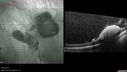
Coats' Disease -51 year old asymptomatic male498 views20/20 vision - had laser to non-perfusion because of proliferation.Jul 27, 2018
|
|

Coats' Disease -51 year old asymptomatic male466 views20/20 vision - had laser to non-perfusion because of proliferation.Jul 27, 2018
|
|

Coats' Disease -51 year old asymptomatic male483 views20/20 vision - had laser to non-perfusion because of proliferation.Jul 27, 2018
|
|

Coats' Disease -51 year old asymptomatic male475 views20/20 vision - had laser to non-perfusion because of proliferation.Jul 27, 2018
|
|

Coats' Disease -51 year old asymptomatic male420 views20/20 vision - had laser to non-perfusion because of proliferation.Jul 27, 2018
|
|
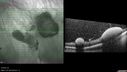
Coats' Disease -51 year old asymptomatic male444 views20/20 vision - had laser to non-perfusion because of proliferation.Jul 27, 2018
|
|
|
|


