Last additions - Congenital Anomalies
|

Coats' Disease - 14 year old - Asymptomatic546 viewsPresented with macular exudates. Temporal macroaneurysms responded to laser. Ultra-wide field FA shows nonperfusionNov 07, 2016
|
|

Coats' Disease - 14 year old - Asymptomatic536 viewsPresented with macular exudates. Temporal macroaneurysms responded to laser. Ultra-wide field FA shows nonperfusionNov 07, 2016
|
|

Coats' Disease - 14 year old - Asymptomatic532 viewsPresented with macular exudates. Temporal macroaneurysms responded to laser. Ultra-wide field FA shows nonperfusionNov 07, 2016
|
|

Coats' Disease - 14 year old - Asymptomatic539 viewsPresented with macular exudates. Temporal macroaneurysms responded to laser. Ultra-wide field FA shows nonperfusionNov 07, 2016
|
|

Coats' Disease - 14 year old - Asymptomatic564 viewsPresented with macular exudates. Temporal macroaneurysms responded to laser. Ultra-wide field FA shows nonperfusionNov 07, 2016
|
|

Coats' Disease - 14 year old - Asymptomatic550 viewsPresented with macular exudates. Temporal macroaneurysms responded to laser. Ultra-wide field FA shows nonperfusionNov 07, 2016
|
|

Coats' Disease - 14 year old - Asymptomatic545 viewsPresented with macular exudates. Temporal macroaneurysms responded to laser. Ultra-wide field FA shows nonperfusionNov 07, 2016
|
|

Coats' Disease - 14 year old - Asymptomatic735 viewsPresented with macular exudates. Temporal macroaneurysms responded to laser. Ultra-wide field FA shows nonperfusionNov 07, 2016
|
|

Sturge Weber SD OCT EDI showing choroidal thickening664 viewsMay 16, 2016
|
|

359 viewsOct 23, 2014
|
|

Coat's Disease493 views47 year old african-american male, presented under the assumption he had Sarcoidosis but appears more like Coat's DiseaseOct 23, 2014
|
|

Coat's Disease OD470 viewsOct 20, 2014
|
|

Hypertrophy of the Retinal Pigment Epithelium606 viewsPatient comes in with vision changes in the right eye. VA is 20/30 in the right eye. Fundus and FAF photos show a large area of Hypertrophy in the right eye.Sep 16, 2014
|
|

Hypertrophy of the Retinal Pigment Epithelium1446 viewsPatient comes in with vision changes in the right eye. VA is 20/30 in the right eye. Fundus and FAF photos show a large area of Hypertrophy in the right eye.Sep 16, 2014
|
|

Retinal Coloboma778 viewsYoung 19-year old female with Charge Syndrome comes in for eye exam. VA is 20/400, right eye, 20/30, left eye. Fundus photography shows coloboma covering the macula and pushing the optic nerve in the right eye. Jun 30, 2014
|
|

Retinal Coloboma1043 viewsYoung 19-year old female with Charge Syndrome comes in for eye exam. VA is 20/400, right eye, 20/30, left eye. Fundus photography shows coloboma covering the macula and pushing the optic nerve in the right eye. Jun 30, 2014
|
|

660 viewsYoung 19-year old female with Charge Syndrome comes in for eye exam. VA is 20/400, right eye, 20/30, left eye. Fundus photography shows coloboma covering the macula and pushing the optic nerve in the right eye. Jun 30, 2014
|
|
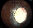
757 viewsYoung 19-year old female with Charge Syndrome comes in for eye exam. VA is 20/400, right eye, 20/30, left eye. Fundus photography shows coloboma covering the macula and pushing the optic nerve in the right eye. Jun 30, 2014
|
|

Retinal Pigment Epithleium Dysgenesis - Autofluorescence610 viewsJun 06, 2014
|
|

Retinal Pigment Epithleium Dysgenesis - SD OCT line Scan527 viewsJun 06, 2014
|
|

Retinal Pigment Epithleium Dysgenesis - SD OCT line scan627 viewsJun 06, 2014
|
|

Retinal Pigment Epithleium Dysgenesis - Autofluorescence663 viewsJun 06, 2014
|
|

Retinal Pigment Epithleium Dysgenesis - Autofluorescence747 viewsJun 06, 2014
|
|

Retinal Pigment Epithleium Dysgenesis - Autofluorescence595 viewsJun 06, 2014
|
|

Retinal Pigment Epithleium Dysgenesis 667 viewsJun 06, 2014
|
|

Retinal Pigment Epithleium Dysgenesis 659 viewsJun 06, 2014
|
|
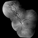
AVM582 viewsRF of AVM in OS of a 48 yr old female.Mar 11, 2014
|
|

AVM1189 viewsFP of AVM in OS of a 48 yr old female.Mar 11, 2014
|
|

AVM581 viewsFA of AVM in OS of a 48 yr old female.Mar 11, 2014
|
|

Choroidal Hemangioma - Diffuse - Enhanced Depth Imaging Spectral Domain OCT Line Scan - Abnormal Eye904 viewsChoroidal Thickness is 697 Microns from Diffuse Choroidal HemangiomaFeb 12, 2014
|
|

Choroidal Hemangioma - Diffuse - Enhanced Depth Imaging Spectral Domain OCT Line Scan - Abnormal Eye651 viewsChoroidal Thickness is 697 Microns from Diffuse Choroidal HemangiomaFeb 12, 2014
|
|

Choroidal Hemangioma - Diffuse - Enhanced Depth Imaging Spectral Domain OCT Line Scan - Normal Eye790 viewsNormal Choroidal Thickness in Right EyeFeb 12, 2014
|
|

Choroidal Hemangioma - Diffuse - Enhanced Depth Imaging Spectral Domain OCT Line Scan - Normal Eye653 viewsNormal Choroidal Thickness in Right EyeFeb 12, 2014
|
|

Coats' Disease with Cryotherapy 1331 viewsYoung male with Coat's Disease with Cryotherapy in the right eye. VA was 20/20, right eye. Left eye was completely normal. Patient will be followed up in 3-months.Aug 14, 2013
|
|

Nearly complete coloboma1286 viewsNearly complete coloboma from necropsy of infant born with multiple congenital anomalies, incompatable with life, including holorprosencephaly. Infant expired on 3rd post-partum dayJul 18, 2013
|
|

50 Year Old woman with Congenital Retinal Vascular Tortuosity - Normal Vision988 viewsApr 08, 2013
|
|

50 Year Old woman with Congenital Retinal Vascular Tortuosity - Normal Vision1063 viewsApr 08, 2013
|
|
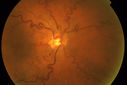
50 Year Old woman with Congenital Retinal Vascular Tortuosity - Normal Vision1122 viewsApr 08, 2013
|
|

50 Year Old woman with Congenital Retinal Vascular Tortuosity - Normal Vision1283 viewsApr 08, 2013
|
|

Coates' Disease524 views38-year old male presents with decreased vision in the left eye. VA was 20/20-right eye and 20/70-left eye with pinhole no improvement. Fundus exam shows exudates near the macula and also exudates with small hemorrhages, superior nasally in the left eye. Mar 28, 2013
|
|
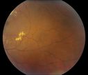
426 views38-year old male presents with decreased vision in the left eye. VA was 20/20-right eye and 20/70-left eye with pinhole no improvement. Fundus exam shows exudates near the macula and also exudates with small hemorrhages, superior nasally in the left eye. Mar 28, 2013
|
|

408 views38-year old male presents with decreased vision in the left eye. VA was 20/20-right eye and 20/70-left eye with pinhole no improvement. Fundus exam shows exudates near the macula and also exudates with small hemorrhages, superior nasally in the left eye. Mar 28, 2013
|
|

494 views38-year old male presents with decreased vision in the left eye. VA was 20/20-right eye and 20/70-left eye with pinhole no improvement. Fundus exam shows exudates near the macula and also exudates with small hemorrhages, superior nasally in the left eye. Mar 28, 2013
|
|

404 views38-year old male presents with decreased vision in the left eye. VA was 20/20-right eye and 20/70-left eye with pinhole no improvement. Fundus exam shows exudates near the macula and also exudates with small hemorrhages, superior nasally in the left eye. Mar 28, 2013
|
|

389 views38-year old male presents with decreased vision in the left eye. VA was 20/20-right eye and 20/70-left eye with pinhole no improvement. Fundus exam shows exudates near the macula and also exudates with small hemorrhages, superior nasally in the left eye. Mar 28, 2013
|
|

398 views38-year old male presents with decreased vision in the left eye. VA was 20/20-right eye and 20/70-left eye with pinhole no improvement. Fundus exam shows exudates near the macula and also exudates with small hemorrhages, superior nasally in the left eye. Mar 28, 2013
|
|

480 views38-year old male presents with decreased vision in the left eye. VA was 20/20-right eye and 20/70-left eye with pinhole no improvement. Fundus exam shows exudates near the macula and also exudates with small hemorrhages, superior nasally in the left eye. Mar 28, 2013
|
|

428 views38-year old male presents with decreased vision in the left eye. VA was 20/20-right eye and 20/70-left eye with pinhole no improvement. Fundus exam shows exudates near the macula and also exudates with small hemorrhages, superior nasally in the left eye. Mar 28, 2013
|
|

466 views38-year old male presents with decreased vision in the left eye. VA was 20/20-right eye and 20/70-left eye with pinhole no improvement. Fundus exam shows exudates near the macula and also exudates with small hemorrhages, superior nasally in the left eye. Mar 28, 2013
|
|

550 views38-year old male presents with decreased vision in the left eye. VA was 20/20-right eye and 20/70-left eye with pinhole no improvement. Fundus exam shows exudates near the macula and also exudates with small hemorrhages, superior nasally in the left eye. Mar 28, 2013
|
|

453 views38-year old male presents with decreased vision in the left eye. VA was 20/20-right eye and 20/70-left eye with pinhole no improvement. Fundus exam shows exudates near the macula and also exudates with small hemorrhages, superior nasally in the left eye. Mar 28, 2013
|
|

479 views38-year old male presents with decreased vision in the left eye. VA was 20/20-right eye and 20/70-left eye with pinhole no improvement. Fundus exam shows exudates near the macula and also exudates with small hemorrhages, superior nasally in the left eye. Mar 28, 2013
|
|

619 views38-year old male presents with decreased vision in the left eye. VA was 20/20-right eye and 20/70-left eye with pinhole no improvement. Fundus exam shows exudates near the macula and also exudates with small hemorrhages, superior nasally in the left eye. Mar 28, 2013
|
|
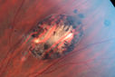
Congenital Hypertrophy of the Retinal Pigment Epithelium CHRPE has Satellite Lesions1045 viewsMar 20, 2013
|
|
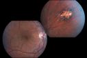
Congenital Hypertrophy of the Retinal Pigment Epithelium CHRPE has Satellite Lesions791 viewsMar 20, 2013
|
|

Congenital Hypertrophy of the Retinal Pigment Epithelium CHRPE has Satellite Lesions664 viewsMar 20, 2013
|
|
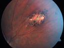
Congenital Hypertrophy of the Retinal Pigment Epithelium CHRPE has Satellite Lesions1051 viewsMar 20, 2013
|
|

OD Albinism with foveal hypoplasia2396 views28 year old female latina with vision of 20/400Mar 05, 2013
|
|

FEVR FA Transit653 viewsFEVR in OS of a 14 yr old girlJan 11, 2013
|
|

640 viewsFEVR in OS of a 14 yr old girlJan 11, 2013
|
|

Retinal Coloboma1392 views14-year old male presents with decreased vision in the left eye. Dx with iris and retinal coloboma in the left eye. Patient VA was 20/20, right eye, 20/100 left eye with pinhole improvement 20\50. Patient was fitted for SCL in the left eye.Aug 16, 2012
|
|

Multifocal CHRPE also called "Bear Tracks"1069 viewsJul 09, 2012
|
|

Chorioretinal Scar1687 views21 year old male with a chorioretinal scar ODJun 21, 2012
|
|

CAVERNOUS HEMONGIOMA2280 views28 year old male. FAApr 20, 2012
|
|

CAVERNOUS HEMONGIOMA2918 views28 year old male w/ 20/200 vision at time of exam. Patient c/o poor vision since childhood. No significant medical history or family medical history. A problem was only noted when patient enlisted in the Army.Apr 20, 2012
|
|

Combined hamartoma of the retina and RPE2838 views16 year old female, diagnosed with a combined hamartoma of the retina and the retinal pigment epithelium.Apr 20, 2012
|
|

Wyburn-Mason Syndrome 2099 views28 year old female, diagnosed with Wyburn-Mason Syndrome at age 7. At time of exam, vision in the left eye was 4/200.Apr 20, 2012
|
|

Coats' Disease in 14 Year Old Boy Treated with Cryotherapy and Avastin727 views14 Year Old Boy with Coats DiseaseFeb 25, 2012
|
|

Coats' Disease in 14 Year Old Boy Treated with Cryotherapy and Avastin797 views14 Year Old Boy with Coats DiseaseFeb 25, 2012
|
|

Coats' Disease in 14 Year Old Boy Treated with Cryotherapy and Avastin854 views14 Year Old Boy with Coats DiseaseFeb 25, 2012
|
|

Coats' Disease in 14 Year Old Boy Treated with Cryotherapy and Avastin613 views14 Year Old Boy with Coats DiseaseFeb 19, 2012
|
|

Coats' Disease in 14 Year Old Boy Treated with Cryotherapy and Avastin693 views14 Year Old Boy with Coats DiseaseFeb 19, 2012
|
|

Coats' Disease in 14 Year Old Boy Treated with Cryotherapy and Avastin1824 views14 Year Old Boy with Coats DiseaseFeb 19, 2012
|
|

Coats' Disease in 14 Year Old Boy Treated with Cryotherapy and Avastin687 views14 Year Old Boy with Coats DiseaseFeb 19, 2012
|
|

Coats' Disease in 14 Year Old Boy Treated with Cryotherapy and Avastin876 views14 Year Old Boy with Coats DiseaseFeb 19, 2012
|
|
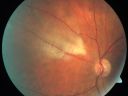
Myelinated Nerve Fiber Layer854 views69-year-old woman OD is 20/30, OS is 20/40. Feb 07, 2012
|
|

Myelinated Nerve Fiber Layer669 views69-year-old woman OD is 20/30, OS is 20/40. Feb 07, 2012
|
|

Myelinated Nerve Fiber Layer1169 views69-year-old woman OD is 20/30, OS is 20/40. Feb 07, 2012
|
|

Myelinated Nerve Fiber Layer862 views69-year-old woman OD is 20/30, OS is 20/40. Feb 07, 2012
|
|

Myelinated Nerve Fiber Layer741 views69-year-old woman OD is 20/30, OS is 20/40. Feb 07, 2012
|
|

wyburn-mason syndrome fundus photos1342 views28 year old female, visual acuity 20/200Jan 10, 2012
|
|

Wyburn Mason Syndrome4040 views28 year old female, visual acuity OS 20/200Jan 10, 2012
|
|

wyburn-mason sydrome1408 views28 year old female, visual acuity OS 20/200Jan 10, 2012
|
|

848 viewsPHPV - Left eye macular area. The patient was diagnosed as having regressed ROP even with a history of full term birth with no antecedent perinatal history of diseaseNov 17, 2011
|
|
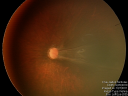
1104 viewsPHPV - Left eye - optic nerveNov 17, 2011
|
|

995 viewsPHPV - right eye. Peripheral extension Nov 17, 2011
|
|
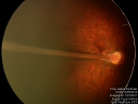
1727 viewsPHPV - right eye - optic nerveNov 17, 2011
|
|
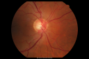
Anomalous Branch Retinal Artery 1049 views59-year-old man OD 20/25, OS 20/25. TORTUOUS RETINAL ARTERY – LEF EYE
(POSSIBLE GROUP 2 WYBURN-MASON SYNDROME
POSSIBLE ACEPHALIC MIGRAINES )Sep 02, 2011
|
|

Anomalous Branch Retinal Artery 710 views59-year-old man OD 20/25, OS 20/25. TORTUOUS RETINAL ARTERY – LEF EYE
(POSSIBLE GROUP 2 WYBURN-MASON SYNDROME
POSSIBLE ACEPHALIC MIGRAINES )Sep 02, 2011
|
|

Anomalous Branch Retinal Artery 926 views59-year-old man OD 20/25, OS 20/25. TORTUOUS RETINAL ARTERY – LEF EYE
(POSSIBLE GROUP 2 WYBURN-MASON SYNDROME
POSSIBLE ACEPHALIC MIGRAINES )Sep 02, 2011
|
|

Anomalous Branch Retinal Artery 728 views59-year-old man OD 20/25, OS 20/25. TORTUOUS RETINAL ARTERY – LEF EYE
(POSSIBLE GROUP 2 WYBURN-MASON SYNDROME
POSSIBLE ACEPHALIC MIGRAINES )Sep 02, 2011
|
|

Anomalous Branch Retinal Artery 1071 views59-year-old man OD 20/25, OS 20/25. TORTUOUS RETINAL ARTERY – LEF EYE
(POSSIBLE GROUP 2 WYBURN-MASON SYNDROME
POSSIBLE ACEPHALIC MIGRAINES )Sep 02, 2011
|
|

Anomalous Branch Retinal Artery 828 views59-year-old man OD 20/25, OS 20/25. TORTUOUS RETINAL ARTERY – LEF EYE
(POSSIBLE GROUP 2 WYBURN-MASON SYNDROME
POSSIBLE ACEPHALIC MIGRAINES )Sep 02, 2011
|
|

Anomalous Branch Retinal Artery 758 views59-year-old man OD 20/25, OS 20/25. TORTUOUS RETINAL ARTERY – LEF EYE
(POSSIBLE GROUP 2 WYBURN-MASON SYNDROME
POSSIBLE ACEPHALIC MIGRAINES )Sep 02, 2011
|
|

Amelanotic - Light Colored CHRPE - Congenital Hypertrophy of the Pigment Epithelium5160 views56-year-old woman nov vision problems. OD is 20/20, OS is 20/16Sep 02, 2011
|
|

Amelanotic - Light Colored CHRPE - Congenital Hypertrophy of the Pigment Epithelium2791 views56-year-old woman nov vision problems. OD is 20/20, OS is 20/16Sep 02, 2011
|
|

CHRPE - Congenital Hypertrophy of the Pigment Epithelium - Peripheral1143 views55-year-old woman OD 20/30, OS 20/25Jul 24, 2011
|
|

CHRPE - Congenital Hypertrophy of the Pigment Epithelium - Peripheral1334 views55-year-old woman OD 20/30, OS 20/25Jul 24, 2011
|
|

Nanopthalmous (Microphthalmous) Chorioretinal Folds and Choroidal Thickening Both Eyes B-Scan Ultrasound1013 viewsJun 28, 2011
|
|

Nanopthalmous (Microphthalmous) Chorioretinal Folds and Choroidal Thickening Both Eyes B-Scan Ultrasound1019 viewsJun 28, 2011
|
|
|
|


