Last additions - Choroidal Melanoma and Choroidal Nevus
|

Pigmented Choroidal Mass Right Eye - Melanoma vs. Nevus vs. Unilateral Melanocytic Proliferation - 4 Month Follow-Up Images523 views71-year-old man his vision 4 month follow-up. Vision is 20/60 and patient sees flashing lights. Lesion has grown slightly in the superior half of the macula.Jan 03, 2012
|
|

Pigmented Choroidal Mass Right Eye - Melanoma vs. Nevus vs. Unilateral Melanocytic Proliferation - 4 Month Follow-Up Images570 views71-year-old man his vision 4 month follow-up. Vision is 20/60 and patient sees flashing lights. Lesion has grown slightly in the superior half of the macula.Jan 03, 2012
|
|

Pigmented Choroidal Mass Right Eye - Melanoma vs. Nevus vs. Unilateral Melanocytic Proliferation - 4 Month Follow-Up Images576 views71-year-old man his vision 4 month follow-up. Vision is 20/60 and patient sees flashing lights. Lesion has grown slightly in the superior half of the macula.Jan 03, 2012
|
|

Pigmented Choroidal Mass Right Eye - Melanoma vs. Nevus vs. Unilateral Melanocytic Proliferation - 4 Month Follow-Up Images499 views71-year-old man his vision 4 month follow-up. Vision is 20/60 and patient sees flashing lights. Lesion has grown slightly in the superior half of the macula.Jan 03, 2012
|
|

Pigmented Choroidal Mass Right Eye - Melanoma vs. Nevus vs. Unilateral Melanocytic Proliferation - 4 Month Follow-Up Images509 views71-year-old man his vision 4 month follow-up. Vision is 20/60 and patient sees flashing lights. Lesion has grown slightly in the superior half of the macula.Jan 03, 2012
|
|

Pigmented Choroidal Mass Right Eye - Melanoma vs. Nevus vs. Unilateral Melanocytic Proliferation - 4 Month Follow-Up Images497 views71-year-old man his vision 4 month follow-up. Vision is 20/60 and patient sees flashing lights. Lesion has grown slightly in the superior half of the macula.Jan 03, 2012
|
|

Pigmented Choroidal Mass Right Eye - Melanoma vs. Nevus vs. Unilateral Melanocytic Proliferation - 4 Month Follow-Up Images506 views71-year-old man his vision 4 month follow-up. Vision is 20/60 and patient sees flashing lights. Lesion has grown slightly in the superior half of the macula.Jan 03, 2012
|
|

Pigmented Choroidal Mass Right Eye - Melanoma vs. Nevus vs. Unilateral Melanocytic Proliferation - 4 Month Follow-Up Images 563 views71-year-old man his vision 4 month follow-up. Vision is 20/60 and patient sees flashing lights. Lesion has grown slightly in the superior half of the macula.Jan 03, 2012
|
|

Pigmented Choroidal Mass Right Eye - Melanoma vs. Nevus vs. Unilateral Melanocytic Proliferation - 4 Month Follow-Up Images534 views71-year-old man his vision 4 month follow-up. Vision is 20/60 and patient sees flashing lights. Lesion has grown slightly in the superior half of the macula.Jan 03, 2012
|
|

Pigmented Choroidal Mass Right Eye - Melanoma vs. Nevus vs. Unilateral Melanocytic Proliferation - 4 Month Follow-Up Images532 views71-year-old man his vision 4 month follow-up. Vision is 20/60 and patient sees flashing lights. Lesion has grown slightly in the superior half of the macula.Jan 03, 2012
|
|

Pigmented Choroidal Mass Right Eye - Melanoma vs. Nevus vs. Unilateral Melanocytic Proliferation - 4 Month Follow-Up Images554 views71-year-old man his vision 4 month follow-up. Vision is 20/60 and patient sees flashing lights. Lesion has grown slightly in the superior half of the macula.Jan 03, 2012
|
|

Pigmented Choroidal Mass Right Eye - Melanoma vs. Nevus vs. Unilateral Melanocytic Proliferation - 4 Month Follow-Up Images562 views71-year-old man his vision 4 month follow-up. Vision is 20/60 and patient sees flashing lights. Lesion has grown slightly in the superior half of the macula.Jan 03, 2012
|
|

Pigmented Choroidal Mass Right Eye - Melanoma vs. Nevus vs. Unilateral Melanocytic Proliferation - 4 Month Follow-Up Images606 views71-year-old man his vision 4 month follow-up. Vision is 20/60 and patient sees flashing lights. Lesion has grown slightly in the superior half of the macula.Jan 03, 2012
|
|

Pigmented Choroidal Mass Right Eye - Melanoma vs. Nevus vs. Unilateral Melanocytic Proliferation - 4 Month Follow-Up Images593 views71-year-old man his vision 4 month follow-up. Vision is 20/60 and patient sees flashing lights. Lesion has grown slightly in the superior half of the macula.Jan 03, 2012
|
|

Pigmented Choroidal Mass Right Eye - Melanoma vs. Nevus vs. Unilateral Melanocytic Proliferation - 4 Month Follow-Up Images562 views71-year-old man his vision 4 month follow-up. Vision is 20/60 and patient sees flashing lights. Lesion has grown slightly in the superior half of the macula.Jan 03, 2012
|
|

Pigmented Choroidal Mass Right Eye - Melanoma vs. Nevus vs. Unilateral Melanocytic Proliferation -4 Month Follow-Up Images559 views71-year-old man his vision 4 month follow-up. Vision is 20/60 and patient sees flashing lights. Lesion has grown slightly in the superior half of the macula.Jan 03, 2012
|
|

Pigmented Choroidal Mass Right Eye - Melanoma vs. Nevus vs. Unilateral Melanocytic Proliferation - 4 Month Follow-Up Images666 views71-year-old man his vision 4 month follow-up. Vision is 20/60 and patient sees flashing lights. Lesion has grown slightly in the superior half of the macula.Jan 03, 2012
|
|
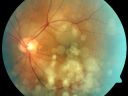
Pigmented Choroidal Mass Right Eye - Melanoma vs. Nevus vs. Unilateral Melanocytic Proliferation - 4 Month Follow-Up Images630 views71-year-old man his vision 4 month follow-up. Vision is 20/60 and patient sees flashing lights. Lesion has grown slightly in the superior half of the macula.Jan 03, 2012
|
|

Pigmented Choroidal Mass Right Eye - Melanoma vs. Nevus vs. Unilateral Melanocytic Proliferation - 4 Month Follow-Up Images619 views71-year-old man his vision 4 month follow-up. Vision is 20/60 and patient sees flashing lights. Lesion has grown slightly in the superior half of the macula.Jan 03, 2012
|
|

Pigmented Choroidal Mass Right Eye - Melanoma vs. Nevus vs. Unilateral Melanocytic Proliferation - 4 Month Follow-Up Images665 views71-year-old man his vision 4 month follow-up. Vision is 20/60 and patient sees flashing lights. Lesion has grown slightly in the superior half of the macula.Jan 03, 2012
|
|

Pigmented Choroidal Mass Right Eye - Melanoma vs. Nevus vs. Unilateral Melanocytic Proliferation - 4 Month Follow-Up Images718 views71-year-old man his vision 4 month follow-up. Vision is 20/60 and patient sees flashing lights. Lesion has grown slightly in the superior half of the macula.Jan 03, 2012
|
|
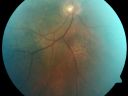
Pigmented Choroidal Mass Right Eye - Melanoma vs. Nevus vs. Unilateral Melanocytic Proliferation - 4 Month Follow-Up Images672 views71-year-old man his vision 4 month follow-up. Vision is 20/60 and patient sees flashing lights. Lesion has grown slightly in the superior half of the macula.Jan 03, 2012
|
|

Pigmented Choroidal Mass Right Eye - Melanoma vs. Nevus vs. Unilateral Melanocytic Proliferation - 4 Month Follow-Up Images731 views71-year-old man his vision 4 month follow-up. Vision is 20/60 and patient sees flashing lights. Lesion has grown slightly in the superior half of the macula.Jan 03, 2012
|
|

Pigmented Choroidal Mass Right Eye - Melanoma vs. Nevus vs. Unilateral Melanocytic Proliferation - 4 Month Follow-Up Images810 views71-year-old man his vision 4 month follow-up. Vision is 20/60 and patient sees flashing lights. Lesion has grown slightly in the superior half of the macula.Jan 03, 2012
|
|

Pigmented Choroidal Mass Right Eye - Melanoma vs. Nevus vs. Unilateral Melanocytic Proliferation - 4 Month Follow-Up Images722 views71-year-old man his vision 4 month follow-up. Vision is 20/60 and patient sees flashing lights. Lesion has grown slightly in the superior half of the macula.Jan 03, 2012
|
|

Pigmented Choroidal Mass Right Eye - Melanoma vs. Nevus vs. Unilateral Melanocytic Proliferation - 4 Month Follow-Up Images753 views71-year-old man his vision 4 month follow-up. Vision is 20/60 and patient sees flashing lights. Lesion has grown slightly in the superior half of the macula.Jan 03, 2012
|
|

Radiation Retinopathy and Macular Pucker 4 years following Brachytherapy (Plaque) for Nasal Melanoma582 views67-year-old woman has a macular pucker in the left eye. She had radiation for a melanoma in that eye 4 years aog. She had cataract surgery 2 years ago and a posterior sub-Tenon Kenalog injection in 7 months ago. Her vision in the left eye is hazy and she has trouble seeing out of it. VA OS 20/70. Sep 20, 2011
|
|

Radiation Retinopathy and Macular Pucker 4 years following Brachytherapy (Plaque) for Nasal Melanoma461 views67-year-old woman has a macular pucker in the left eye. She had radiation for a melanoma in that eye 4 years aog. She had cataract surgery 2 years ago and a posterior sub-Tenon Kenalog injection in 7 months ago. Her vision in the left eye is hazy and she has trouble seeing out of it. VA OS 20/70. Sep 20, 2011
|
|

Radiation Retinopathy and Macular Pucker 4 years following Brachytherapy (Plaque) for Nasal Melanoma548 views67-year-old woman has a macular pucker in the left eye. She had radiation for a melanoma in that eye 4 years aog. She had cataract surgery 2 years ago and a posterior sub-Tenon Kenalog injection in 7 months ago. Her vision in the left eye is hazy and she has trouble seeing out of it. VA OS 20/70. Sep 20, 2011
|
|

Radiation Retinopathy and Macular Pucker 4 years following Brachytherapy (Plaque) for Nasal Melanoma542 views67-year-old woman has a macular pucker in the left eye. She had radiation for a melanoma in that eye 4 years aog. She had cataract surgery 2 years ago and a posterior sub-Tenon Kenalog injection in 7 months ago. Her vision in the left eye is hazy and she has trouble seeing out of it. VA OS 20/70. Sep 20, 2011
|
|

Radiation Retinopathy and Macular Pucker 4 years following Brachytherapy (Plaque) for Nasal Melanoma534 views67-year-old woman has a macular pucker in the left eye. She had radiation for a melanoma in that eye 4 years aog. She had cataract surgery 2 years ago and a posterior sub-Tenon Kenalog injection in 7 months ago. Her vision in the left eye is hazy and she has trouble seeing out of it. VA OS 20/70. Sep 20, 2011
|
|

Radiation Retinopathy and Macular Pucker 4 years following Brachytherapy (Plaque) for Nasal Melanoma476 views67-year-old woman has a macular pucker in the left eye. She had radiation for a melanoma in that eye 4 years aog. She had cataract surgery 2 years ago and a posterior sub-Tenon Kenalog injection in 7 months ago. Her vision in the left eye is hazy and she has trouble seeing out of it. VA OS 20/70. Sep 20, 2011
|
|
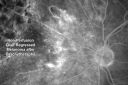
Radiation Retinopathy and Macular Pucker 4 years following Brachytherapy (Plaque) for Nasal Melanoma499 views67-year-old woman has a macular pucker in the left eye. She had radiation for a melanoma in that eye 4 years aog. She had cataract surgery 2 years ago and a posterior sub-Tenon Kenalog injection in 7 months ago. Her vision in the left eye is hazy and she has trouble seeing out of it. VA OS 20/70. Sep 20, 2011
|
|

Radiation Retinopathy and Macular Pucker 4 years following Brachytherapy (Plaque) for Nasal Melanoma420 views67-year-old woman has a macular pucker in the left eye. She had radiation for a melanoma in that eye 4 years aog. She had cataract surgery 2 years ago and a posterior sub-Tenon Kenalog injection in 7 months ago. Her vision in the left eye is hazy and she has trouble seeing out of it. VA OS 20/70. Sep 20, 2011
|
|

Radiation Retinopathy and Macular Pucker 4 years following Brachytherapy (Plaque) for Nasal Melanoma509 views67-year-old woman has a macular pucker in the left eye. She had radiation for a melanoma in that eye 4 years aog. She had cataract surgery 2 years ago and a posterior sub-Tenon Kenalog injection in 7 months ago. Her vision in the left eye is hazy and she has trouble seeing out of it. VA OS 20/70. Sep 20, 2011
|
|
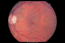
Radiation Retinopathy and Macular Pucker 4 years following Brachytherapy (Plaque) for Nasal Melanoma530 views67-year-old woman has a macular pucker in the left eye. She had radiation for a melanoma in that eye 4 years aog. She had cataract surgery 2 years ago and a posterior sub-Tenon Kenalog injection in 7 months ago. Her vision in the left eye is hazy and she has trouble seeing out of it. VA OS 20/70. Sep 20, 2011
|
|
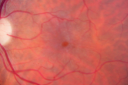
Radiation Retinopathy and Macular Pucker 4 years following Brachytherapy (Plaque) for Nasal Melanoma560 views67-year-old woman has a macular pucker in the left eye. She had radiation for a melanoma in that eye 4 years aog. She had cataract surgery 2 years ago and a posterior sub-Tenon Kenalog injection in 7 months ago. Her vision in the left eye is hazy and she has trouble seeing out of it. VA OS 20/70. Sep 20, 2011
|
|

Radiation Retinopathy and Macular Pucker 4 years following Brachytherapy (Plaque) for Nasal Melanoma778 views67-year-old woman has a macular pucker in the left eye. She had radiation for a melanoma in that eye 4 years aog. She had cataract surgery 2 years ago and a posterior sub-Tenon Kenalog injection in 7 months ago. Her vision in the left eye is hazy and she has trouble seeing out of it. VA OS 20/70. Sep 20, 2011
|
|

Peripapillary Choroidal Nevus1083 views37-year-old woman was seen in the office on July 27, 2011. She has photographs from 2009 and 2010 showing a peripapillary choroidal nevus in the right eye. VISUAL ACUITY: OD 20/20Sep 02, 2011
|
|
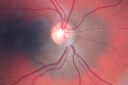
Peripapillary Choroidal Nevus1095 views37-year-old woman was seen in the office on July 27, 2011. She has photographs from 2009 and 2010 showing a peripapillary choroidal nevus in the right eye. VISUAL ACUITY: OD 20/20Sep 02, 2011
|
|
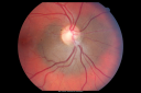
Peripapillary Choroidal Nevus942 views37-year-old woman was seen in the office on July 27, 2011. She has photographs from 2009 and 2010 showing a peripapillary choroidal nevus in the right eye. VISUAL ACUITY: OD 20/20Sep 02, 2011
|
|

Peripapillary Choroidal Nevus1275 views37-year-old woman was seen in the office on July 27, 2011. She has photographs from 2009 and 2010 showing a peripapillary choroidal nevus in the right eye. VISUAL ACUITY: OD 20/20Sep 02, 2011
|
|

Choroidal Nevus with Overlying Drusen3317 views48-year-old woman Vision OD is 20/16, OS is 20/20.Jul 24, 2011
|
|

Choroidal Nevus with Overlying Drusen1067 views48-year-old woman Vision OD is 20/16, OS is 20/20.Jul 24, 2011
|
|

Choroidal Nevus with Overlying Drusen1680 views48-year-old woman Vision OD is 20/16, OS is 20/20.Jul 24, 2011
|
|

Choroidal Melanoma - Exudative Retinal Detachment 82 Year Old Man B Scan - Transverse1353 views82-year-old man who had 3 falls 2 weeks ago. After the falls he started checking his vision and noticed there was a veil over the left eye, which he had seen for about two weeks in the superior visual field. OD 20/25, OS 20/40
(patient was medically never well enough for brachytherapy and died 6 months later from heart disease) Jul 07, 2011
|
|

Choroidal Melanoma - Exudative Retinal Detachment 82 Year Old Man B Scan Ultrasound Transverce954 views82-year-old man who had 3 falls 2 weeks ago. After the falls he started checking his vision and noticed there was a veil over the left eye, which he had seen for about two weeks in the superior visual field. OD 20/25, OS 20/40
(patient was medically never well enough for brachytherapy and died 6 months later from heart disease) Jul 07, 2011
|
|
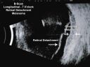
Choroidal Melanoma - Exudative Retinal Detachment 82 Year Old Man B Scan Ultrasound Longitudinal1236 views82-year-old man who had 3 falls 2 weeks ago. After the falls he started checking his vision and noticed there was a veil over the left eye, which he had seen for about two weeks in the superior visual field. OD 20/25, OS 20/40
(patient was medically never well enough for brachytherapy and died 6 months later from heart disease) Jul 07, 2011
|
|

Choroidal Melanoma - Exudative Retinal Detachment 82 Year Old Man B Scan Ultrasound Longitudinal873 views82-year-old man who had 3 falls 2 weeks ago. After the falls he started checking his vision and noticed there was a veil over the left eye, which he had seen for about two weeks in the superior visual field. OD 20/25, OS 20/40
(patient was medically never well enough for brachytherapy and died 6 months later from heart disease) Jul 07, 2011
|
|

Choroidal Melanoma - Exudative Retinal Detachment 82 Year Old Man 584 views82-year-old man who had 3 falls 2 weeks ago. After the falls he started checking his vision and noticed there was a veil over the left eye, which he had seen for about two weeks in the superior visual field. OD 20/25, OS 20/40
(patient was medically never well enough for brachytherapy and died 6 months later from heart disease) Jun 28, 2011
|
|

Choroidal Melanoma - Exudative Retinal Detachment 82 Year Old Man 567 views82-year-old man who had 3 falls 2 weeks ago. After the falls he started checking his vision and noticed there was a veil over the left eye, which he had seen for about two weeks in the superior visual field. OD 20/25, OS 20/40
(patient was medically never well enough for brachytherapy and died 6 months later from heart disease) Jun 28, 2011
|
|

Choroidal Melanoma - Exudative Retinal Detachment 82 Year Old Man 531 views82-year-old man who had 3 falls 2 weeks ago. After the falls he started checking his vision and noticed there was a veil over the left eye, which he had seen for about two weeks in the superior visual field. OD 20/25, OS 20/40
(patient was medically never well enough for brachytherapy and died 6 months later from heart disease) Jun 28, 2011
|
|

Choroidal Melanoma - Exudative Retinal Detachment 82 Year Old Man 520 views82-year-old man who had 3 falls 2 weeks ago. After the falls he started checking his vision and noticed there was a veil over the left eye, which he had seen for about two weeks in the superior visual field. OD 20/25, OS 20/40
(patient was medically never well enough for brachytherapy and died 6 months later from heart disease) Jun 28, 2011
|
|
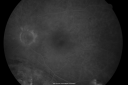
Choroidal Melanoma - Exudative Retinal Detachment 82 Year Old Man 484 views82-year-old man who had 3 falls 2 weeks ago. After the falls he started checking his vision and noticed there was a veil over the left eye, which he had seen for about two weeks in the superior visual field. OD 20/25, OS 20/40
(patient was medically never well enough for brachytherapy and died 6 months later from heart disease) Jun 28, 2011
|
|
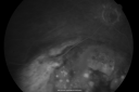
Choroidal Melanoma - Exudative Retinal Detachment 82 Year Old Man 485 views82-year-old man who had 3 falls 2 weeks ago. After the falls he started checking his vision and noticed there was a veil over the left eye, which he had seen for about two weeks in the superior visual field. OD 20/25, OS 20/40
(patient was medically never well enough for brachytherapy and died 6 months later from heart disease) Jun 28, 2011
|
|

Choroidal Melanoma - Exudative Retinal Detachment 82 Year Old Man 521 views82-year-old man who had 3 falls 2 weeks ago. After the falls he started checking his vision and noticed there was a veil over the left eye, which he had seen for about two weeks in the superior visual field. OD 20/25, OS 20/40
(patient was medically never well enough for brachytherapy and died 6 months later from heart disease) Jun 28, 2011
|
|

Choroidal Melanoma - Exudative Retinal Detachment 82 Year Old Man 543 views82-year-old man who had 3 falls 2 weeks ago. After the falls he started checking his vision and noticed there was a veil over the left eye, which he had seen for about two weeks in the superior visual field. OD 20/25, OS 20/40
(patient was medically never well enough for brachytherapy and died 6 months later from heart disease) Jun 28, 2011
|
|
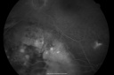
Choroidal Melanoma - Exudative Retinal Detachment 82 Year Old Man 510 views82-year-old man who had 3 falls 2 weeks ago. After the falls he started checking his vision and noticed there was a veil over the left eye, which he had seen for about two weeks in the superior visual field. OD 20/25, OS 20/40
(patient was medically never well enough for brachytherapy and died 6 months later from heart disease) Jun 28, 2011
|
|

Choroidal Melanoma - Exudative Retinal Detachment 82 Year Old Man 545 views82-year-old man who had 3 falls 2 weeks ago. After the falls he started checking his vision and noticed there was a veil over the left eye, which he had seen for about two weeks in the superior visual field. OD 20/25, OS 20/40
(patient was medically never well enough for brachytherapy and died 6 months later from heart disease) Jun 28, 2011
|
|

Choroidal Melanoma - Exudative Retinal Detachment 82 Year Old Man 549 views82-year-old man who had 3 falls 2 weeks ago. After the falls he started checking his vision and noticed there was a veil over the left eye, which he had seen for about two weeks in the superior visual field. OD 20/25, OS 20/40
(patient was medically never well enough for brachytherapy and died 6 months later from heart disease) Jun 28, 2011
|
|

Choroidal Melanoma - Exudative Retinal Detachment 82 Year Old Man 534 views82-year-old man who had 3 falls 2 weeks ago. After the falls he started checking his vision and noticed there was a veil over the left eye, which he had seen for about two weeks in the superior visual field. OD 20/25, OS 20/40
(patient was medically never well enough for brachytherapy and died 6 months later from heart disease) Jun 28, 2011
|
|

Choroidal Melanoma - Exudative Retinal Detachment 82 Year Old Man 567 views82-year-old man who had 3 falls 2 weeks ago. After the falls he started checking his vision and noticed there was a veil over the left eye, which he had seen for about two weeks in the superior visual field. OD 20/25, OS 20/40
(patient was medically never well enough for brachytherapy and died 6 months later from heart disease) Jun 28, 2011
|
|

Choroidal Melanoma - Exudative Retinal Detachment 82 Year Old Man 530 views82-year-old man who had 3 falls 2 weeks ago. After the falls he started checking his vision and noticed there was a veil over the left eye, which he had seen for about two weeks in the superior visual field. OD 20/25, OS 20/40
(patient was medically never well enough for brachytherapy and died 6 months later from heart disease) Jun 28, 2011
|
|

Choroidal Melanoma - Exudative Retinal Detachment 82 Year Old Man 607 views82-year-old man who had 3 falls 2 weeks ago. After the falls he started checking his vision and noticed there was a veil over the left eye, which he had seen for about two weeks in the superior visual field. OD 20/25, OS 20/40
(patient was medically never well enough for brachytherapy and died 6 months later from heart disease) Jun 28, 2011
|
|

Choroidal Melanoma - Exudative Retinal Detachment 82 Year Old Man 613 views82-year-old man who had 3 falls 2 weeks ago. After the falls he started checking his vision and noticed there was a veil over the left eye, which he had seen for about two weeks in the superior visual field. OD 20/25, OS 20/40
(patient was medically never well enough for brachytherapy and died 6 months later from heart disease) Jun 28, 2011
|
|
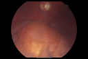
Choroidal Melanoma - Exudative Retinal Detachment 82 Year Old Man 700 views82-year-old man who had 3 falls 2 weeks ago. After the falls he started checking his vision and noticed there was a veil over the left eye, which he had seen for about two weeks in the superior visual field. OD 20/25, OS 20/40
(patient was medically never well enough for brachytherapy and died 6 months later from heart disease) Jun 28, 2011
|
|
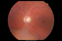
Choroidal Melanoma - Exudative Retinal Detachment 82 Year Old Man 771 views82-year-old man who had 3 falls 2 weeks ago. After the falls he started checking his vision and noticed there was a veil over the left eye, which he had seen for about two weeks in the superior visual field. OD 20/25, OS 20/40
(patient was medically never well enough for brachytherapy and died 6 months later from heart disease) Jun 28, 2011
|
|

Choroidal Melanoma - Exudative Retinal Detachment 82 Year Old Man 732 views82-year-old man who had 3 falls 2 weeks ago. After the falls he started checking his vision and noticed there was a veil over the left eye, which he had seen for about two weeks in the superior visual field. OD 20/25, OS 20/40
(patient was medically never well enough for brachytherapy and died 6 months later from heart disease) Jun 28, 2011
|
|

Choroidal Melanoma - Exudative Retinal Detachment 82 Year Old Man 695 views82-year-old man who had 3 falls 2 weeks ago. After the falls he started checking his vision and noticed there was a veil over the left eye, which he had seen for about two weeks in the superior visual field. OD 20/25, OS 20/40
(patient was medically never well enough for brachytherapy and died 6 months later from heart disease) Jun 28, 2011
|
|

CHRPE - Gardner Syndrome - FAMILIAL COLORECTAL POLYPOSIS - 11 year old female2134 views11-year-old child with CHRPE OS has six undescended adult teeth and also she has had a cyst removed from her arm pit and she has three more in her arm pit and one on her face and she is seeing a dermatologist for that in the near future. The mother reports no health problems, however, the patient’s grandmother does have a history of intestinal polyps, as does the patient’s grandmother’s brothers son. Jun 14, 2011
|
|

CHRPE - Gardner Syndrome - FAMILIAL COLORECTAL POLYPOSIS - 11 year old female1726 views11-year-old child with CHRPE OS has six undescended adult teeth and also she has had a cyst removed from her arm pit and she has three more in her arm pit and one on her face and she is seeing a dermatologist for that in the near future. The mother reports no health problems, however, the patient’s grandmother does have a history of intestinal polyps, as does the patient’s grandmother’s brothers son. Jun 14, 2011
|
|

CHRPE - Gardner Syndrome - FAMILIAL COLORECTAL POLYPOSIS - 11 year old female4095 views11-year-old child with CHRPE OS has six undescended adult teeth and also she has had a cyst removed from her arm pit and she has three more in her arm pit and one on her face and she is seeing a dermatologist for that in the near future. The mother reports no health problems, however, the patient’s grandmother does have a history of intestinal polyps, as does the patient’s grandmother’s brothers son. Jun 14, 2011
|
|

CHRPE - Gardner Syndrome - FAMILIAL COLORECTAL POLYPOSIS - 11 year old female1673 views11-year-old child with CHRPE OS has six undescended adult teeth and also she has had a cyst removed from her arm pit and she has three more in her arm pit and one on her face and she is seeing a dermatologist for that in the near future. The mother reports no health problems, however, the patient’s grandmother does have a history of intestinal polyps, as does the patient’s grandmother’s brothers son. Jun 14, 2011
|
|

525 viewsMay 14, 2011
|
|
522 viewsMay 14, 2011
|
|

563 viewsMay 14, 2011
|
|

531 viewsMay 14, 2011
|
|

563 viewsMay 14, 2011
|
|

573 viewsMay 14, 2011
|
|

561 viewsMay 14, 2011
|
|

549 viewsMay 14, 2011
|
|
481 viewsMay 14, 2011
|
|

509 viewsMay 14, 2011
|
|

505 viewsMay 14, 2011
|
|

478 viewsMay 14, 2011
|
|

513 viewsMay 14, 2011
|
|

537 viewsMay 14, 2011
|
|
529 viewsMay 14, 2011
|
|

592 viewsMay 14, 2011
|
|

611 viewsMay 14, 2011
|
|

704 viewsMay 14, 2011
|
|

835 viewsMay 14, 2011
|
|

Amelanotic Choroidal Nevus 1145 views45-year-old man OD 20/20, OS 20/20. 5x5 mm flat, yellow choroidal lesion just inferonasal to the optic nerve, two disc-diameters from the nerve with scalloped edges. It looks like a confluent drusen, but it is not elevated. Dec 13, 2010
|
|

Amelanotic Choroidal Nevus 1026 views45-year-old man OD 20/20, OS 20/20. 5x5 mm flat, yellow choroidal lesion just inferonasal to the optic nerve, two disc-diameters from the nerve with scalloped edges. It looks like a confluent drusen, but it is not elevated. Dec 13, 2010
|
|

Amelanotic Choroidal Nevus 1071 views45-year-old man OD 20/20, OS 20/20. 5x5 mm flat, yellow choroidal lesion just inferonasal to the optic nerve, two disc-diameters from the nerve with scalloped edges. It looks like a confluent drusen, but it is not elevated. Dec 13, 2010
|
|

Amelanotic Choroidal Nevus 2097 views45-year-old man OD 20/20, OS 20/20. 5x5 mm flat, yellow choroidal lesion just inferonasal to the optic nerve, two disc-diameters from the nerve with scalloped edges. It looks like a confluent drusen, but it is not elevated. Dec 13, 2010
|
|

Amelanotic Choroidal Nevus 1730 views45-year-old man OD 20/20, OS 20/20. 5x5 mm flat, yellow choroidal lesion just inferonasal to the optic nerve, two disc-diameters from the nerve with scalloped edges. It looks like a confluent drusen, but it is not elevated. Dec 13, 2010
|
|

Choroidal Nevus in Macula 926 viewsNov 29, 2010
|
|
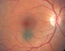
Choroidal Nevus in Macula 900 viewsNov 29, 2010
|
|

Radioactive Plaque Placement for Choroidal Melanoma Lead Patch after Surgery995 viewsNov 25, 2010
|
|
|
|


