neueste Dateien - Choroidal Melanoma and Choroidal Nevus
|

Radioactive Plaque Placement for Choroidal Melanoma Eye After Surgery with Conjunctiva Closed1114 x angesehen25. November 2010
|
|

Radioactive Plaque Placement for Choroidal Melanoma Radioactive Plaque Placement for Choroidal Melanoma 1136 x angesehen25. November 2010
|
|

Radioactive Plaque Placement for Choroidal Melanoma Plaque Sutured to Globe1136 x angesehen25. November 2010
|
|

Radioactive Plaque Placement for Choroidal Melanoma Plaque Sutured to Globe999 x angesehen25. November 2010
|
|

Radioactive Plaque Placement for Choroidal Melanoma Plaque Placed but not Sutured to Eye Yet1285 x angesehen25. November 2010
|
|

Radioactive Plaque Placement for Choroidal Melanoma Plaque Placed but not Sutured to Eye Ye1103 x angesehen25. November 2010
|
|

Radioactive Plaque Placement for Choroidal Melanoma Plaque Placed but not Sutured to Eye Ye1055 x angesehen25. November 2010
|
|

Radioactive Plaque Placement for Choroidal Melanoma Iodine Radioactive Seeds in Plaque1236 x angesehen25. November 2010
|
|

Radioactive Plaque Placement for Choroidal Melanoma Gold Plaque about the size of a Quarter999 x angesehen25. November 2010
|
|

Radioactive Plaque Placement for Choroidal Melanoma Transillumination of Globe to Localize Melanoma 988 x angesehen25. November 2010
|
|

Radioactive Plaque Placement for Choroidal Melanoma Transillumination of Globe to Localize Melanoma Arrow Points to Tumor Shadow906 x angesehen25. November 2010
|
|

Radioactive Plaque Placement for Choroidal Melanoma Transillumination of Globe to Localize Melanoma Arrow Points to Tumor Shadow1024 x angesehen25. November 2010
|
|

Radioactive Plaque Placement for Choroidal Melanoma Transillumination of Globe to Localize Melanoma 1009 x angesehen25. November 2010
|
|

Radioactive Plaque Placement for Choroidal Melanoma Transillumination of Globe to Localize Melanoma 979 x angesehen25. November 2010
|
|

Radioactive Plaque Placement for Choroidal Melanoma Eye is Anesthetized Prior to Surgery1211 x angesehen25. November 2010
|
|
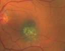
Macular Choroidal Nevus1725 x angesehen78-year-old woman has choroidal nevus in the macula of the left eye. You asked her to come in here for an evaluation and nevus was noted in the past about 7 years ago. She has not noticed any change in vision in that eye.
VISUAL ACUITY: OU 20/30. 12. November 2010
|
|

Choroidal Melanoma Medium599 x angesehen33-year-old man with a history of decreased in the right eye. He had blurring in the eye about a year ago. He reports that it improved spontaneously until it was “nearly resolvedâ€. He said he had some persistent distortion, but that over the past couple of weeks he has noticed this dark spot recurring in the right eye, the left eye is unaffected. He does not have any flashes, floaters or change in his peripheral visual acuity. 20/80 OD, 20/16 OS. No improvement with pinhole. IOP: 12 OD, 11 OS.
POSTERIOR SEGMENT EXAMINATION: Shows a clear view with no PVD in either eye. C/D ratio is 0.2 OU. Large retinal vessels are unremarkable. The macula of the right eye has a brown choroidal lesion with pigment overlying the surface and orange spots along its lateral surface. The left macula is normal.
B-SCAN: B-scan ecography of the ultrasound of the right eye shows a mass about 5 mm high with a moderate to low internal reflectivity on A-scan echogram. There is serous fluid inferiorly on the echogram07. November 2010
|
|

Choroidal Melanoma Medium745 x angesehen33-year-old man with a history of decreased in the right eye. He had blurring in the eye about a year ago. He reports that it improved spontaneously until it was “nearly resolvedâ€. He said he had some persistent distortion, but that over the past couple of weeks he has noticed this dark spot recurring in the right eye, the left eye is unaffected. He does not have any flashes, floaters or change in his peripheral visual acuity. 20/80 OD, 20/16 OS. No improvement with pinhole. IOP: 12 OD, 11 OS.
POSTERIOR SEGMENT EXAMINATION: Shows a clear view with no PVD in either eye. C/D ratio is 0.2 OU. Large retinal vessels are unremarkable. The macula of the right eye has a brown choroidal lesion with pigment overlying the surface and orange spots along its lateral surface. The left macula is normal.
B-SCAN: B-scan ecography of the ultrasound of the right eye shows a mass about 5 mm high with a moderate to low internal reflectivity on A-scan echogram. There is serous fluid inferiorly on the echogram07. November 2010
|
|
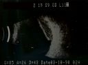
Choroidal Melanoma Medium683 x angesehen33-year-old man with a history of decreased in the right eye. He had blurring in the eye about a year ago. He reports that it improved spontaneously until it was “nearly resolvedâ€. He said he had some persistent distortion, but that over the past couple of weeks he has noticed this dark spot recurring in the right eye, the left eye is unaffected. He does not have any flashes, floaters or change in his peripheral visual acuity. 20/80 OD, 20/16 OS. No improvement with pinhole. IOP: 12 OD, 11 OS.
POSTERIOR SEGMENT EXAMINATION: Shows a clear view with no PVD in either eye. C/D ratio is 0.2 OU. Large retinal vessels are unremarkable. The macula of the right eye has a brown choroidal lesion with pigment overlying the surface and orange spots along its lateral surface. The left macula is normal.
B-SCAN: B-scan ecography of the ultrasound of the right eye shows a mass about 5 mm high with a moderate to low internal reflectivity on A-scan echogram. There is serous fluid inferiorly on the echogram07. November 2010
|
|

Choroidal Melanoma Medium776 x angesehen33-year-old man with a history of decreased in the right eye. He had blurring in the eye about a year ago. He reports that it improved spontaneously until it was “nearly resolvedâ€. He said he had some persistent distortion, but that over the past couple of weeks he has noticed this dark spot recurring in the right eye, the left eye is unaffected. He does not have any flashes, floaters or change in his peripheral visual acuity. 20/80 OD, 20/16 OS. No improvement with pinhole. IOP: 12 OD, 11 OS.
POSTERIOR SEGMENT EXAMINATION: Shows a clear view with no PVD in either eye. C/D ratio is 0.2 OU. Large retinal vessels are unremarkable. The macula of the right eye has a brown choroidal lesion with pigment overlying the surface and orange spots along its lateral surface. The left macula is normal.
B-SCAN: B-scan ecography of the ultrasound of the right eye shows a mass about 5 mm high with a moderate to low internal reflectivity on A-scan echogram. There is serous fluid inferiorly on the echogram07. November 2010
|
|

Choroidal Melanoma Medium515 x angesehen33-year-old man with a history of decreased in the right eye. He had blurring in the eye about a year ago. He reports that it improved spontaneously until it was “nearly resolvedâ€. He said he had some persistent distortion, but that over the past couple of weeks he has noticed this dark spot recurring in the right eye, the left eye is unaffected. He does not have any flashes, floaters or change in his peripheral visual acuity. 20/80 OD, 20/16 OS. No improvement with pinhole. IOP: 12 OD, 11 OS.
POSTERIOR SEGMENT EXAMINATION: Shows a clear view with no PVD in either eye. C/D ratio is 0.2 OU. Large retinal vessels are unremarkable. The macula of the right eye has a brown choroidal lesion with pigment overlying the surface and orange spots along its lateral surface. The left macula is normal.
B-SCAN: B-scan ecography of the ultrasound of the right eye shows a mass about 5 mm high with a moderate to low internal reflectivity on A-scan echogram. There is serous fluid inferiorly on the echogram07. November 2010
|
|
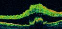
Choroidal Melanoma Medium603 x angesehen33-year-old man with a history of decreased in the right eye. He had blurring in the eye about a year ago. He reports that it improved spontaneously until it was “nearly resolvedâ€. He said he had some persistent distortion, but that over the past couple of weeks he has noticed this dark spot recurring in the right eye, the left eye is unaffected. He does not have any flashes, floaters or change in his peripheral visual acuity. 20/80 OD, 20/16 OS. No improvement with pinhole. IOP: 12 OD, 11 OS.
POSTERIOR SEGMENT EXAMINATION: Shows a clear view with no PVD in either eye. C/D ratio is 0.2 OU. Large retinal vessels are unremarkable. The macula of the right eye has a brown choroidal lesion with pigment overlying the surface and orange spots along its lateral surface. The left macula is normal.
B-SCAN: B-scan ecography of the ultrasound of the right eye shows a mass about 5 mm high with a moderate to low internal reflectivity on A-scan echogram. There is serous fluid inferiorly on the echogram07. November 2010
|
|

Choroidal Melanoma Medium519 x angesehen33-year-old man with a history of decreased in the right eye. He had blurring in the eye about a year ago. He reports that it improved spontaneously until it was “nearly resolvedâ€. He said he had some persistent distortion, but that over the past couple of weeks he has noticed this dark spot recurring in the right eye, the left eye is unaffected. He does not have any flashes, floaters or change in his peripheral visual acuity. 20/80 OD, 20/16 OS. No improvement with pinhole. IOP: 12 OD, 11 OS.
POSTERIOR SEGMENT EXAMINATION: Shows a clear view with no PVD in either eye. C/D ratio is 0.2 OU. Large retinal vessels are unremarkable. The macula of the right eye has a brown choroidal lesion with pigment overlying the surface and orange spots along its lateral surface. The left macula is normal.
B-SCAN: B-scan ecography of the ultrasound of the right eye shows a mass about 5 mm high with a moderate to low internal reflectivity on A-scan echogram. There is serous fluid inferiorly on the echogram07. November 2010
|
|

Choroidal Melanoma Medium493 x angesehen33-year-old man with a history of decreased in the right eye. He had blurring in the eye about a year ago. He reports that it improved spontaneously until it was “nearly resolvedâ€. He said he had some persistent distortion, but that over the past couple of weeks he has noticed this dark spot recurring in the right eye, the left eye is unaffected. He does not have any flashes, floaters or change in his peripheral visual acuity. 20/80 OD, 20/16 OS. No improvement with pinhole. IOP: 12 OD, 11 OS.
POSTERIOR SEGMENT EXAMINATION: Shows a clear view with no PVD in either eye. C/D ratio is 0.2 OU. Large retinal vessels are unremarkable. The macula of the right eye has a brown choroidal lesion with pigment overlying the surface and orange spots along its lateral surface. The left macula is normal.
B-SCAN: B-scan ecography of the ultrasound of the right eye shows a mass about 5 mm high with a moderate to low internal reflectivity on A-scan echogram. There is serous fluid inferiorly on the echogram07. November 2010
|
|

Choroidal Melanoma Medium525 x angesehen33-year-old man with a history of decreased in the right eye. He had blurring in the eye about a year ago. He reports that it improved spontaneously until it was “nearly resolvedâ€. He said he had some persistent distortion, but that over the past couple of weeks he has noticed this dark spot recurring in the right eye, the left eye is unaffected. He does not have any flashes, floaters or change in his peripheral visual acuity. 20/80 OD, 20/16 OS. No improvement with pinhole. IOP: 12 OD, 11 OS.
POSTERIOR SEGMENT EXAMINATION: Shows a clear view with no PVD in either eye. C/D ratio is 0.2 OU. Large retinal vessels are unremarkable. The macula of the right eye has a brown choroidal lesion with pigment overlying the surface and orange spots along its lateral surface. The left macula is normal.
B-SCAN: B-scan ecography of the ultrasound of the right eye shows a mass about 5 mm high with a moderate to low internal reflectivity on A-scan echogram. There is serous fluid inferiorly on the echogram07. November 2010
|
|

Choroidal Melanoma Medium506 x angesehen33-year-old man with a history of decreased in the right eye. He had blurring in the eye about a year ago. He reports that it improved spontaneously until it was “nearly resolvedâ€. He said he had some persistent distortion, but that over the past couple of weeks he has noticed this dark spot recurring in the right eye, the left eye is unaffected. He does not have any flashes, floaters or change in his peripheral visual acuity. 20/80 OD, 20/16 OS. No improvement with pinhole. IOP: 12 OD, 11 OS.
POSTERIOR SEGMENT EXAMINATION: Shows a clear view with no PVD in either eye. C/D ratio is 0.2 OU. Large retinal vessels are unremarkable. The macula of the right eye has a brown choroidal lesion with pigment overlying the surface and orange spots along its lateral surface. The left macula is normal.
B-SCAN: B-scan ecography of the ultrasound of the right eye shows a mass about 5 mm high with a moderate to low internal reflectivity on A-scan echogram. There is serous fluid inferiorly on the echogram07. November 2010
|
|

Choroidal Melanoma Medium479 x angesehen33-year-old man with a history of decreased in the right eye. He had blurring in the eye about a year ago. He reports that it improved spontaneously until it was “nearly resolvedâ€. He said he had some persistent distortion, but that over the past couple of weeks he has noticed this dark spot recurring in the right eye, the left eye is unaffected. He does not have any flashes, floaters or change in his peripheral visual acuity. 20/80 OD, 20/16 OS. No improvement with pinhole. IOP: 12 OD, 11 OS.
POSTERIOR SEGMENT EXAMINATION: Shows a clear view with no PVD in either eye. C/D ratio is 0.2 OU. Large retinal vessels are unremarkable. The macula of the right eye has a brown choroidal lesion with pigment overlying the surface and orange spots along its lateral surface. The left macula is normal.
B-SCAN: B-scan ecography of the ultrasound of the right eye shows a mass about 5 mm high with a moderate to low internal reflectivity on A-scan echogram. There is serous fluid inferiorly on the echogram07. November 2010
|
|

Choroidal Melanoma Medium497 x angesehen33-year-old man with a history of decreased in the right eye. He had blurring in the eye about a year ago. He reports that it improved spontaneously until it was “nearly resolvedâ€. He said he had some persistent distortion, but that over the past couple of weeks he has noticed this dark spot recurring in the right eye, the left eye is unaffected. He does not have any flashes, floaters or change in his peripheral visual acuity. 20/80 OD, 20/16 OS. No improvement with pinhole. IOP: 12 OD, 11 OS.
POSTERIOR SEGMENT EXAMINATION: Shows a clear view with no PVD in either eye. C/D ratio is 0.2 OU. Large retinal vessels are unremarkable. The macula of the right eye has a brown choroidal lesion with pigment overlying the surface and orange spots along its lateral surface. The left macula is normal.
B-SCAN: B-scan ecography of the ultrasound of the right eye shows a mass about 5 mm high with a moderate to low internal reflectivity on A-scan echogram. There is serous fluid inferiorly on the echogram07. November 2010
|
|

Choroidal Melanoma Medium508 x angesehen33-year-old man with a history of decreased in the right eye. He had blurring in the eye about a year ago. He reports that it improved spontaneously until it was “nearly resolvedâ€. He said he had some persistent distortion, but that over the past couple of weeks he has noticed this dark spot recurring in the right eye, the left eye is unaffected. He does not have any flashes, floaters or change in his peripheral visual acuity. 20/80 OD, 20/16 OS. No improvement with pinhole. IOP: 12 OD, 11 OS.
POSTERIOR SEGMENT EXAMINATION: Shows a clear view with no PVD in either eye. C/D ratio is 0.2 OU. Large retinal vessels are unremarkable. The macula of the right eye has a brown choroidal lesion with pigment overlying the surface and orange spots along its lateral surface. The left macula is normal.
B-SCAN: B-scan ecography of the ultrasound of the right eye shows a mass about 5 mm high with a moderate to low internal reflectivity on A-scan echogram. There is serous fluid inferiorly on the echogram07. November 2010
|
|

Choroidal Melanoma Medium509 x angesehen33-year-old man with a history of decreased in the right eye. He had blurring in the eye about a year ago. He reports that it improved spontaneously until it was “nearly resolvedâ€. He said he had some persistent distortion, but that over the past couple of weeks he has noticed this dark spot recurring in the right eye, the left eye is unaffected. He does not have any flashes, floaters or change in his peripheral visual acuity. 20/80 OD, 20/16 OS. No improvement with pinhole. IOP: 12 OD, 11 OS.
POSTERIOR SEGMENT EXAMINATION: Shows a clear view with no PVD in either eye. C/D ratio is 0.2 OU. Large retinal vessels are unremarkable. The macula of the right eye has a brown choroidal lesion with pigment overlying the surface and orange spots along its lateral surface. The left macula is normal.
B-SCAN: B-scan ecography of the ultrasound of the right eye shows a mass about 5 mm high with a moderate to low internal reflectivity on A-scan echogram. There is serous fluid inferiorly on the echogram07. November 2010
|
|

Choroidal Melanoma Medium517 x angesehen33-year-old man with a history of decreased in the right eye. He had blurring in the eye about a year ago. He reports that it improved spontaneously until it was “nearly resolvedâ€. He said he had some persistent distortion, but that over the past couple of weeks he has noticed this dark spot recurring in the right eye, the left eye is unaffected. He does not have any flashes, floaters or change in his peripheral visual acuity. 20/80 OD, 20/16 OS. No improvement with pinhole. IOP: 12 OD, 11 OS.
POSTERIOR SEGMENT EXAMINATION: Shows a clear view with no PVD in either eye. C/D ratio is 0.2 OU. Large retinal vessels are unremarkable. The macula of the right eye has a brown choroidal lesion with pigment overlying the surface and orange spots along its lateral surface. The left macula is normal.
B-SCAN: B-scan ecography of the ultrasound of the right eye shows a mass about 5 mm high with a moderate to low internal reflectivity on A-scan echogram. There is serous fluid inferiorly on the echogram07. November 2010
|
|

Choroidal Melanoma Medium509 x angesehen33-year-old man with a history of decreased in the right eye. He had blurring in the eye about a year ago. He reports that it improved spontaneously until it was “nearly resolvedâ€. He said he had some persistent distortion, but that over the past couple of weeks he has noticed this dark spot recurring in the right eye, the left eye is unaffected. He does not have any flashes, floaters or change in his peripheral visual acuity. 20/80 OD, 20/16 OS. No improvement with pinhole. IOP: 12 OD, 11 OS.
POSTERIOR SEGMENT EXAMINATION: Shows a clear view with no PVD in either eye. C/D ratio is 0.2 OU. Large retinal vessels are unremarkable. The macula of the right eye has a brown choroidal lesion with pigment overlying the surface and orange spots along its lateral surface. The left macula is normal.
B-SCAN: B-scan ecography of the ultrasound of the right eye shows a mass about 5 mm high with a moderate to low internal reflectivity on A-scan echogram. There is serous fluid inferiorly on the echogram07. November 2010
|
|

Choroidal Melanoma Medium559 x angesehen33-year-old man with a history of decreased in the right eye. He had blurring in the eye about a year ago. He reports that it improved spontaneously until it was “nearly resolvedâ€. He said he had some persistent distortion, but that over the past couple of weeks he has noticed this dark spot recurring in the right eye, the left eye is unaffected. He does not have any flashes, floaters or change in his peripheral visual acuity. 20/80 OD, 20/16 OS. No improvement with pinhole. IOP: 12 OD, 11 OS.
POSTERIOR SEGMENT EXAMINATION: Shows a clear view with no PVD in either eye. C/D ratio is 0.2 OU. Large retinal vessels are unremarkable. The macula of the right eye has a brown choroidal lesion with pigment overlying the surface and orange spots along its lateral surface. The left macula is normal.
B-SCAN: B-scan ecography of the ultrasound of the right eye shows a mass about 5 mm high with a moderate to low internal reflectivity on A-scan echogram. There is serous fluid inferiorly on the echogram07. November 2010
|
|

Choroidal Melanoma Medium579 x angesehen33-year-old man with a history of decreased in the right eye. He had blurring in the eye about a year ago. He reports that it improved spontaneously until it was “nearly resolvedâ€. He said he had some persistent distortion, but that over the past couple of weeks he has noticed this dark spot recurring in the right eye, the left eye is unaffected. He does not have any flashes, floaters or change in his peripheral visual acuity. 20/80 OD, 20/16 OS. No improvement with pinhole. IOP: 12 OD, 11 OS.
POSTERIOR SEGMENT EXAMINATION: Shows a clear view with no PVD in either eye. C/D ratio is 0.2 OU. Large retinal vessels are unremarkable. The macula of the right eye has a brown choroidal lesion with pigment overlying the surface and orange spots along its lateral surface. The left macula is normal.
B-SCAN: B-scan ecography of the ultrasound of the right eye shows a mass about 5 mm high with a moderate to low internal reflectivity on A-scan echogram. There is serous fluid inferiorly on the echogram07. November 2010
|
|

571 x angesehen33-year-old man with a history of decreased in the right eye. He had blurring in the eye about a year ago. He reports that it improved spontaneously until it was “nearly resolvedâ€. He said he had some persistent distortion, but that over the past couple of weeks he has noticed this dark spot recurring in the right eye, the left eye is unaffected. He does not have any flashes, floaters or change in his peripheral visual acuity. 20/80 OD, 20/16 OS. No improvement with pinhole. IOP: 12 OD, 11 OS.
POSTERIOR SEGMENT EXAMINATION: Shows a clear view with no PVD in either eye. C/D ratio is 0.2 OU. Large retinal vessels are unremarkable. The macula of the right eye has a brown choroidal lesion with pigment overlying the surface and orange spots along its lateral surface. The left macula is normal.
B-SCAN: B-scan ecography of the ultrasound of the right eye shows a mass about 5 mm high with a moderate to low internal reflectivity on A-scan echogram. There is serous fluid inferiorly on the echogram07. November 2010
|
|

Choroidal Melanoma Medium516 x angesehen33-year-old man with a history of decreased in the right eye. He had blurring in the eye about a year ago. He reports that it improved spontaneously until it was “nearly resolvedâ€. He said he had some persistent distortion, but that over the past couple of weeks he has noticed this dark spot recurring in the right eye, the left eye is unaffected. He does not have any flashes, floaters or change in his peripheral visual acuity. 20/80 OD, 20/16 OS. No improvement with pinhole. IOP: 12 OD, 11 OS.
POSTERIOR SEGMENT EXAMINATION: Shows a clear view with no PVD in either eye. C/D ratio is 0.2 OU. Large retinal vessels are unremarkable. The macula of the right eye has a brown choroidal lesion with pigment overlying the surface and orange spots along its lateral surface. The left macula is normal.
B-SCAN: B-scan ecography of the ultrasound of the right eye shows a mass about 5 mm high with a moderate to low internal reflectivity on A-scan echogram. There is serous fluid inferiorly on the echogram07. November 2010
|
|

Choroidal Melanoma Medium551 x angesehen33-year-old man with a history of decreased in the right eye. He had blurring in the eye about a year ago. He reports that it improved spontaneously until it was “nearly resolvedâ€. He said he had some persistent distortion, but that over the past couple of weeks he has noticed this dark spot recurring in the right eye, the left eye is unaffected. He does not have any flashes, floaters or change in his peripheral visual acuity. 20/80 OD, 20/16 OS. No improvement with pinhole. IOP: 12 OD, 11 OS.
POSTERIOR SEGMENT EXAMINATION: Shows a clear view with no PVD in either eye. C/D ratio is 0.2 OU. Large retinal vessels are unremarkable. The macula of the right eye has a brown choroidal lesion with pigment overlying the surface and orange spots along its lateral surface. The left macula is normal.
B-SCAN: B-scan ecography of the ultrasound of the right eye shows a mass about 5 mm high with a moderate to low internal reflectivity on A-scan echogram. There is serous fluid inferiorly on the echogram07. November 2010
|
|

Choroidal Melanoma Medium670 x angesehen33-year-old man with a history of decreased in the right eye. He had blurring in the eye about a year ago. He reports that it improved spontaneously until it was “nearly resolvedâ€. He said he had some persistent distortion, but that over the past couple of weeks he has noticed this dark spot recurring in the right eye, the left eye is unaffected. He does not have any flashes, floaters or change in his peripheral visual acuity. 20/80 OD, 20/16 OS. No improvement with pinhole. IOP: 12 OD, 11 OS.
POSTERIOR SEGMENT EXAMINATION: Shows a clear view with no PVD in either eye. C/D ratio is 0.2 OU. Large retinal vessels are unremarkable. The macula of the right eye has a brown choroidal lesion with pigment overlying the surface and orange spots along its lateral surface. The left macula is normal.
B-SCAN: B-scan ecography of the ultrasound of the right eye shows a mass about 5 mm high with a moderate to low internal reflectivity on A-scan echogram. There is serous fluid inferiorly on the echogram07. November 2010
|
|

Melanoma - Large Ciliary Body A scan shows medium to low internal reflectivity828 x angesehen30. Oktober 2010
|
|
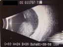
Melanoma - Large Ciliary Body B-Scan ultrasound shows tumore filling much of the eye972 x angesehen30. Oktober 2010
|
|

Melanoma - Large Ciliary Body Photo shows tumor visible throught the pupil953 x angesehen30. Oktober 2010
|
|

Melanoma - Large Ciliary Body Photo shows tumor visible through the pupil1132 x angesehen30. Oktober 2010
|
|

Melanoma - Large Ciliary Body B Scan Ultrasound740 x angesehen30. Oktober 2010
|
|
|
| 543 Dateien auf 6 Seite(n) |
 |
|
|
6 |
|


