Last additions - Tumors of the Retina and Retinal Pigment Epithelium
|

Astrocytoma (Astrocytic Hamartoma) no calcification201 views38 year old man with no symptomsApr 14, 2025
|
|

Astrocytoma (Astrocytic Hamartoma) no calcification202 views38 year old man with no symptomsApr 14, 2025
|
|
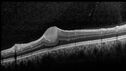
Astrocytoma (Astrocytic Hamartoma) no calcification234 views38 year old man with no symptomsApr 14, 2025
|
|

Astrocytoma (Astrocytic Hamartoma) no calcification169 views38 year old man with no symptomsApr 14, 2025
|
|
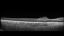
Astrocytoma (Astrocytic Hamartoma) no calcification154 views38 year old man with no symptomsApr 14, 2025
|
|

Advanced age-related macular degeneration288 views87 year old female. Severe macular bleed 15 years ago OD. Now with disciform scar. Left eye has progressing geographic atrophy.
VA OD: HM, OS: 5/200. IOP normal OUMar 17, 2024
|
|

Advanced age-related macular degeneration158 views87 year old female. Severe macular bleed 15 years ago OD. Now with disciform scar. Left eye has progressing geographic atrophy.
VA OD: HM, OS: 5/200. IOP normal OUMar 17, 2024
|
|

Advanced age-related macular degeneration169 views87 year old female. Severe macular bleed 15 years ago OD. Now with disciform scar. Left eye has progressing geographic atrophy.
VA OD: HM, OS: 5/200. IOP normal OUMar 17, 2024
|
|

Advanced age-related macular degeneration162 views87 year old female. Severe macular bleed 15 years ago OD. Now with disciform scar. Left eye has progressing geographic atrophy.
VA OD: HM, OS: 5/200. IOP normal OUMar 17, 2024
|
|

Advanced age-related macular degeneration134 views87 year old female. Severe macular bleed 15 years ago OD. Now with disciform scar. Left eye has progressing geographic atrophy.
VA OD: HM, OS: 5/200. IOP normal OUMar 17, 2024
|
|

Advanced age-related macular degeneration172 views87 year old female. Severe macular bleed 15 years ago OD. Now with disciform scar. Left eye has progressing geographic atrophy.
VA OD: HM, OS: 5/200. IOP normal OUMar 17, 2024
|
|

Advanced age-related macular degeneration158 views87 year old female. Severe macular bleed 15 years ago OD. Now with disciform scar. Left eye has progressing geographic atrophy.
VA OD: HM, OS: 5/200. IOP normal OUMar 17, 2024
|
|

Advanced age-related macular degeneration137 views87 year old female. Severe macular bleed 15 years ago OD. Now with disciform scar. Left eye has progressing geographic atrophy.
VA OD: HM, OS: 5/200. IOP normal OUMar 17, 2024
|
|

Advanced age-related macular degeneration158 views87 year old female. Severe macular bleed 15 years ago OD. Now with disciform scar. Left eye has progressing geographic atrophy.
VA OD: HM, OS: 5/200. IOP normal OUMar 17, 2024
|
|

Retinal Angioma155 views46 year old female - Her original reason for an eye examination was that she had been in a car accident. Then she saw that the vision was blurry in just the left eye. Prior to that she had no known vision changes. Since the accident she notices that her vision is better and her headaches are getting less bothersome.
PMhx benign, Meds: none
VA OD: Dcc20/20
VA OS: Dcc20/25Mar 11, 2024
|
|

Retinal Angioma152 views46 year old female - Her original reason for an eye examination was that she had been in a car accident. Then she saw that the vision was blurry in just the left eye. Prior to that she had no known vision changes. Since the accident she notices that her vision is better and her headaches are getting less bothersome.
PMhx benign, Meds: none
VA OD: Dcc20/20
VA OS: Dcc20/25Mar 11, 2024
|
|

Retinal Angioma171 views46 year old female - Her original reason for an eye examination was that she had been in a car accident. Then she saw that the vision was blurry in just the left eye. Prior to that she had no known vision changes. Since the accident she notices that her vision is better and her headaches are getting less bothersome.
PMhx benign, Meds: none
VA OD: Dcc20/20
VA OS: Dcc20/25Mar 11, 2024
|
|

Retinal Angioma143 views46 year old female - Her original reason for an eye examination was that she had been in a car accident. Then she saw that the vision was blurry in just the left eye. Prior to that she had no known vision changes. Since the accident she notices that her vision is better and her headaches are getting less bothersome.
PMhx benign, Meds: none
VA OD: Dcc20/20
VA OS: Dcc20/25Mar 11, 2024
|
|

Retinal Angioma167 views46 year old female - Her original reason for an eye examination was that she had been in a car accident. Then she saw that the vision was blurry in just the left eye. Prior to that she had no known vision changes. Since the accident she notices that her vision is better and her headaches are getting less bothersome.
PMhx benign, Meds: none
VA OD: Dcc20/20
VA OS: Dcc20/25Mar 11, 2024
|
|

Retinal Angioma157 views46 year old female - Her original reason for an eye examination was that she had been in a car accident. Then she saw that the vision was blurry in just the left eye. Prior to that she had no known vision changes. Since the accident she notices that her vision is better and her headaches are getting less bothersome.
PMhx benign, Meds: none
VA OD: Dcc20/20
VA OS: Dcc20/25Mar 11, 2024
|
|

Retinal Angioma142 views46 year old female - Her original reason for an eye examination was that she had been in a car accident. Then she saw that the vision was blurry in just the left eye. Prior to that she had no known vision changes. Since the accident she notices that her vision is better and her headaches are getting less bothersome.
PMhx benign, Meds: none
VA OD: Dcc20/20
VA OS: Dcc20/25Mar 11, 2024
|
|

Retinal Angioma157 views46 year old female - Her original reason for an eye examination was that she had been in a car accident. Then she saw that the vision was blurry in just the left eye. Prior to that she had no known vision changes. Since the accident she notices that her vision is better and her headaches are getting less bothersome.
PMhx benign, Meds: none
VA OD: Dcc20/20
VA OS: Dcc20/25Mar 11, 2024
|
|

Retinal Angioma140 views46 year old female - Her original reason for an eye examination was that she had been in a car accident. Then she saw that the vision was blurry in just the left eye. Prior to that she had no known vision changes. Since the accident she notices that her vision is better and her headaches are getting less bothersome.
PMhx benign, Meds: none
VA OD: Dcc20/20
VA OS: Dcc20/25Mar 11, 2024
|
|

Retinal Angioma129 views46 year old female - Her original reason for an eye examination was that she had been in a car accident. Then she saw that the vision was blurry in just the left eye. Prior to that she had no known vision changes. Since the accident she notices that her vision is better and her headaches are getting less bothersome.
PMhx benign, Meds: none
VA OD: Dcc20/20
VA OS: Dcc20/25Mar 11, 2024
|
|

Retinal Angioma144 views46 year old female - Her original reason for an eye examination was that she had been in a car accident. Then she saw that the vision was blurry in just the left eye. Prior to that she had no known vision changes. Since the accident she notices that her vision is better and her headaches are getting less bothersome.
PMhx benign, Meds: none
VA OD: Dcc20/20
VA OS: Dcc20/25Mar 11, 2024
|
|

Retinal Angioma135 views46 year old female - Her original reason for an eye examination was that she had been in a car accident. Then she saw that the vision was blurry in just the left eye. Prior to that she had no known vision changes. Since the accident she notices that her vision is better and her headaches are getting less bothersome.
PMhx benign, Meds: none
VA OD: Dcc20/20
VA OS: Dcc20/25Mar 11, 2024
|
|

Retinal Angioma146 views46 year old female - Her original reason for an eye examination was that she had been in a car accident. Then she saw that the vision was blurry in just the left eye. Prior to that she had no known vision changes. Since the accident she notices that her vision is better and her headaches are getting less bothersome.
PMhx benign, Meds: none
VA OD: Dcc20/20
VA OS: Dcc20/25Mar 11, 2024
|
|

Radiation Retinopathy Both Eyes - 2023 - Two years after radiation for ocular lymphoma 240 viewsAug 21, 2023
|
|

Radiation Retinopathy Both Eyes - 2023 - Two years after radiation for ocular lymphoma (24)266 viewsAug 21, 2023
|
|

Radiation Retinopathy Both Eyes - 2023 - Two years after radiation for ocular lymphoma (23)228 viewsAug 21, 2023
|
|

Radiation Retinopathy Both Eyes - 2023 - Two years after radiation for ocular lymphoma (22)238 viewsAug 21, 2023
|
|

Radiation Retinopathy Both Eyes - 2023 - Two years after radiation for ocular lymphoma (21)292 viewsAug 21, 2023
|
|
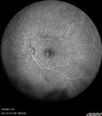
Radiation Retinopathy Both Eyes - 2023 - Two years after radiation for ocular lymphoma (20)286 viewsAug 21, 2023
|
|

Radiation Retinopathy Both Eyes - 2023 - Two years after radiation for ocular lymphoma (19)229 viewsAug 21, 2023
|
|
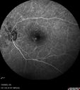
Radiation Retinopathy Both Eyes - 2023 - Two years after radiation for ocular lymphoma (18)239 viewsAug 21, 2023
|
|

Radiation Retinopathy Both Eyes - 2023 - Two years after radiation for ocular lymphoma (1)244 viewsAug 21, 2023
|
|
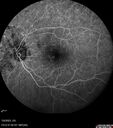
Radiation Retinopathy Both Eyes - 2023 - Two years after radiation for ocular lymphoma (17)251 viewsAug 21, 2023
|
|

Radiation Retinopathy Both Eyes - 2023 - Two years after radiation for ocular lymphoma (16)235 viewsAug 21, 2023
|
|
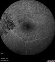
Radiation Retinopathy Both Eyes - 2023 - Two years after radiation for ocular lymphoma (15)233 viewsAug 21, 2023
|
|

Radiation Retinopathy Both Eyes - 2023 - Two years after radiation for ocular lymphoma (14)253 viewsAug 21, 2023
|
|

Radiation Retinopathy Both Eyes - 2023 - Two years after radiation for ocular lymphoma (13)261 viewsAug 21, 2023
|
|

Radiation Retinopathy Both Eyes - 2023 - Two years after radiation for ocular lymphoma (12)245 viewsAug 21, 2023
|
|

Radiation Retinopathy Both Eyes - 2023 - Two years after radiation for ocular lymphoma (11)274 viewsAug 21, 2023
|
|

Radiation Retinopathy Both Eyes - 2023 - Two years after radiation for ocular lymphoma (10)241 viewsAug 21, 2023
|
|

Radiation Retinopathy Both Eyes - 2023 - Two years after radiation for ocular lymphoma (9)255 viewsAug 21, 2023
|
|

Radiation Retinopathy Both Eyes - 2023 - Two years after radiation for ocular lymphoma (8)264 viewsAug 21, 2023
|
|

Radiation Retinopathy Both Eyes - 2023 - Two years after radiation for ocular lymphoma (6)221 viewsAug 21, 2023
|
|

Radiation Retinopathy Both Eyes - 2023 - Two years after radiation for ocular lymphoma (7)240 viewsAug 21, 2023
|
|
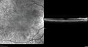
Radiation Retinopathy Both Eyes - 2023 - Two years after radiation for ocular lymphoma (5)251 viewsAug 21, 2023
|
|

Radiation Retinopathy Both Eyes - 2023 - Two years after radiation for ocular lymphoma (4)237 viewsAug 21, 2023
|
|

Radiation Retinopathy Both Eyes - 2023 - Two years after radiation for ocular lymphoma (3)239 viewsAug 21, 2023
|
|

Radiation Retinopathy Both Eyes - 2023 - Two years after radiation for ocular lymphoma (2)262 viewsAug 21, 2023
|
|

Retinal Cavernous Hemangioma336 views27 year old female with normal vision - MRI was normalFeb 04, 2022
|
|

Retinal Cavernous Hemangioma322 views27 year old female with normal vision - MRI was normalFeb 04, 2022
|
|

Retinal Cavernous Hemangioma338 views27 year old female with normal vision - MRI was normalFeb 04, 2022
|
|

Retinal Cavernous Hemangioma304 views27 year old female with normal vision - MRI was normalFeb 04, 2022
|
|

Ocular lymphoma - better following intravitreal methotrexate321 viewsVA improved from LP to 20/200 after 4 injections of methotrexateJan 06, 2022
|
|

Ocular Lymphoma - Flare up despite chemotherapy and radiation therapy - This was treated with intravitreal methotrexate385 viewsVision in the left eye has declined to LP in this image following ocular recurrence of lymphomaJan 06, 2022
|
|

Ocular Lymphoma - Initially presented like auto-immune retinopathy367 views43 year old man: initial presentation - The vision in the left eye is blurry and opaque for about 2 weeks. In the central of the vision, when he covers the right eye, in the center of the left eye there is a film with a light in the middle. This has never happened before. He has not been sick for the last few months. A few weeks ago he saw waves on the floor when looking down but that has gone away. Since the vision loss started the vision has not changed much. The central vision is not getting better. Â
PMHx – Borderline HTN
VA OD: Dcc20/20
VA OS: Dcc20/40-1 IOP: TP: OD:15 OS:19
1+ NS
No uveitis
Labs normal. One positive antiretinal antibody - Arrestin
VA improved over 2 months to normal. Then the patient had 2 more episodes of self-limited vision loss (no treatment). Then 2 years later he presented with a diagnosis of CNS lymphoma and later developed subretinal deposits (last images)Oct 25, 2021
|
|
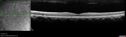
Ocular Lymphoma - Initially presented like auto-immune retinopathy345 views43 year old man: initial presentation - The vision in the left eye is blurry and opaque for about 2 weeks. In the central of the vision, when he covers the right eye, in the center of the left eye there is a film with a light in the middle. This has never happened before. He has not been sick for the last few months. A few weeks ago he saw waves on the floor when looking down but that has gone away. Since the vision loss started the vision has not changed much. The central vision is not getting better. Â
PMHx – Borderline HTN
VA OD: Dcc20/20
VA OS: Dcc20/40-1 IOP: TP: OD:15 OS:19
1+ NS
No uveitis
Labs normal. One positive antiretinal antibody - Arrestin
VA improved over 2 months to normal. Then the patient had 2 more episodes of self-limited vision loss (no treatment). Then 2 years later he presented with a diagnosis of CNS lymphoma and later developed subretinal deposits (last images)Oct 25, 2021
|
|

Ocular Lymphoma - Initially presented like auto-immune retinopathy385 views43 year old man: initial presentation - The vision in the left eye is blurry and opaque for about 2 weeks. In the central of the vision, when he covers the right eye, in the center of the left eye there is a film with a light in the middle. This has never happened before. He has not been sick for the last few months. A few weeks ago he saw waves on the floor when looking down but that has gone away. Since the vision loss started the vision has not changed much. The central vision is not getting better. Â
PMHx – Borderline HTN
VA OD: Dcc20/20
VA OS: Dcc20/40-1 IOP: TP: OD:15 OS:19
1+ NS
No uveitis
Labs normal. One positive antiretinal antibody - Arrestin
VA improved over 2 months to normal. Then the patient had 2 more episodes of self-limited vision loss (no treatment). Then 2 years later he presented with a diagnosis of CNS lymphoma and later developed subretinal deposits (last images)Oct 25, 2021
|
|

Ocular Lymphoma - Initially presented like auto-immune retinopathy350 views43 year old man: initial presentation - The vision in the left eye is blurry and opaque for about 2 weeks. In the central of the vision, when he covers the right eye, in the center of the left eye there is a film with a light in the middle. This has never happened before. He has not been sick for the last few months. A few weeks ago he saw waves on the floor when looking down but that has gone away. Since the vision loss started the vision has not changed much. The central vision is not getting better. Â
PMHx – Borderline HTN
VA OD: Dcc20/20
VA OS: Dcc20/40-1 IOP: TP: OD:15 OS:19
1+ NS
No uveitis
Labs normal. One positive antiretinal antibody - Arrestin
VA improved over 2 months to normal. Then the patient had 2 more episodes of self-limited vision loss (no treatment). Then 2 years later he presented with a diagnosis of CNS lymphoma and later developed subretinal deposits (last images)Oct 25, 2021
|
|

Ocular Lymphoma - Initially presented like auto-immune retinopathy321 views43 year old man: initial presentation - The vision in the left eye is blurry and opaque for about 2 weeks. In the central of the vision, when he covers the right eye, in the center of the left eye there is a film with a light in the middle. This has never happened before. He has not been sick for the last few months. A few weeks ago he saw waves on the floor when looking down but that has gone away. Since the vision loss started the vision has not changed much. The central vision is not getting better. Â
PMHx – Borderline HTN
VA OD: Dcc20/20
VA OS: Dcc20/40-1 IOP: TP: OD:15 OS:19
1+ NS
No uveitis
Labs normal. One positive antiretinal antibody - Arrestin
VA improved over 2 months to normal. Then the patient had 2 more episodes of self-limited vision loss (no treatment). Then 2 years later he presented with a diagnosis of CNS lymphoma and later developed subretinal deposits (last images)Oct 25, 2021
|
|

Ocular Lymphoma - Initially presented like auto-immune retinopathy316 views43 year old man: initial presentation - The vision in the left eye is blurry and opaque for about 2 weeks. In the central of the vision, when he covers the right eye, in the center of the left eye there is a film with a light in the middle. This has never happened before. He has not been sick for the last few months. A few weeks ago he saw waves on the floor when looking down but that has gone away. Since the vision loss started the vision has not changed much. The central vision is not getting better. Â
PMHx – Borderline HTN
VA OD: Dcc20/20
VA OS: Dcc20/40-1 IOP: TP: OD:15 OS:19
1+ NS
No uveitis
Labs normal. One positive antiretinal antibody - Arrestin
VA improved over 2 months to normal. Then the patient had 2 more episodes of self-limited vision loss (no treatment). Then 2 years later he presented with a diagnosis of CNS lymphoma and later developed subretinal deposits (last images)Oct 25, 2021
|
|

Ocular Lymphoma - Initially presented like auto-immune retinopathy298 views43 year old man: initial presentation - The vision in the left eye is blurry and opaque for about 2 weeks. In the central of the vision, when he covers the right eye, in the center of the left eye there is a film with a light in the middle. This has never happened before. He has not been sick for the last few months. A few weeks ago he saw waves on the floor when looking down but that has gone away. Since the vision loss started the vision has not changed much. The central vision is not getting better. Â
PMHx – Borderline HTN
VA OD: Dcc20/20
VA OS: Dcc20/40-1 IOP: TP: OD:15 OS:19
1+ NS
No uveitis
Labs normal. One positive antiretinal antibody - Arrestin
VA improved over 2 months to normal. Then the patient had 2 more episodes of self-limited vision loss (no treatment). Then 2 years later he presented with a diagnosis of CNS lymphoma and later developed subretinal deposits (last images)Oct 25, 2021
|
|

Ocular Lymphoma - Initially presented like auto-immune retinopathy332 views43 year old man: initial presentation - The vision in the left eye is blurry and opaque for about 2 weeks. In the central of the vision, when he covers the right eye, in the center of the left eye there is a film with a light in the middle. This has never happened before. He has not been sick for the last few months. A few weeks ago he saw waves on the floor when looking down but that has gone away. Since the vision loss started the vision has not changed much. The central vision is not getting better. Â
PMHx – Borderline HTN
VA OD: Dcc20/20
VA OS: Dcc20/40-1 IOP: TP: OD:15 OS:19
1+ NS
No uveitis
Labs normal. One positive antiretinal antibody - Arrestin
VA improved over 2 months to normal. Then the patient had 2 more episodes of self-limited vision loss (no treatment). Then 2 years later he presented with a diagnosis of CNS lymphoma and later developed subretinal deposits (last images)Oct 25, 2021
|
|

Ocular Lymphoma - Initially presented like auto-immune retinopathy306 views43 year old man: initial presentation - The vision in the left eye is blurry and opaque for about 2 weeks. In the central of the vision, when he covers the right eye, in the center of the left eye there is a film with a light in the middle. This has never happened before. He has not been sick for the last few months. A few weeks ago he saw waves on the floor when looking down but that has gone away. Since the vision loss started the vision has not changed much. The central vision is not getting better. Â
PMHx – Borderline HTN
VA OD: Dcc20/20
VA OS: Dcc20/40-1 IOP: TP: OD:15 OS:19
1+ NS
No uveitis
Labs normal. One positive antiretinal antibody - Arrestin
VA improved over 2 months to normal. Then the patient had 2 more episodes of self-limited vision loss (no treatment). Then 2 years later he presented with a diagnosis of CNS lymphoma and later developed subretinal deposits (last images)Oct 25, 2021
|
|

Ocular Lymphoma - Initially presented like auto-immune retinopathy296 views43 year old man: initial presentation - The vision in the left eye is blurry and opaque for about 2 weeks. In the central of the vision, when he covers the right eye, in the center of the left eye there is a film with a light in the middle. This has never happened before. He has not been sick for the last few months. A few weeks ago he saw waves on the floor when looking down but that has gone away. Since the vision loss started the vision has not changed much. The central vision is not getting better. Â
PMHx – Borderline HTN
VA OD: Dcc20/20
VA OS: Dcc20/40-1 IOP: TP: OD:15 OS:19
1+ NS
No uveitis
Labs normal. One positive antiretinal antibody - Arrestin
VA improved over 2 months to normal. Then the patient had 2 more episodes of self-limited vision loss (no treatment). Then 2 years later he presented with a diagnosis of CNS lymphoma and later developed subretinal deposits (last images)Oct 25, 2021
|
|

Ocular Lymphoma - Initially presented like auto-immune retinopathy325 views43 year old man: initial presentation - The vision in the left eye is blurry and opaque for about 2 weeks. In the central of the vision, when he covers the right eye, in the center of the left eye there is a film with a light in the middle. This has never happened before. He has not been sick for the last few months. A few weeks ago he saw waves on the floor when looking down but that has gone away. Since the vision loss started the vision has not changed much. The central vision is not getting better. Â
PMHx – Borderline HTN
VA OD: Dcc20/20
VA OS: Dcc20/40-1 IOP: TP: OD:15 OS:19
1+ NS
No uveitis
Labs normal. One positive antiretinal antibody - Arrestin
VA improved over 2 months to normal. Then the patient had 2 more episodes of self-limited vision loss (no treatment). Then 2 years later he presented with a diagnosis of CNS lymphoma and later developed subretinal deposits (last images)Oct 25, 2021
|
|

Ocular Lymphoma - Initially presented like auto-immune retinopathy333 views43 year old man: initial presentation - The vision in the left eye is blurry and opaque for about 2 weeks. In the central of the vision, when he covers the right eye, in the center of the left eye there is a film with a light in the middle. This has never happened before. He has not been sick for the last few months. A few weeks ago he saw waves on the floor when looking down but that has gone away. Since the vision loss started the vision has not changed much. The central vision is not getting better. Â
PMHx – Borderline HTN
VA OD: Dcc20/20
VA OS: Dcc20/40-1 IOP: TP: OD:15 OS:19
1+ NS
No uveitis
Labs normal. One positive antiretinal antibody - Arrestin
VA improved over 2 months to normal. Then the patient had 2 more episodes of self-limited vision loss (no treatment). Then 2 years later he presented with a diagnosis of CNS lymphoma and later developed subretinal deposits (last images)Oct 25, 2021
|
|

Ocular Lymphoma - Initially presented like auto-immune retinopathy333 views43 year old man: initial presentation - The vision in the left eye is blurry and opaque for about 2 weeks. In the central of the vision, when he covers the right eye, in the center of the left eye there is a film with a light in the middle. This has never happened before. He has not been sick for the last few months. A few weeks ago he saw waves on the floor when looking down but that has gone away. Since the vision loss started the vision has not changed much. The central vision is not getting better. Â
PMHx – Borderline HTN
VA OD: Dcc20/20
VA OS: Dcc20/40-1 IOP: TP: OD:15 OS:19
1+ NS
No uveitis
Labs normal. One positive antiretinal antibody - Arrestin
VA improved over 2 months to normal. Then the patient had 2 more episodes of self-limited vision loss (no treatment). Then 2 years later he presented with a diagnosis of CNS lymphoma and later developed subretinal deposits (last images)Oct 25, 2021
|
|

Ocular Lymphoma - Initially presented like auto-immune retinopathy324 views43 year old man: initial presentation - The vision in the left eye is blurry and opaque for about 2 weeks. In the central of the vision, when he covers the right eye, in the center of the left eye there is a film with a light in the middle. This has never happened before. He has not been sick for the last few months. A few weeks ago he saw waves on the floor when looking down but that has gone away. Since the vision loss started the vision has not changed much. The central vision is not getting better. Â
PMHx – Borderline HTN
VA OD: Dcc20/20
VA OS: Dcc20/40-1 IOP: TP: OD:15 OS:19
1+ NS
No uveitis
Labs normal. One positive antiretinal antibody - Arrestin
VA improved over 2 months to normal. Then the patient had 2 more episodes of self-limited vision loss (no treatment). Then 2 years later he presented with a diagnosis of CNS lymphoma and later developed subretinal deposits (last images)Oct 25, 2021
|
|

Ocular Lymphoma - Initially presented like auto-immune retinopathy309 views43 year old man: initial presentation - The vision in the left eye is blurry and opaque for about 2 weeks. In the central of the vision, when he covers the right eye, in the center of the left eye there is a film with a light in the middle. This has never happened before. He has not been sick for the last few months. A few weeks ago he saw waves on the floor when looking down but that has gone away. Since the vision loss started the vision has not changed much. The central vision is not getting better. Â
PMHx – Borderline HTN
VA OD: Dcc20/20
VA OS: Dcc20/40-1 IOP: TP: OD:15 OS:19
1+ NS
No uveitis
Labs normal. One positive antiretinal antibody - Arrestin
VA improved over 2 months to normal. Then the patient had 2 more episodes of self-limited vision loss (no treatment). Then 2 years later he presented with a diagnosis of CNS lymphoma and later developed subretinal deposits (last images)Oct 25, 2021
|
|

Ocular Lymphoma - Initially presented like auto-immune retinopathy323 views43 year old man: initial presentation - The vision in the left eye is blurry and opaque for about 2 weeks. In the central of the vision, when he covers the right eye, in the center of the left eye there is a film with a light in the middle. This has never happened before. He has not been sick for the last few months. A few weeks ago he saw waves on the floor when looking down but that has gone away. Since the vision loss started the vision has not changed much. The central vision is not getting better. Â
PMHx – Borderline HTN
VA OD: Dcc20/20
VA OS: Dcc20/40-1 IOP: TP: OD:15 OS:19
1+ NS
No uveitis
Labs normal. One positive antiretinal antibody - Arrestin
VA improved over 2 months to normal. Then the patient had 2 more episodes of self-limited vision loss (no treatment). Then 2 years later he presented with a diagnosis of CNS lymphoma and later developed subretinal deposits (last images)Oct 25, 2021
|
|
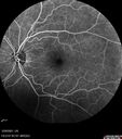
Ocular Lymphoma - Initially presented like auto-immune retinopathy322 views43 year old man: initial presentation - The vision in the left eye is blurry and opaque for about 2 weeks. In the central of the vision, when he covers the right eye, in the center of the left eye there is a film with a light in the middle. This has never happened before. He has not been sick for the last few months. A few weeks ago he saw waves on the floor when looking down but that has gone away. Since the vision loss started the vision has not changed much. The central vision is not getting better. Â
PMHx – Borderline HTN
VA OD: Dcc20/20
VA OS: Dcc20/40-1 IOP: TP: OD:15 OS:19
1+ NS
No uveitis
Labs normal. One positive antiretinal antibody - Arrestin
VA improved over 2 months to normal. Then the patient had 2 more episodes of self-limited vision loss (no treatment). Then 2 years later he presented with a diagnosis of CNS lymphoma and later developed subretinal deposits (last images)Oct 25, 2021
|
|

Ocular Lymphoma - Initially presented like auto-immune retinopathy306 views43 year old man: initial presentation - The vision in the left eye is blurry and opaque for about 2 weeks. In the central of the vision, when he covers the right eye, in the center of the left eye there is a film with a light in the middle. This has never happened before. He has not been sick for the last few months. A few weeks ago he saw waves on the floor when looking down but that has gone away. Since the vision loss started the vision has not changed much. The central vision is not getting better. Â
PMHx – Borderline HTN
VA OD: Dcc20/20
VA OS: Dcc20/40-1 IOP: TP: OD:15 OS:19
1+ NS
No uveitis
Labs normal. One positive antiretinal antibody - Arrestin
VA improved over 2 months to normal. Then the patient had 2 more episodes of self-limited vision loss (no treatment). Then 2 years later he presented with a diagnosis of CNS lymphoma and later developed subretinal deposits (last images)Oct 25, 2021
|
|

Ocular Lymphoma - Initially presented like auto-immune retinopathy333 views43 year old man: initial presentation - The vision in the left eye is blurry and opaque for about 2 weeks. In the central of the vision, when he covers the right eye, in the center of the left eye there is a film with a light in the middle. This has never happened before. He has not been sick for the last few months. A few weeks ago he saw waves on the floor when looking down but that has gone away. Since the vision loss started the vision has not changed much. The central vision is not getting better. Â
PMHx – Borderline HTN
VA OD: Dcc20/20
VA OS: Dcc20/40-1 IOP: TP: OD:15 OS:19
1+ NS
No uveitis
Labs normal. One positive antiretinal antibody - Arrestin
VA improved over 2 months to normal. Then the patient had 2 more episodes of self-limited vision loss (no treatment). Then 2 years later he presented with a diagnosis of CNS lymphoma and later developed subretinal deposits (last images)Oct 25, 2021
|
|

Ocular Lymphoma - Initially presented like auto-immune retinopathy307 views43 year old man: initial presentation - The vision in the left eye is blurry and opaque for about 2 weeks. In the central of the vision, when he covers the right eye, in the center of the left eye there is a film with a light in the middle. This has never happened before. He has not been sick for the last few months. A few weeks ago he saw waves on the floor when looking down but that has gone away. Since the vision loss started the vision has not changed much. The central vision is not getting better. Â
PMHx – Borderline HTN
VA OD: Dcc20/20
VA OS: Dcc20/40-1 IOP: TP: OD:15 OS:19
1+ NS
No uveitis
Labs normal. One positive antiretinal antibody - Arrestin
VA improved over 2 months to normal. Then the patient had 2 more episodes of self-limited vision loss (no treatment). Then 2 years later he presented with a diagnosis of CNS lymphoma and later developed subretinal deposits (last images)Oct 25, 2021
|
|

Ocular Lymphoma - Initially presented like auto-immune retinopathy362 views43 year old man: initial presentation - The vision in the left eye is blurry and opaque for about 2 weeks. In the central of the vision, when he covers the right eye, in the center of the left eye there is a film with a light in the middle. This has never happened before. He has not been sick for the last few months. A few weeks ago he saw waves on the floor when looking down but that has gone away. Since the vision loss started the vision has not changed much. The central vision is not getting better. Â
PMHx – Borderline HTN
VA OD: Dcc20/20
VA OS: Dcc20/40-1 IOP: TP: OD:15 OS:19
1+ NS
No uveitis
Labs normal. One positive antiretinal antibody - Arrestin
VA improved over 2 months to normal. Then the patient had 2 more episodes of self-limited vision loss (no treatment). Then 2 years later he presented with a diagnosis of CNS lymphoma and later developed subretinal deposits (last images)Oct 25, 2021
|
|

Ocular Lymphoma - Initially presented like auto-immune retinopathy322 views43 year old man: initial presentation - The vision in the left eye is blurry and opaque for about 2 weeks. In the central of the vision, when he covers the right eye, in the center of the left eye there is a film with a light in the middle. This has never happened before. He has not been sick for the last few months. A few weeks ago he saw waves on the floor when looking down but that has gone away. Since the vision loss started the vision has not changed much. The central vision is not getting better. Â
PMHx – Borderline HTN
VA OD: Dcc20/20
VA OS: Dcc20/40-1 IOP: TP: OD:15 OS:19
1+ NS
No uveitis
Labs normal. One positive antiretinal antibody - Arrestin
VA improved over 2 months to normal. Then the patient had 2 more episodes of self-limited vision loss (no treatment). Then 2 years later he presented with a diagnosis of CNS lymphoma and later developed subretinal deposits (last images)Oct 25, 2021
|
|

Ocular Lymphoma - Initially presented like auto-immune retinopathy330 views43 year old man: initial presentation - The vision in the left eye is blurry and opaque for about 2 weeks. In the central of the vision, when he covers the right eye, in the center of the left eye there is a film with a light in the middle. This has never happened before. He has not been sick for the last few months. A few weeks ago he saw waves on the floor when looking down but that has gone away. Since the vision loss started the vision has not changed much. The central vision is not getting better. Â
PMHx – Borderline HTN
VA OD: Dcc20/20
VA OS: Dcc20/40-1 IOP: TP: OD:15 OS:19
1+ NS
No uveitis
Labs normal. One positive antiretinal antibody - Arrestin
VA improved over 2 months to normal. Then the patient had 2 more episodes of self-limited vision loss (no treatment). Then 2 years later he presented with a diagnosis of CNS lymphoma and later developed subretinal deposits (last images)Oct 25, 2021
|
|

Ocular Lymphoma - Initially presented like auto-immune retinopathy340 views43 year old man: initial presentation - The vision in the left eye is blurry and opaque for about 2 weeks. In the central of the vision, when he covers the right eye, in the center of the left eye there is a film with a light in the middle. This has never happened before. He has not been sick for the last few months. A few weeks ago he saw waves on the floor when looking down but that has gone away. Since the vision loss started the vision has not changed much. The central vision is not getting better. Â
PMHx – Borderline HTN
VA OD: Dcc20/20
VA OS: Dcc20/40-1 IOP: TP: OD:15 OS:19
1+ NS
No uveitis
Labs normal. One positive antiretinal antibody - Arrestin
VA improved over 2 months to normal. Then the patient had 2 more episodes of self-limited vision loss (no treatment). Then 2 years later he presented with a diagnosis of CNS lymphoma and later developed subretinal deposits (last images)Oct 25, 2021
|
|

Ocular Lymphoma - Initially presented like auto-immune retinopathy345 views43 year old man: initial presentation - The vision in the left eye is blurry and opaque for about 2 weeks. In the central of the vision, when he covers the right eye, in the center of the left eye there is a film with a light in the middle. This has never happened before. He has not been sick for the last few months. A few weeks ago he saw waves on the floor when looking down but that has gone away. Since the vision loss started the vision has not changed much. The central vision is not getting better. Â
PMHx – Borderline HTN
VA OD: Dcc20/20
VA OS: Dcc20/40-1 IOP: TP: OD:15 OS:19
1+ NS
No uveitis
Labs normal. One positive antiretinal antibody - Arrestin
VA improved over 2 months to normal. Then the patient had 2 more episodes of self-limited vision loss (no treatment). Then 2 years later he presented with a diagnosis of CNS lymphoma and later developed subretinal deposits (last images)Oct 25, 2021
|
|

Ocular Lymphoma - Initially presented like auto-immune retinopathy321 views43 year old man: initial presentation - The vision in the left eye is blurry and opaque for about 2 weeks. In the central of the vision, when he covers the right eye, in the center of the left eye there is a film with a light in the middle. This has never happened before. He has not been sick for the last few months. A few weeks ago he saw waves on the floor when looking down but that has gone away. Since the vision loss started the vision has not changed much. The central vision is not getting better. Â
PMHx – Borderline HTN
VA OD: Dcc20/20
VA OS: Dcc20/40-1 IOP: TP: OD:15 OS:19
1+ NS
No uveitis
Labs normal. One positive antiretinal antibody - Arrestin
VA improved over 2 months to normal. Then the patient had 2 more episodes of self-limited vision loss (no treatment). Then 2 years later he presented with a diagnosis of CNS lymphoma and later developed subretinal deposits (last images)Oct 25, 2021
|
|

Ocular Lymphoma - Initially presented like auto-immune retinopathy316 views43 year old man: initial presentation - The vision in the left eye is blurry and opaque for about 2 weeks. In the central of the vision, when he covers the right eye, in the center of the left eye there is a film with a light in the middle. This has never happened before. He has not been sick for the last few months. A few weeks ago he saw waves on the floor when looking down but that has gone away. Since the vision loss started the vision has not changed much. The central vision is not getting better. Â
PMHx – Borderline HTN
VA OD: Dcc20/20
VA OS: Dcc20/40-1 IOP: TP: OD:15 OS:19
1+ NS
No uveitis
Labs normal. One positive antiretinal antibody - Arrestin
VA improved over 2 months to normal. Then the patient had 2 more episodes of self-limited vision loss (no treatment). Then 2 years later he presented with a diagnosis of CNS lymphoma and later developed subretinal deposits (last images)Oct 25, 2021
|
|

Ocular Lymphoma - Initially presented like auto-immune retinopathy321 views43 year old man: initial presentation - The vision in the left eye is blurry and opaque for about 2 weeks. In the central of the vision, when he covers the right eye, in the center of the left eye there is a film with a light in the middle. This has never happened before. He has not been sick for the last few months. A few weeks ago he saw waves on the floor when looking down but that has gone away. Since the vision loss started the vision has not changed much. The central vision is not getting better. Â
PMHx – Borderline HTN
VA OD: Dcc20/20
VA OS: Dcc20/40-1 IOP: TP: OD:15 OS:19
1+ NS
No uveitis
Labs normal. One positive antiretinal antibody - Arrestin
VA improved over 2 months to normal. Then the patient had 2 more episodes of self-limited vision loss (no treatment). Then 2 years later he presented with a diagnosis of CNS lymphoma and later developed subretinal deposits (last images)Oct 25, 2021
|
|

Ocular Lymphoma - Initially presented like auto-immune retinopathy311 views43 year old man: initial presentation - The vision in the left eye is blurry and opaque for about 2 weeks. In the central of the vision, when he covers the right eye, in the center of the left eye there is a film with a light in the middle. This has never happened before. He has not been sick for the last few months. A few weeks ago he saw waves on the floor when looking down but that has gone away. Since the vision loss started the vision has not changed much. The central vision is not getting better. Â
PMHx – Borderline HTN
VA OD: Dcc20/20
VA OS: Dcc20/40-1 IOP: TP: OD:15 OS:19
1+ NS
No uveitis
Labs normal. One positive antiretinal antibody - Arrestin
VA improved over 2 months to normal. Then the patient had 2 more episodes of self-limited vision loss (no treatment). Then 2 years later he presented with a diagnosis of CNS lymphoma and later developed subretinal deposits (last images)Oct 25, 2021
|
|

Ocular Lymphoma - Initially presented like auto-immune retinopathy296 views43 year old man: initial presentation - The vision in the left eye is blurry and opaque for about 2 weeks. In the central of the vision, when he covers the right eye, in the center of the left eye there is a film with a light in the middle. This has never happened before. He has not been sick for the last few months. A few weeks ago he saw waves on the floor when looking down but that has gone away. Since the vision loss started the vision has not changed much. The central vision is not getting better. Â
PMHx – Borderline HTN
VA OD: Dcc20/20
VA OS: Dcc20/40-1 IOP: TP: OD:15 OS:19
1+ NS
No uveitis
Labs normal. One positive antiretinal antibody - Arrestin
VA improved over 2 months to normal. Then the patient had 2 more episodes of self-limited vision loss (no treatment). Then 2 years later he presented with a diagnosis of CNS lymphoma and later developed subretinal deposits (last images)Oct 25, 2021
|
|

Ocular Lymphoma - Initially presented like auto-immune retinopathy316 views43 year old man: initial presentation - The vision in the left eye is blurry and opaque for about 2 weeks. In the central of the vision, when he covers the right eye, in the center of the left eye there is a film with a light in the middle. This has never happened before. He has not been sick for the last few months. A few weeks ago he saw waves on the floor when looking down but that has gone away. Since the vision loss started the vision has not changed much. The central vision is not getting better. Â
PMHx – Borderline HTN
VA OD: Dcc20/20
VA OS: Dcc20/40-1 IOP: TP: OD:15 OS:19
1+ NS
No uveitis
Labs normal. One positive antiretinal antibody - Arrestin
VA improved over 2 months to normal. Then the patient had 2 more episodes of self-limited vision loss (no treatment). Then 2 years later he presented with a diagnosis of CNS lymphoma and later developed subretinal deposits (last images)Oct 25, 2021
|
|

Ocular Lymphoma - Initially presented like auto-immune retinopathy321 views43 year old man: initial presentation - The vision in the left eye is blurry and opaque for about 2 weeks. In the central of the vision, when he covers the right eye, in the center of the left eye there is a film with a light in the middle. This has never happened before. He has not been sick for the last few months. A few weeks ago he saw waves on the floor when looking down but that has gone away. Since the vision loss started the vision has not changed much. The central vision is not getting better. Â
PMHx – Borderline HTN
VA OD: Dcc20/20
VA OS: Dcc20/40-1 IOP: TP: OD:15 OS:19
1+ NS
No uveitis
Labs normal. One positive antiretinal antibody - Arrestin
VA improved over 2 months to normal. Then the patient had 2 more episodes of self-limited vision loss (no treatment). Then 2 years later he presented with a diagnosis of CNS lymphoma and later developed subretinal deposits (last images)Oct 25, 2021
|
|
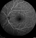
Ocular Lymphoma - Initially presented like auto-immune retinopathy371 views43 year old man: initial presentation - The vision in the left eye is blurry and opaque for about 2 weeks. In the central of the vision, when he covers the right eye, in the center of the left eye there is a film with a light in the middle. This has never happened before. He has not been sick for the last few months. A few weeks ago he saw waves on the floor when looking down but that has gone away. Since the vision loss started the vision has not changed much. The central vision is not getting better. Â
PMHx – Borderline HTN
VA OD: Dcc20/20
VA OS: Dcc20/40-1 IOP: TP: OD:15 OS:19
1+ NS
No uveitis
Labs normal. One positive antiretinal antibody - Arrestin
VA improved over 2 months to normal. Then the patient had 2 more episodes of self-limited vision loss (no treatment). Then 2 years later he presented with a diagnosis of CNS lymphoma and later developed subretinal deposits (last images)Oct 25, 2021
|
|

Ocular Lymphoma - Initially presented like auto-immune retinopathy336 views43 year old man: initial presentation - The vision in the left eye is blurry and opaque for about 2 weeks. In the central of the vision, when he covers the right eye, in the center of the left eye there is a film with a light in the middle. This has never happened before. He has not been sick for the last few months. A few weeks ago he saw waves on the floor when looking down but that has gone away. Since the vision loss started the vision has not changed much. The central vision is not getting better. Â
PMHx – Borderline HTN
VA OD: Dcc20/20
VA OS: Dcc20/40-1 IOP: TP: OD:15 OS:19
1+ NS
No uveitis
Labs normal. One positive antiretinal antibody - Arrestin
VA improved over 2 months to normal. Then the patient had 2 more episodes of self-limited vision loss (no treatment). Then 2 years later he presented with a diagnosis of CNS lymphoma and later developed subretinal deposits (last images)Oct 25, 2021
|
|

Ocular Lymphoma - Initially presented like auto-immune retinopathy355 views43 year old man: initial presentation - The vision in the left eye is blurry and opaque for about 2 weeks. In the central of the vision, when he covers the right eye, in the center of the left eye there is a film with a light in the middle. This has never happened before. He has not been sick for the last few months. A few weeks ago he saw waves on the floor when looking down but that has gone away. Since the vision loss started the vision has not changed much. The central vision is not getting better. Â
PMHx – Borderline HTN
VA OD: Dcc20/20
VA OS: Dcc20/40-1 IOP: TP: OD:15 OS:19
1+ NS
No uveitis
Labs normal. One positive antiretinal antibody - Arrestin
VA improved over 2 months to normal. Then the patient had 2 more episodes of self-limited vision loss (no treatment). Then 2 years later he presented with a diagnosis of CNS lymphoma and later developed subretinal deposits (last images)Oct 25, 2021
|
|

Ocular Lymphoma - Initially presented like auto-immune retinopathy338 views43 year old man: initial presentation - The vision in the left eye is blurry and opaque for about 2 weeks. In the central of the vision, when he covers the right eye, in the center of the left eye there is a film with a light in the middle. This has never happened before. He has not been sick for the last few months. A few weeks ago he saw waves on the floor when looking down but that has gone away. Since the vision loss started the vision has not changed much. The central vision is not getting better. Â
PMHx – Borderline HTN
VA OD: Dcc20/20
VA OS: Dcc20/40-1 IOP: TP: OD:15 OS:19
1+ NS
No uveitis
Labs normal. One positive antiretinal antibody - Arrestin
VA improved over 2 months to normal. Then the patient had 2 more episodes of self-limited vision loss (no treatment). Then 2 years later he presented with a diagnosis of CNS lymphoma and later developed subretinal deposits (last images)Oct 25, 2021
|
|

Ocular Lymphoma - Initially presented like auto-immune retinopathy327 views43 year old man: initial presentation - The vision in the left eye is blurry and opaque for about 2 weeks. In the central of the vision, when he covers the right eye, in the center of the left eye there is a film with a light in the middle. This has never happened before. He has not been sick for the last few months. A few weeks ago he saw waves on the floor when looking down but that has gone away. Since the vision loss started the vision has not changed much. The central vision is not getting better. Â
PMHx – Borderline HTN
VA OD: Dcc20/20
VA OS: Dcc20/40-1 IOP: TP: OD:15 OS:19
1+ NS
No uveitis
Labs normal. One positive antiretinal antibody - Arrestin
VA improved over 2 months to normal. Then the patient had 2 more episodes of self-limited vision loss (no treatment). Then 2 years later he presented with a diagnosis of CNS lymphoma and later developed subretinal deposits (last images)Oct 25, 2021
|
|

Ocular Lymphoma - Initially presented like auto-immune retinopathy344 views43 year old man: initial presentation - The vision in the left eye is blurry and opaque for about 2 weeks. In the central of the vision, when he covers the right eye, in the center of the left eye there is a film with a light in the middle. This has never happened before. He has not been sick for the last few months. A few weeks ago he saw waves on the floor when looking down but that has gone away. Since the vision loss started the vision has not changed much. The central vision is not getting better. Â
PMHx – Borderline HTN
VA OD: Dcc20/20
VA OS: Dcc20/40-1 IOP: TP: OD:15 OS:19
1+ NS
No uveitis
Labs normal. One positive antiretinal antibody - Arrestin
VA improved over 2 months to normal. Then the patient had 2 more episodes of self-limited vision loss (no treatment). Then 2 years later he presented with a diagnosis of CNS lymphoma and later developed subretinal deposits (last images)Oct 25, 2021
|
|
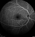
Ocular Lymphoma - Initially presented like auto-immune retinopathy332 views43 year old man: initial presentation - The vision in the left eye is blurry and opaque for about 2 weeks. In the central of the vision, when he covers the right eye, in the center of the left eye there is a film with a light in the middle. This has never happened before. He has not been sick for the last few months. A few weeks ago he saw waves on the floor when looking down but that has gone away. Since the vision loss started the vision has not changed much. The central vision is not getting better. Â
PMHx – Borderline HTN
VA OD: Dcc20/20
VA OS: Dcc20/40-1 IOP: TP: OD:15 OS:19
1+ NS
No uveitis
Labs normal. One positive antiretinal antibody - Arrestin
VA improved over 2 months to normal. Then the patient had 2 more episodes of self-limited vision loss (no treatment). Then 2 years later he presented with a diagnosis of CNS lymphoma and later developed subretinal deposits (last images)Oct 25, 2021
|
|

Ocular Lymphoma - Initially presented like auto-immune retinopathy334 views43 year old man: initial presentation - The vision in the left eye is blurry and opaque for about 2 weeks. In the central of the vision, when he covers the right eye, in the center of the left eye there is a film with a light in the middle. This has never happened before. He has not been sick for the last few months. A few weeks ago he saw waves on the floor when looking down but that has gone away. Since the vision loss started the vision has not changed much. The central vision is not getting better. Â
PMHx – Borderline HTN
VA OD: Dcc20/20
VA OS: Dcc20/40-1 IOP: TP: OD:15 OS:19
1+ NS
No uveitis
Labs normal. One positive antiretinal antibody - Arrestin
VA improved over 2 months to normal. Then the patient had 2 more episodes of self-limited vision loss (no treatment). Then 2 years later he presented with a diagnosis of CNS lymphoma and later developed subretinal deposits (last images)Oct 25, 2021
|
|

Ocular Lymphoma - Initially presented like auto-immune retinopathy331 views43 year old man: initial presentation - The vision in the left eye is blurry and opaque for about 2 weeks. In the central of the vision, when he covers the right eye, in the center of the left eye there is a film with a light in the middle. This has never happened before. He has not been sick for the last few months. A few weeks ago he saw waves on the floor when looking down but that has gone away. Since the vision loss started the vision has not changed much. The central vision is not getting better. Â
PMHx – Borderline HTN
VA OD: Dcc20/20
VA OS: Dcc20/40-1 IOP: TP: OD:15 OS:19
1+ NS
No uveitis
Labs normal. One positive antiretinal antibody - Arrestin
VA improved over 2 months to normal. Then the patient had 2 more episodes of self-limited vision loss (no treatment). Then 2 years later he presented with a diagnosis of CNS lymphoma and later developed subretinal deposits (last images)Oct 25, 2021
|
|
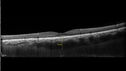
Ocular Lymphoma - Initially presented like auto-immune retinopathy343 views43 year old man: initial presentation - The vision in the left eye is blurry and opaque for about 2 weeks. In the central of the vision, when he covers the right eye, in the center of the left eye there is a film with a light in the middle. This has never happened before. He has not been sick for the last few months. A few weeks ago he saw waves on the floor when looking down but that has gone away. Since the vision loss started the vision has not changed much. The central vision is not getting better. Â
PMHx – Borderline HTN
VA OD: Dcc20/20
VA OS: Dcc20/40-1 IOP: TP: OD:15 OS:19
1+ NS
No uveitis
Labs normal. One positive antiretinal antibody - Arrestin
VA improved over 2 months to normal. Then the patient had 2 more episodes of self-limited vision loss (no treatment). Then 2 years later he presented with a diagnosis of CNS lymphoma and later developed subretinal deposits (last images)Oct 25, 2021
|
|
|
|


