Derniers ajouts - Iatrogenic Diseases
|

Phototoxicity following cataract surgeryvu 154 fois70 year old female with nasal spot in vision since cataract surgery 3 years ago. VA 20/20 OU.14 Février 2024
|
|

Phototoxicity following cataract surgeryvu 180 fois70 year old female with nasal spot in vision since cataract surgery 3 years ago. VA 20/20 OU.14 Février 2024
|
|

Phototoxicity following cataract surgeryvu 207 fois70 year old female with nasal spot in vision since cataract surgery 3 years ago. VA 20/20 OU.14 Février 2024
|
|

Phototoxicity following cataract surgeryvu 168 fois70 year old female with nasal spot in vision since cataract surgery 3 years ago. VA 20/20 OU.14 Février 2024
|
|

Phototoxicity following cataract surgeryvu 198 fois70 year old female with nasal spot in vision since cataract surgery 3 years ago. VA 20/20 OU.14 Février 2024
|
|

Phototoxicity following cataract surgeryvu 156 fois70 year old female with nasal spot in vision since cataract surgery 3 years ago. VA 20/20 OU.14 Février 2024
|
|

Phototoxicity following cataract surgeryvu 179 fois70 year old female with nasal spot in vision since cataract surgery 3 years ago. VA 20/20 OU.14 Février 2024
|
|

Phototoxicity following cataract surgeryvu 189 fois70 year old female with nasal spot in vision since cataract surgery 3 years ago. VA 20/20 OU.14 Février 2024
|
|

Phototoxicity following cataract surgeryvu 160 fois70 year old female with nasal spot in vision since cataract surgery 3 years ago. VA 20/20 OU.14 Février 2024
|
|

Phototoxicity following cataract surgeryvu 146 fois70 year old female with nasal spot in vision since cataract surgery 3 years ago. VA 20/20 OU.14 Février 2024
|
|

Phototoxicity following cataract surgeryvu 180 fois70 year old female with nasal spot in vision since cataract surgery 3 years ago. VA 20/20 OU.14 Février 2024
|
|

Phototoxicity following cataract surgeryvu 186 fois70 year old female with nasal spot in vision since cataract surgery 3 years ago. VA 20/20 OU.14 Février 2024
|
|

Phototoxicity following cataract surgeryvu 148 fois70 year old female with nasal spot in vision since cataract surgery 3 years ago. VA 20/20 OU.14 Février 2024
|
|

Phototoxicity following cataract surgeryvu 203 fois70 year old female with nasal spot in vision since cataract surgery 3 years ago. VA 20/20 OU.14 Février 2024
|
|

Phototoxicity following cataract surgeryvu 174 fois70 year old female with nasal spot in vision since cataract surgery 3 years ago. VA 20/20 OU.14 Février 2024
|
|

Phototoxicity following cataract surgeryvu 179 fois70 year old female with nasal spot in vision since cataract surgery 3 years ago. VA 20/20 OU.14 Février 2024
|
|

Phototoxicity following cataract surgeryvu 170 fois70 year old female with nasal spot in vision since cataract surgery 3 years ago. VA 20/20 OU.14 Février 2024
|
|

Phototoxicity following cataract surgeryvu 171 fois70 year old female with nasal spot in vision since cataract surgery 3 years ago. VA 20/20 OU.14 Février 2024
|
|

Phototoxicity following cataract surgeryvu 171 fois70 year old female with nasal spot in vision since cataract surgery 3 years ago. VA 20/20 OU.14 Février 2024
|
|

Phototoxicity following cataract surgeryvu 200 fois70 year old female with nasal spot in vision since cataract surgery 3 years ago. VA 20/20 OU.14 Février 2024
|
|

Uveitis Glaucoma Hyphema (UGH) syndromevu 319 fois65 year old female with intraocular lens half in the sulcus, iris atrophy, and recurrent uveitis since cataract surgery. She has a mild case and has been managed with topical steroids as needed.02 Janvier 2023
|
|

Uveitis Glaucoma Hyphema (UGH) syndromevu 313 fois65 year old female with intraocular lens half in the sulcus, iris atrophy, and recurrent uveitis since cataract surgery. She has a mild case and has been managed with topical steroids as needed.02 Janvier 2023
|
|
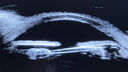
Uveitis Glaucoma Hyphema (UGH) syndromevu 299 fois65 year old female with intraocular lens half in the sulcus, iris atrophy, and recurrent uveitis since cataract surgery. She has a mild case and has been managed with topical steroids as needed.02 Janvier 2023
|
|
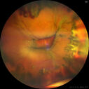
Choroidal Effusions following IOL exchangevu 299 fois67 year old man 10 years ago had retinal detachment with buckle, vitrectomy, laser, gas. Vision was 20/25 post-op. 2 months ago had sudden vision loss in the right eye from a dislocated PCIOL (in the bag). 3 weeks ago had a vitrectomy, remove IOL and scleral suture fixation of Akreos AO60 PCIOL with gortex sutures. IOP was 12 on post-op day 1 with a vitreous hemorrhage. Post-op day 7, vitreous hemorrhage cleared.
PMhx: HTN, High Cholesterol.
Med: Amlodipine, ASA, Atorvastatin, Metoprolol
Vision 20/200,09 Mars 2022
|
|

Choroidal Effusions following IOL exchangevu 303 fois67 year old man 10 years ago had retinal detachment with buckle, vitrectomy, laser, gas. Vision was 20/25 post-op. 2 months ago had sudden vision loss in the right eye from a dislocated PCIOL (in the bag). 3 weeks ago had a vitrectomy, remove IOL and scleral suture fixation of Akreos AO60 PCIOL with gortex sutures. IOP was 12 on post-op day 1 with a vitreous hemorrhage. Post-op day 7, vitreous hemorrhage cleared.
PMhx: HTN, High Cholesterol.
Med: Amlodipine, ASA, Atorvastatin, Metoprolol
Vision 20/200,09 Mars 2022
|
|

Choroidal Effusions following IOL exchangevu 310 fois67 year old man 10 years ago had retinal detachment with buckle, vitrectomy, laser, gas. Vision was 20/25 post-op. 2 months ago had sudden vision loss in the right eye from a dislocated PCIOL (in the bag). 3 weeks ago had a vitrectomy, remove IOL and scleral suture fixation of Akreos AO60 PCIOL with gortex sutures. IOP was 12 on post-op day 1 with a vitreous hemorrhage. Post-op day 7, vitreous hemorrhage cleared.
PMhx: HTN, High Cholesterol.
Med: Amlodipine, ASA, Atorvastatin, Metoprolol
Vision 20/200,09 Mars 2022
|
|

Choroidal Effusions following IOL exchangevu 280 fois67 year old man 10 years ago had retinal detachment with buckle, vitrectomy, laser, gas. Vision was 20/25 post-op. 2 months ago had sudden vision loss in the right eye from a dislocated PCIOL (in the bag). 3 weeks ago had a vitrectomy, remove IOL and scleral suture fixation of Akreos AO60 PCIOL with gortex sutures. IOP was 12 on post-op day 1 with a vitreous hemorrhage. Post-op day 7, vitreous hemorrhage cleared.
PMhx: HTN, High Cholesterol.
Med: Amlodipine, ASA, Atorvastatin, Metoprolol
Vision 20/200,09 Mars 2022
|
|

Choroidal Effusions following IOL exchangevu 299 fois67 year old man 10 years ago had retinal detachment with buckle, vitrectomy, laser, gas. Vision was 20/25 post-op. 2 months ago had sudden vision loss in the right eye from a dislocated PCIOL (in the bag). 3 weeks ago had a vitrectomy, remove IOL and scleral suture fixation of Akreos AO60 PCIOL with gortex sutures. IOP was 12 on post-op day 1 with a vitreous hemorrhage. Post-op day 7, vitreous hemorrhage cleared.
PMhx: HTN, High Cholesterol.
Med: Amlodipine, ASA, Atorvastatin, Metoprolol
Vision 20/200,09 Mars 2022
|
|

Choroidal Effusions following IOL exchangevu 316 fois67 year old man 10 years ago had retinal detachment with buckle, vitrectomy, laser, gas. Vision was 20/25 post-op. 2 months ago had sudden vision loss in the right eye from a dislocated PCIOL (in the bag). 3 weeks ago had a vitrectomy, remove IOL and scleral suture fixation of Akreos AO60 PCIOL with gortex sutures. IOP was 12 on post-op day 1 with a vitreous hemorrhage. Post-op day 7, vitreous hemorrhage cleared.
PMhx: HTN, High Cholesterol.
Med: Amlodipine, ASA, Atorvastatin, Metoprolol
Vision 20/200,09 Mars 2022
|
|

Choroidal Effusions following IOL exchangevu 297 fois67 year old man 10 years ago had retinal detachment with buckle, vitrectomy, laser, gas. Vision was 20/25 post-op. 2 months ago had sudden vision loss in the right eye from a dislocated PCIOL (in the bag). 3 weeks ago had a vitrectomy, remove IOL and scleral suture fixation of Akreos AO60 PCIOL with gortex sutures. IOP was 12 on post-op day 1 with a vitreous hemorrhage. Post-op day 7, vitreous hemorrhage cleared.
PMhx: HTN, High Cholesterol.
Med: Amlodipine, ASA, Atorvastatin, Metoprolol
Vision 20/200,09 Mars 2022
|
|

vu 393 fois30 Septembre 2020
|
|

vu 438 fois30 Septembre 2020
|
|

vu 394 fois30 Septembre 2020
|
|

vu 414 fois30 Septembre 2020
|
|

vu 388 fois30 Septembre 2020
|
|

vu 410 fois30 Septembre 2020
|
|

Upside Down Lens Syndrome - Anterior Chamber IOL Upside Downvu 692 fois77 year old female who had vitrectomy for retained lens fragments and has had difficulty with recurrent uveitis since her cataract surgery. The anterior chamber intraocular lens is upside down. A lens exchange is planned.07 Mars 2020
|
|

Choroidal Effusions following Glaucoma Surgery 9 months agovu 410 fois67 year old female with glaucoma surgery 9 month ago. Her IOP is 12. She has peripheral choroidal effusions. VA 20/100 ph 20/3211 Février 2020
|
|

Choroidal Effusions following Glaucoma Surgery 9 months agovu 444 fois67 year old female with glaucoma surgery 9 month ago. Her IOP is 12. She has peripheral choroidal effusions. VA 20/100 ph 20/3211 Février 2020
|
|

Choroidal Effusions following Glaucoma Surgery 9 months agovu 428 fois67 year old female with glaucoma surgery 9 month ago. Her IOP is 12. She has peripheral choroidal effusions. VA 20/100 ph 20/3211 Février 2020
|
|
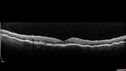
Choroidal Effusions following Glaucoma Surgery 9 months agovu 474 fois67 year old female with glaucoma surgery 9 month ago. Her IOP is 12. She has peripheral choroidal effusions. VA 20/100 ph 20/3211 Février 2020
|
|

Choroidal Effusions following Glaucoma Surgery 9 months agovu 436 fois67 year old female with glaucoma surgery 9 month ago. Her IOP is 12. She has peripheral choroidal effusions. VA 20/100 ph 20/3211 Février 2020
|
|

Choroidal Effusions following Glaucoma Surgery 9 months agovu 424 fois67 year old female with glaucoma surgery 9 month ago. Her IOP is 12. She has peripheral choroidal effusions. VA 20/100 ph 20/3211 Février 2020
|
|
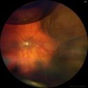
Choroidal Effusion One Day following macular pucker surgeryvu 409 fois70 year old female one day following macular pucker surgery. Her VA was 20/100, IOP was 12. The air bubble was to pressurize the eye at the conclusion of the 25 gauge vitrectomy. The choroidal effusion is in the periphery.06 Février 2020
|
|

Perforating Needle injury retrobulbar anesthetic - excellent final visionvu 428 fois71 year old man who had a vitrectomy for macular pucker and then developed a retinal detachment. There was an accidental needle stick during a retrobulbar anesthetic. The posterior puncture site is just inferior to the macula. The patient had a vitrectomy the next day for the retinal detachment and then 3 months later a vitrectomy to remove the fibrous proliferation from the puncture site. The final visual acuity was 20/20.07 Août 2019
|
|

Perforating Needle injury retrobulbar anesthetic - excellent final visionvu 464 fois71 year old man who had a vitrectomy for macular pucker and then developed a retinal detachment. There was an accidental needle stick during a retrobulbar anesthetic. The posterior puncture site is just inferior to the macula. The patient had a vitrectomy the next day for the retinal detachment and then 3 months later a vitrectomy to remove the fibrous proliferation from the puncture site. The final visual acuity was 20/20.07 Août 2019
|
|

Perforating Needle injury retrobulbar anesthetic - excellent final visionvu 592 fois71 year old man who had a vitrectomy for macular pucker and then developed a retinal detachment. There was an accidental needle stick during a retrobulbar anesthetic. The posterior puncture site is just inferior to the macula. The patient had a vitrectomy the next day for the retinal detachment and then 3 months later a vitrectomy to remove the fibrous proliferation from the puncture site. The final visual acuity was 20/20.07 Août 2019
|
|

Perforating Needle injury retrobulbar anesthetic - excellent final visionvu 594 fois71 year old man who had a vitrectomy for macular pucker and then developed a retinal detachment. There was an accidental needle stick during a retrobulbar anesthetic. The posterior puncture site is just inferior to the macula. The patient had a vitrectomy the next day for the retinal detachment and then 3 months later a vitrectomy to remove the fibrous proliferation from the puncture site. The final visual acuity was 20/20.07 Août 2019
|
|

Perforating Needle injury retrobulbar anesthetic - excellent final visionvu 541 fois71 year old man who had a vitrectomy for macular pucker and then developed a retinal detachment. There was an accidental needle stick during a retrobulbar anesthetic. The posterior puncture site is just inferior to the macula. The patient had a vitrectomy the next day for the retinal detachment and then 3 months later a vitrectomy to remove the fibrous proliferation from the puncture site. The final visual acuity was 20/20.07 Août 2019
|
|
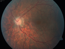
Outer retinal holes and cataract from YAG laser vitreolysisvu 494 fois44 year old man with 7 year history of multiple lasers for floaters. FAF and OCT show outer retinal holes and lens has focal defects26 Mars 2019
|
|

Outer retinal holes and cataract from YAG laser vitreolysisvu 478 fois44 year old man with 7 year history of multiple lasers for floaters. FAF and OCT show outer retinal holes and lens has focal defects26 Mars 2019
|
|

Outer retinal holes and cataract from YAG laser vitreolysisvu 459 fois44 year old man with 7 year history of multiple lasers for floaters. FAF and OCT show outer retinal holes and lens has focal defects26 Mars 2019
|
|

Outer retinal holes and cataract from YAG laser vitreolysisvu 537 fois44 year old man with 7 year history of multiple lasers for floaters. FAF and OCT show outer retinal holes and lens has focal defects26 Mars 2019
|
|

Outer retinal holes and cataract from YAG laser vitreolysisvu 481 fois44 year old man with 7 year history of multiple lasers for floaters. FAF and OCT show outer retinal holes and lens has focal defects26 Mars 2019
|
|

Outer retinal holes and cataract from YAG laser vitreolysisvu 445 fois44 year old man with 7 year history of multiple lasers for floaters. FAF and OCT show outer retinal holes and lens has focal defects26 Mars 2019
|
|

Outer retinal holes and cataract from YAG laser vitreolysisvu 435 fois44 year old man with 7 year history of multiple lasers for floaters. FAF and OCT show outer retinal holes and lens has focal defects26 Mars 2019
|
|

Outer retinal holes and cataract from YAG laser vitreolysisvu 486 fois44 year old man with 7 year history of multiple lasers for floaters. FAF and OCT show outer retinal holes and lens has focal defects26 Mars 2019
|
|

Outer retinal holes and cataract from YAG laser vitreolysisvu 418 fois44 year old man with 7 year history of multiple lasers for floaters. FAF and OCT show outer retinal holes and lens has focal defects26 Mars 2019
|
|

Outer retinal holes and cataract from YAG laser vitreolysisvu 415 fois44 year old man with 7 year history of multiple lasers for floaters. FAF and OCT show outer retinal holes and lens has focal defects26 Mars 2019
|
|

Outer retinal holes and cataract from YAG laser vitreolysisvu 489 fois44 year old man with 7 year history of multiple lasers for floaters. FAF and OCT show outer retinal holes and lens has focal defects26 Mars 2019
|
|

Outer retinal holes and cataract from YAG laser vitreolysisvu 464 fois44 year old man with 7 year history of multiple lasers for floaters. FAF and OCT show outer retinal holes and lens has focal defects26 Mars 2019
|
|

Outer retinal holes and cataract from YAG laser vitreolysisvu 504 fois44 year old man with 7 year history of multiple lasers for floaters. FAF and OCT show outer retinal holes and lens has focal defects26 Mars 2019
|
|

Outer retinal holes and cataract from YAG laser vitreolysisvu 480 fois44 year old man with 7 year history of multiple lasers for floaters. FAF and OCT show outer retinal holes and lens has focal defects26 Mars 2019
|
|

Outer retinal holes and cataract from YAG laser vitreolysisvu 482 fois44 year old man with 7 year history of multiple lasers for floaters. FAF and OCT show outer retinal holes and lens has focal defects26 Mars 2019
|
|

Photic Maculopathy Following Cataract Surgeryvu 420 foisInitial images are from 1 week and subsequent images from 1 month post surgery.18 Décembre 2018
|
|

Photic Maculopathy Following Cataract Surgeryvu 497 foisInitial images are from 1 week and subsequent images from 1 month post surgery.18 Décembre 2018
|
|

Photic Maculopathy Following Cataract Surgeryvu 438 foisInitial images are from 1 week and subsequent images from 1 month post surgery.18 Décembre 2018
|
|

Photic Maculopathy Following Cataract Surgeryvu 509 foisInitial images are from 1 week and subsequent images from 1 month post surgery.18 Décembre 2018
|
|
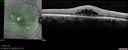
Photic Maculopathy Following Cataract Surgeryvu 493 foisInitial images are from 1 week and subsequent images from 1 month post surgery.18 Décembre 2018
|
|
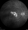
Photic Maculopathy Following Cataract Surgeryvu 446 foisInitial images are from 1 week and subsequent images from 1 month post surgery.18 Décembre 2018
|
|
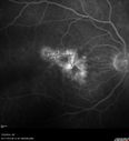
Photic Maculopathy Following Cataract Surgeryvu 577 foisInitial images are from 1 week and subsequent images from 1 month post surgery.18 Décembre 2018
|
|

Photic Maculopathy Following Cataract Surgeryvu 432 foisInitial images are from 1 week and subsequent images from 1 month post surgery.18 Décembre 2018
|
|

Photic Maculopathy Following Cataract Surgeryvu 540 foisInitial images are from 1 week and subsequent images from 1 month post surgery.18 Décembre 2018
|
|

Photic Maculopathy Following Cataract Surgeryvu 588 foisInitial images are from 1 week and subsequent images from 1 month post surgery.18 Décembre 2018
|
|

Photic Maculopathy Following Cataract Surgeryvu 494 foisInitial images are from 1 week and subsequent images from 1 month post surgery.18 Décembre 2018
|
|

Photic Maculopathy Following Cataract Surgeryvu 474 foisInitial images are from 1 week and subsequent images from 1 month post surgery.18 Décembre 2018
|
|

Photic Maculopathy Following Cataract Surgeryvu 530 foisInitial images are from 1 week and subsequent images from 1 month post surgery.18 Décembre 2018
|
|

Macular Damage post Macular Hole Surgeryvu 513 foisThere is a huge hole in the middle of the macula and several small holes in the posterior pole. The patient had 2 surgeries for an idiopathic stage II hole and then was offered a third procedure by the same surgeon and sought a second opinion (and was told to not have anything more done)03 Juillet 2018
|
|
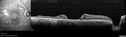
Macular Damage post Macular Hole Surgeryvu 453 foisThere is a huge hole in the middle of the macula and several small holes in the posterior pole. The patient had 2 surgeries for an idiopathic stage II hole and then was offered a third procedure by the same surgeon and sought a second opinion (and was told to not have anything more done)03 Juillet 2018
|
|

Macular Damage post Macular Hole Surgeryvu 499 foisThere is a huge hole in the middle of the macula and several small holes in the posterior pole. The patient had 2 surgeries for an idiopathic stage II hole and then was offered a third procedure by the same surgeon and sought a second opinion (and was told to not have anything more done)03 Juillet 2018
|
|

Macular Damage post Macular Hole Surgeryvu 479 foisThere is a huge hole in the middle of the macula and several small holes in the posterior pole. The patient had 2 surgeries for an idiopathic stage II hole and then was offered a third procedure by the same surgeon and sought a second opinion (and was told to not have anything more done)03 Juillet 2018
|
|

Macular Damage post Macular Hole Surgeryvu 522 foisThere is a huge hole in the middle of the macula and several small holes in the posterior pole. The patient had 2 surgeries for an idiopathic stage II hole and then was offered a third procedure by the same surgeon and sought a second opinion (and was told to not have anything more done)03 Juillet 2018
|
|

Macular Damage post Macular Hole Surgeryvu 479 foisThere is a huge hole in the middle of the macula and several small holes in the posterior pole. The patient had 2 surgeries for an idiopathic stage II hole and then was offered a third procedure by the same surgeon and sought a second opinion (and was told to not have anything more done)03 Juillet 2018
|
|

Macular Damage post Macular Hole Surgeryvu 519 foisThere is a huge hole in the middle of the macula and several small holes in the posterior pole. The patient had 2 surgeries for an idiopathic stage II hole and then was offered a third procedure by the same surgeon and sought a second opinion (and was told to not have anything more done)03 Juillet 2018
|
|

Macular Laser Scarsvu 893 fois86 year old man who had Laser done from 2010 - 2018 for diabetic retinopathy. The right eye lost vision immediately after laser was done a few years ago. VA 1/200 OD; 20/25 OS12 Mars 2018
|
|

Macular Laser Scarsvu 574 fois86 year old man who had Laser done from 2010 - 2018 for diabetic retinopathy. The right eye lost vision immediately after laser was done a few years ago. VA 1/200 OD; 20/25 OS12 Mars 2018
|
|

Macular Laser Scarsvu 628 fois86 year old man who had Laser done from 2010 - 2018 for diabetic retinopathy. The right eye lost vision immediately after laser was done a few years ago. VA 1/200 OD; 20/25 OS12 Mars 2018
|
|

Macular Laser Scarsvu 576 fois86 year old man who had Laser done from 2010 - 2018 for diabetic retinopathy. The right eye lost vision immediately after laser was done a few years ago. VA 1/200 OD; 20/25 OS12 Mars 2018
|
|

Absent Maculavu 576 foisRetina is balled up and dragged superiorly under silicone oil. This patient started with macular pucker surgery for a mild chronic pucker. She developed 2 retinal detachments. The OCT scan is through the center of the macula under oil. There is no retina present on the scan02 Octobre 2017
|
|

Macular Laser Scars Left Eye - 5/200 Visionvu 515 fois83 year old woman who had extensive macular laser in the left eye about 15 years ago for DME. Vision is now 20/50 right eye and 3/200 left eye04 Mai 2017
|
|
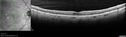
Macular Laser Scars Left Eye - 5/200 Visionvu 491 fois83 year old woman who had extensive macular laser in the left eye about 15 years ago for DME. Vision is now 20/50 right eye and 3/200 left eye04 Mai 2017
|
|

Macular Laser Scars Left Eye - 5/200 Visionvu 551 fois83 year old woman who had extensive macular laser in the left eye about 15 years ago for DME. Vision is now 20/50 right eye and 3/200 left eye04 Mai 2017
|
|

Macular Laser Scars Left Eye - 5/200 Visionvu 557 fois83 year old woman who had extensive macular laser in the left eye about 15 years ago for DME. Vision is now 20/50 right eye and 3/200 left eye04 Mai 2017
|
|

Macular Laser Scars Left Eye - 5/200 Visionvu 502 fois83 year old woman who had extensive macular laser in the left eye about 15 years ago for DME. Vision is now 20/50 right eye and 3/200 left eye04 Mai 2017
|
|

Macular Laser Scars Left Eye - 5/200 Visionvu 546 fois83 year old woman who had extensive macular laser in the left eye about 15 years ago for DME. Vision is now 20/50 right eye and 3/200 left eye04 Mai 2017
|
|

Macular Laser Scars Left Eye - 5/200 Visionvu 543 fois83 year old woman who had extensive macular laser in the left eye about 15 years ago for DME. Vision is now 20/50 right eye and 3/200 left eye04 Mai 2017
|
|

Macular Laser Scars Left Eye - 5/200 Visionvu 564 fois83 year old woman who had extensive macular laser in the left eye about 15 years ago for DME. Vision is now 20/50 right eye and 3/200 left eye04 Mai 2017
|
|

Macular Atrophy - Phototoxicity - Macular Hole Surgery - Late ICG imagevu 675 fois25 years ago patient suffered immediate severe vision loss following macular hole surgery. You can almost see light pipe tracks in the atrophy in the macula. Vision is 5/20007 Novembre 2016
|
|

Macular Atrophy - Phototoxicity - Macular Hole Surgeryvu 624 fois25 years ago patient suffered immediate severe vision loss following macular hole surgery. You can almost see light pipe tracks in the atrophy in the macula. Vision is 5/20007 Novembre 2016
|
|

Macular Atrophy - Phototoxicity - Macular Hole Surgeryvu 683 fois25 years ago patient suffered immediate severe vision loss following macular hole surgery. You can almost see light pipe tracks in the atrophy in the macula. Vision is 5/20007 Novembre 2016
|
|
| 153 photos sur 2 page(s) |
1 |
|


