neueste Dateien - Iatrogenic Diseases
|

Phototoxicity following cataract surgery154 x angesehen70 year old female with nasal spot in vision since cataract surgery 3 years ago. VA 20/20 OU.14. Februar 2024
|
|

Phototoxicity following cataract surgery180 x angesehen70 year old female with nasal spot in vision since cataract surgery 3 years ago. VA 20/20 OU.14. Februar 2024
|
|

Phototoxicity following cataract surgery207 x angesehen70 year old female with nasal spot in vision since cataract surgery 3 years ago. VA 20/20 OU.14. Februar 2024
|
|

Phototoxicity following cataract surgery168 x angesehen70 year old female with nasal spot in vision since cataract surgery 3 years ago. VA 20/20 OU.14. Februar 2024
|
|

Phototoxicity following cataract surgery198 x angesehen70 year old female with nasal spot in vision since cataract surgery 3 years ago. VA 20/20 OU.14. Februar 2024
|
|

Phototoxicity following cataract surgery156 x angesehen70 year old female with nasal spot in vision since cataract surgery 3 years ago. VA 20/20 OU.14. Februar 2024
|
|

Phototoxicity following cataract surgery178 x angesehen70 year old female with nasal spot in vision since cataract surgery 3 years ago. VA 20/20 OU.14. Februar 2024
|
|

Phototoxicity following cataract surgery188 x angesehen70 year old female with nasal spot in vision since cataract surgery 3 years ago. VA 20/20 OU.14. Februar 2024
|
|

Phototoxicity following cataract surgery160 x angesehen70 year old female with nasal spot in vision since cataract surgery 3 years ago. VA 20/20 OU.14. Februar 2024
|
|

Phototoxicity following cataract surgery146 x angesehen70 year old female with nasal spot in vision since cataract surgery 3 years ago. VA 20/20 OU.14. Februar 2024
|
|

Phototoxicity following cataract surgery180 x angesehen70 year old female with nasal spot in vision since cataract surgery 3 years ago. VA 20/20 OU.14. Februar 2024
|
|

Phototoxicity following cataract surgery185 x angesehen70 year old female with nasal spot in vision since cataract surgery 3 years ago. VA 20/20 OU.14. Februar 2024
|
|

Phototoxicity following cataract surgery147 x angesehen70 year old female with nasal spot in vision since cataract surgery 3 years ago. VA 20/20 OU.14. Februar 2024
|
|

Phototoxicity following cataract surgery202 x angesehen70 year old female with nasal spot in vision since cataract surgery 3 years ago. VA 20/20 OU.14. Februar 2024
|
|

Phototoxicity following cataract surgery172 x angesehen70 year old female with nasal spot in vision since cataract surgery 3 years ago. VA 20/20 OU.14. Februar 2024
|
|

Phototoxicity following cataract surgery178 x angesehen70 year old female with nasal spot in vision since cataract surgery 3 years ago. VA 20/20 OU.14. Februar 2024
|
|

Phototoxicity following cataract surgery170 x angesehen70 year old female with nasal spot in vision since cataract surgery 3 years ago. VA 20/20 OU.14. Februar 2024
|
|

Phototoxicity following cataract surgery171 x angesehen70 year old female with nasal spot in vision since cataract surgery 3 years ago. VA 20/20 OU.14. Februar 2024
|
|

Phototoxicity following cataract surgery171 x angesehen70 year old female with nasal spot in vision since cataract surgery 3 years ago. VA 20/20 OU.14. Februar 2024
|
|

Phototoxicity following cataract surgery198 x angesehen70 year old female with nasal spot in vision since cataract surgery 3 years ago. VA 20/20 OU.14. Februar 2024
|
|

Uveitis Glaucoma Hyphema (UGH) syndrome318 x angesehen65 year old female with intraocular lens half in the sulcus, iris atrophy, and recurrent uveitis since cataract surgery. She has a mild case and has been managed with topical steroids as needed.02. Januar 2023
|
|

Uveitis Glaucoma Hyphema (UGH) syndrome312 x angesehen65 year old female with intraocular lens half in the sulcus, iris atrophy, and recurrent uveitis since cataract surgery. She has a mild case and has been managed with topical steroids as needed.02. Januar 2023
|
|
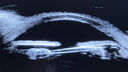
Uveitis Glaucoma Hyphema (UGH) syndrome298 x angesehen65 year old female with intraocular lens half in the sulcus, iris atrophy, and recurrent uveitis since cataract surgery. She has a mild case and has been managed with topical steroids as needed.02. Januar 2023
|
|
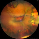
Choroidal Effusions following IOL exchange299 x angesehen67 year old man 10 years ago had retinal detachment with buckle, vitrectomy, laser, gas. Vision was 20/25 post-op. 2 months ago had sudden vision loss in the right eye from a dislocated PCIOL (in the bag). 3 weeks ago had a vitrectomy, remove IOL and scleral suture fixation of Akreos AO60 PCIOL with gortex sutures. IOP was 12 on post-op day 1 with a vitreous hemorrhage. Post-op day 7, vitreous hemorrhage cleared.
PMhx: HTN, High Cholesterol.
Med: Amlodipine, ASA, Atorvastatin, Metoprolol
Vision 20/200,09. März 2022
|
|

Choroidal Effusions following IOL exchange303 x angesehen67 year old man 10 years ago had retinal detachment with buckle, vitrectomy, laser, gas. Vision was 20/25 post-op. 2 months ago had sudden vision loss in the right eye from a dislocated PCIOL (in the bag). 3 weeks ago had a vitrectomy, remove IOL and scleral suture fixation of Akreos AO60 PCIOL with gortex sutures. IOP was 12 on post-op day 1 with a vitreous hemorrhage. Post-op day 7, vitreous hemorrhage cleared.
PMhx: HTN, High Cholesterol.
Med: Amlodipine, ASA, Atorvastatin, Metoprolol
Vision 20/200,09. März 2022
|
|

Choroidal Effusions following IOL exchange309 x angesehen67 year old man 10 years ago had retinal detachment with buckle, vitrectomy, laser, gas. Vision was 20/25 post-op. 2 months ago had sudden vision loss in the right eye from a dislocated PCIOL (in the bag). 3 weeks ago had a vitrectomy, remove IOL and scleral suture fixation of Akreos AO60 PCIOL with gortex sutures. IOP was 12 on post-op day 1 with a vitreous hemorrhage. Post-op day 7, vitreous hemorrhage cleared.
PMhx: HTN, High Cholesterol.
Med: Amlodipine, ASA, Atorvastatin, Metoprolol
Vision 20/200,09. März 2022
|
|

Choroidal Effusions following IOL exchange280 x angesehen67 year old man 10 years ago had retinal detachment with buckle, vitrectomy, laser, gas. Vision was 20/25 post-op. 2 months ago had sudden vision loss in the right eye from a dislocated PCIOL (in the bag). 3 weeks ago had a vitrectomy, remove IOL and scleral suture fixation of Akreos AO60 PCIOL with gortex sutures. IOP was 12 on post-op day 1 with a vitreous hemorrhage. Post-op day 7, vitreous hemorrhage cleared.
PMhx: HTN, High Cholesterol.
Med: Amlodipine, ASA, Atorvastatin, Metoprolol
Vision 20/200,09. März 2022
|
|

Choroidal Effusions following IOL exchange299 x angesehen67 year old man 10 years ago had retinal detachment with buckle, vitrectomy, laser, gas. Vision was 20/25 post-op. 2 months ago had sudden vision loss in the right eye from a dislocated PCIOL (in the bag). 3 weeks ago had a vitrectomy, remove IOL and scleral suture fixation of Akreos AO60 PCIOL with gortex sutures. IOP was 12 on post-op day 1 with a vitreous hemorrhage. Post-op day 7, vitreous hemorrhage cleared.
PMhx: HTN, High Cholesterol.
Med: Amlodipine, ASA, Atorvastatin, Metoprolol
Vision 20/200,09. März 2022
|
|

Choroidal Effusions following IOL exchange316 x angesehen67 year old man 10 years ago had retinal detachment with buckle, vitrectomy, laser, gas. Vision was 20/25 post-op. 2 months ago had sudden vision loss in the right eye from a dislocated PCIOL (in the bag). 3 weeks ago had a vitrectomy, remove IOL and scleral suture fixation of Akreos AO60 PCIOL with gortex sutures. IOP was 12 on post-op day 1 with a vitreous hemorrhage. Post-op day 7, vitreous hemorrhage cleared.
PMhx: HTN, High Cholesterol.
Med: Amlodipine, ASA, Atorvastatin, Metoprolol
Vision 20/200,09. März 2022
|
|

Choroidal Effusions following IOL exchange296 x angesehen67 year old man 10 years ago had retinal detachment with buckle, vitrectomy, laser, gas. Vision was 20/25 post-op. 2 months ago had sudden vision loss in the right eye from a dislocated PCIOL (in the bag). 3 weeks ago had a vitrectomy, remove IOL and scleral suture fixation of Akreos AO60 PCIOL with gortex sutures. IOP was 12 on post-op day 1 with a vitreous hemorrhage. Post-op day 7, vitreous hemorrhage cleared.
PMhx: HTN, High Cholesterol.
Med: Amlodipine, ASA, Atorvastatin, Metoprolol
Vision 20/200,09. März 2022
|
|

393 x angesehen30. September 2020
|
|

438 x angesehen30. September 2020
|
|

393 x angesehen30. September 2020
|
|

414 x angesehen30. September 2020
|
|

387 x angesehen30. September 2020
|
|

410 x angesehen30. September 2020
|
|

Upside Down Lens Syndrome - Anterior Chamber IOL Upside Down692 x angesehen77 year old female who had vitrectomy for retained lens fragments and has had difficulty with recurrent uveitis since her cataract surgery. The anterior chamber intraocular lens is upside down. A lens exchange is planned.07. März 2020
|
|

Choroidal Effusions following Glaucoma Surgery 9 months ago409 x angesehen67 year old female with glaucoma surgery 9 month ago. Her IOP is 12. She has peripheral choroidal effusions. VA 20/100 ph 20/3211. Februar 2020
|
|

Choroidal Effusions following Glaucoma Surgery 9 months ago443 x angesehen67 year old female with glaucoma surgery 9 month ago. Her IOP is 12. She has peripheral choroidal effusions. VA 20/100 ph 20/3211. Februar 2020
|
|

Choroidal Effusions following Glaucoma Surgery 9 months ago428 x angesehen67 year old female with glaucoma surgery 9 month ago. Her IOP is 12. She has peripheral choroidal effusions. VA 20/100 ph 20/3211. Februar 2020
|
|
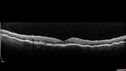
Choroidal Effusions following Glaucoma Surgery 9 months ago474 x angesehen67 year old female with glaucoma surgery 9 month ago. Her IOP is 12. She has peripheral choroidal effusions. VA 20/100 ph 20/3211. Februar 2020
|
|

Choroidal Effusions following Glaucoma Surgery 9 months ago434 x angesehen67 year old female with glaucoma surgery 9 month ago. Her IOP is 12. She has peripheral choroidal effusions. VA 20/100 ph 20/3211. Februar 2020
|
|

Choroidal Effusions following Glaucoma Surgery 9 months ago424 x angesehen67 year old female with glaucoma surgery 9 month ago. Her IOP is 12. She has peripheral choroidal effusions. VA 20/100 ph 20/3211. Februar 2020
|
|
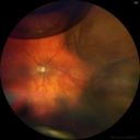
Choroidal Effusion One Day following macular pucker surgery408 x angesehen70 year old female one day following macular pucker surgery. Her VA was 20/100, IOP was 12. The air bubble was to pressurize the eye at the conclusion of the 25 gauge vitrectomy. The choroidal effusion is in the periphery.06. Februar 2020
|
|

Perforating Needle injury retrobulbar anesthetic - excellent final vision428 x angesehen71 year old man who had a vitrectomy for macular pucker and then developed a retinal detachment. There was an accidental needle stick during a retrobulbar anesthetic. The posterior puncture site is just inferior to the macula. The patient had a vitrectomy the next day for the retinal detachment and then 3 months later a vitrectomy to remove the fibrous proliferation from the puncture site. The final visual acuity was 20/20.07. August 2019
|
|

Perforating Needle injury retrobulbar anesthetic - excellent final vision463 x angesehen71 year old man who had a vitrectomy for macular pucker and then developed a retinal detachment. There was an accidental needle stick during a retrobulbar anesthetic. The posterior puncture site is just inferior to the macula. The patient had a vitrectomy the next day for the retinal detachment and then 3 months later a vitrectomy to remove the fibrous proliferation from the puncture site. The final visual acuity was 20/20.07. August 2019
|
|

Perforating Needle injury retrobulbar anesthetic - excellent final vision592 x angesehen71 year old man who had a vitrectomy for macular pucker and then developed a retinal detachment. There was an accidental needle stick during a retrobulbar anesthetic. The posterior puncture site is just inferior to the macula. The patient had a vitrectomy the next day for the retinal detachment and then 3 months later a vitrectomy to remove the fibrous proliferation from the puncture site. The final visual acuity was 20/20.07. August 2019
|
|

Perforating Needle injury retrobulbar anesthetic - excellent final vision594 x angesehen71 year old man who had a vitrectomy for macular pucker and then developed a retinal detachment. There was an accidental needle stick during a retrobulbar anesthetic. The posterior puncture site is just inferior to the macula. The patient had a vitrectomy the next day for the retinal detachment and then 3 months later a vitrectomy to remove the fibrous proliferation from the puncture site. The final visual acuity was 20/20.07. August 2019
|
|

Perforating Needle injury retrobulbar anesthetic - excellent final vision541 x angesehen71 year old man who had a vitrectomy for macular pucker and then developed a retinal detachment. There was an accidental needle stick during a retrobulbar anesthetic. The posterior puncture site is just inferior to the macula. The patient had a vitrectomy the next day for the retinal detachment and then 3 months later a vitrectomy to remove the fibrous proliferation from the puncture site. The final visual acuity was 20/20.07. August 2019
|
|
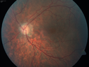
Outer retinal holes and cataract from YAG laser vitreolysis493 x angesehen44 year old man with 7 year history of multiple lasers for floaters. FAF and OCT show outer retinal holes and lens has focal defects26. März 2019
|
|

Outer retinal holes and cataract from YAG laser vitreolysis478 x angesehen44 year old man with 7 year history of multiple lasers for floaters. FAF and OCT show outer retinal holes and lens has focal defects26. März 2019
|
|

Outer retinal holes and cataract from YAG laser vitreolysis458 x angesehen44 year old man with 7 year history of multiple lasers for floaters. FAF and OCT show outer retinal holes and lens has focal defects26. März 2019
|
|

Outer retinal holes and cataract from YAG laser vitreolysis537 x angesehen44 year old man with 7 year history of multiple lasers for floaters. FAF and OCT show outer retinal holes and lens has focal defects26. März 2019
|
|

Outer retinal holes and cataract from YAG laser vitreolysis481 x angesehen44 year old man with 7 year history of multiple lasers for floaters. FAF and OCT show outer retinal holes and lens has focal defects26. März 2019
|
|

Outer retinal holes and cataract from YAG laser vitreolysis444 x angesehen44 year old man with 7 year history of multiple lasers for floaters. FAF and OCT show outer retinal holes and lens has focal defects26. März 2019
|
|

Outer retinal holes and cataract from YAG laser vitreolysis435 x angesehen44 year old man with 7 year history of multiple lasers for floaters. FAF and OCT show outer retinal holes and lens has focal defects26. März 2019
|
|

Outer retinal holes and cataract from YAG laser vitreolysis485 x angesehen44 year old man with 7 year history of multiple lasers for floaters. FAF and OCT show outer retinal holes and lens has focal defects26. März 2019
|
|

Outer retinal holes and cataract from YAG laser vitreolysis418 x angesehen44 year old man with 7 year history of multiple lasers for floaters. FAF and OCT show outer retinal holes and lens has focal defects26. März 2019
|
|

Outer retinal holes and cataract from YAG laser vitreolysis414 x angesehen44 year old man with 7 year history of multiple lasers for floaters. FAF and OCT show outer retinal holes and lens has focal defects26. März 2019
|
|

Outer retinal holes and cataract from YAG laser vitreolysis488 x angesehen44 year old man with 7 year history of multiple lasers for floaters. FAF and OCT show outer retinal holes and lens has focal defects26. März 2019
|
|

Outer retinal holes and cataract from YAG laser vitreolysis464 x angesehen44 year old man with 7 year history of multiple lasers for floaters. FAF and OCT show outer retinal holes and lens has focal defects26. März 2019
|
|

Outer retinal holes and cataract from YAG laser vitreolysis502 x angesehen44 year old man with 7 year history of multiple lasers for floaters. FAF and OCT show outer retinal holes and lens has focal defects26. März 2019
|
|

Outer retinal holes and cataract from YAG laser vitreolysis479 x angesehen44 year old man with 7 year history of multiple lasers for floaters. FAF and OCT show outer retinal holes and lens has focal defects26. März 2019
|
|

Outer retinal holes and cataract from YAG laser vitreolysis482 x angesehen44 year old man with 7 year history of multiple lasers for floaters. FAF and OCT show outer retinal holes and lens has focal defects26. März 2019
|
|

Photic Maculopathy Following Cataract Surgery420 x angesehenInitial images are from 1 week and subsequent images from 1 month post surgery.18. Dezember 2018
|
|

Photic Maculopathy Following Cataract Surgery497 x angesehenInitial images are from 1 week and subsequent images from 1 month post surgery.18. Dezember 2018
|
|

Photic Maculopathy Following Cataract Surgery435 x angesehenInitial images are from 1 week and subsequent images from 1 month post surgery.18. Dezember 2018
|
|

Photic Maculopathy Following Cataract Surgery507 x angesehenInitial images are from 1 week and subsequent images from 1 month post surgery.18. Dezember 2018
|
|
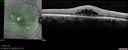
Photic Maculopathy Following Cataract Surgery491 x angesehenInitial images are from 1 week and subsequent images from 1 month post surgery.18. Dezember 2018
|
|
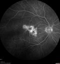
Photic Maculopathy Following Cataract Surgery444 x angesehenInitial images are from 1 week and subsequent images from 1 month post surgery.18. Dezember 2018
|
|
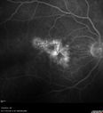
Photic Maculopathy Following Cataract Surgery576 x angesehenInitial images are from 1 week and subsequent images from 1 month post surgery.18. Dezember 2018
|
|

Photic Maculopathy Following Cataract Surgery430 x angesehenInitial images are from 1 week and subsequent images from 1 month post surgery.18. Dezember 2018
|
|

Photic Maculopathy Following Cataract Surgery540 x angesehenInitial images are from 1 week and subsequent images from 1 month post surgery.18. Dezember 2018
|
|

Photic Maculopathy Following Cataract Surgery588 x angesehenInitial images are from 1 week and subsequent images from 1 month post surgery.18. Dezember 2018
|
|

Photic Maculopathy Following Cataract Surgery494 x angesehenInitial images are from 1 week and subsequent images from 1 month post surgery.18. Dezember 2018
|
|

Photic Maculopathy Following Cataract Surgery474 x angesehenInitial images are from 1 week and subsequent images from 1 month post surgery.18. Dezember 2018
|
|

Photic Maculopathy Following Cataract Surgery530 x angesehenInitial images are from 1 week and subsequent images from 1 month post surgery.18. Dezember 2018
|
|

Macular Damage post Macular Hole Surgery513 x angesehenThere is a huge hole in the middle of the macula and several small holes in the posterior pole. The patient had 2 surgeries for an idiopathic stage II hole and then was offered a third procedure by the same surgeon and sought a second opinion (and was told to not have anything more done)03. Juli 2018
|
|
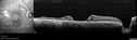
Macular Damage post Macular Hole Surgery453 x angesehenThere is a huge hole in the middle of the macula and several small holes in the posterior pole. The patient had 2 surgeries for an idiopathic stage II hole and then was offered a third procedure by the same surgeon and sought a second opinion (and was told to not have anything more done)03. Juli 2018
|
|

Macular Damage post Macular Hole Surgery497 x angesehenThere is a huge hole in the middle of the macula and several small holes in the posterior pole. The patient had 2 surgeries for an idiopathic stage II hole and then was offered a third procedure by the same surgeon and sought a second opinion (and was told to not have anything more done)03. Juli 2018
|
|

Macular Damage post Macular Hole Surgery479 x angesehenThere is a huge hole in the middle of the macula and several small holes in the posterior pole. The patient had 2 surgeries for an idiopathic stage II hole and then was offered a third procedure by the same surgeon and sought a second opinion (and was told to not have anything more done)03. Juli 2018
|
|

Macular Damage post Macular Hole Surgery522 x angesehenThere is a huge hole in the middle of the macula and several small holes in the posterior pole. The patient had 2 surgeries for an idiopathic stage II hole and then was offered a third procedure by the same surgeon and sought a second opinion (and was told to not have anything more done)03. Juli 2018
|
|

Macular Damage post Macular Hole Surgery479 x angesehenThere is a huge hole in the middle of the macula and several small holes in the posterior pole. The patient had 2 surgeries for an idiopathic stage II hole and then was offered a third procedure by the same surgeon and sought a second opinion (and was told to not have anything more done)03. Juli 2018
|
|

Macular Damage post Macular Hole Surgery519 x angesehenThere is a huge hole in the middle of the macula and several small holes in the posterior pole. The patient had 2 surgeries for an idiopathic stage II hole and then was offered a third procedure by the same surgeon and sought a second opinion (and was told to not have anything more done)03. Juli 2018
|
|

Macular Laser Scars893 x angesehen86 year old man who had Laser done from 2010 - 2018 for diabetic retinopathy. The right eye lost vision immediately after laser was done a few years ago. VA 1/200 OD; 20/25 OS12. März 2018
|
|

Macular Laser Scars574 x angesehen86 year old man who had Laser done from 2010 - 2018 for diabetic retinopathy. The right eye lost vision immediately after laser was done a few years ago. VA 1/200 OD; 20/25 OS12. März 2018
|
|

Macular Laser Scars628 x angesehen86 year old man who had Laser done from 2010 - 2018 for diabetic retinopathy. The right eye lost vision immediately after laser was done a few years ago. VA 1/200 OD; 20/25 OS12. März 2018
|
|

Macular Laser Scars574 x angesehen86 year old man who had Laser done from 2010 - 2018 for diabetic retinopathy. The right eye lost vision immediately after laser was done a few years ago. VA 1/200 OD; 20/25 OS12. März 2018
|
|

Absent Macula576 x angesehenRetina is balled up and dragged superiorly under silicone oil. This patient started with macular pucker surgery for a mild chronic pucker. She developed 2 retinal detachments. The OCT scan is through the center of the macula under oil. There is no retina present on the scan02. Oktober 2017
|
|

Macular Laser Scars Left Eye - 5/200 Vision515 x angesehen83 year old woman who had extensive macular laser in the left eye about 15 years ago for DME. Vision is now 20/50 right eye and 3/200 left eye04. Mai 2017
|
|
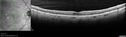
Macular Laser Scars Left Eye - 5/200 Vision490 x angesehen83 year old woman who had extensive macular laser in the left eye about 15 years ago for DME. Vision is now 20/50 right eye and 3/200 left eye04. Mai 2017
|
|

Macular Laser Scars Left Eye - 5/200 Vision551 x angesehen83 year old woman who had extensive macular laser in the left eye about 15 years ago for DME. Vision is now 20/50 right eye and 3/200 left eye04. Mai 2017
|
|

Macular Laser Scars Left Eye - 5/200 Vision557 x angesehen83 year old woman who had extensive macular laser in the left eye about 15 years ago for DME. Vision is now 20/50 right eye and 3/200 left eye04. Mai 2017
|
|

Macular Laser Scars Left Eye - 5/200 Vision502 x angesehen83 year old woman who had extensive macular laser in the left eye about 15 years ago for DME. Vision is now 20/50 right eye and 3/200 left eye04. Mai 2017
|
|

Macular Laser Scars Left Eye - 5/200 Vision546 x angesehen83 year old woman who had extensive macular laser in the left eye about 15 years ago for DME. Vision is now 20/50 right eye and 3/200 left eye04. Mai 2017
|
|

Macular Laser Scars Left Eye - 5/200 Vision543 x angesehen83 year old woman who had extensive macular laser in the left eye about 15 years ago for DME. Vision is now 20/50 right eye and 3/200 left eye04. Mai 2017
|
|

Macular Laser Scars Left Eye - 5/200 Vision564 x angesehen83 year old woman who had extensive macular laser in the left eye about 15 years ago for DME. Vision is now 20/50 right eye and 3/200 left eye04. Mai 2017
|
|

Macular Atrophy - Phototoxicity - Macular Hole Surgery - Late ICG image674 x angesehen25 years ago patient suffered immediate severe vision loss following macular hole surgery. You can almost see light pipe tracks in the atrophy in the macula. Vision is 5/20007. November 2016
|
|

Macular Atrophy - Phototoxicity - Macular Hole Surgery624 x angesehen25 years ago patient suffered immediate severe vision loss following macular hole surgery. You can almost see light pipe tracks in the atrophy in the macula. Vision is 5/20007. November 2016
|
|

Macular Atrophy - Phototoxicity - Macular Hole Surgery683 x angesehen25 years ago patient suffered immediate severe vision loss following macular hole surgery. You can almost see light pipe tracks in the atrophy in the macula. Vision is 5/20007. November 2016
|
|
| 153 Dateien auf 2 Seite(n) |
1 |
|


