Last additions - Tumors, Masses, Pigmented Lesions
|

Retinoma - Retinocytoma - B Scan Ultrasound883 viewsUltrasound shows shadowing from the calcifications in the tumor.Feb 03, 2011
|
|

Retinocytoma - Retinoma - Regressed Retinoblastoma 43 year old male incidental finding 652 viewsFeb 03, 2011
|
|

Retinocytoma - Retinoma - Regressed Retinoblastoma 43 year old male incidental finding 607 viewsFeb 03, 2011
|
|
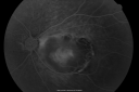
Retinocytoma - Retinoma - Regressed Retinoblastoma 43 year old male incidental finding 607 viewsFeb 03, 2011
|
|

Retinocytoma - Retinoma - Regressed Retinoblastoma 43 year old male incidental finding 715 viewsFeb 03, 2011
|
|

Retinocytoma - Retinoma - Regressed Retinoblastoma 43 year old male incidental finding 584 viewsFeb 03, 2011
|
|

Retinocytoma - Retinoma - Regressed Retinoblastoma 43 year old male incidental finding 642 viewsFeb 03, 2011
|
|

Retinocytoma - Retinoma - Regressed Retinoblastoma 43 year old male incidental finding 650 viewsFeb 03, 2011
|
|

Retinocytoma - Retinoma - Regressed Retinoblastoma 43 year old male incidental finding 713 viewsFeb 03, 2011
|
|

Retinocytoma - Retinoma - Regressed Retinoblastoma 43 year old male incidental finding 736 viewsFeb 03, 2011
|
|

Retinocytoma - Retinoma - Regressed Retinoblastoma 43 year old male incidental finding 667 viewsFeb 03, 2011
|
|

Retinocytoma - Retinoma - Regressed Retinoblastoma 43 year old male incidental finding 740 viewsFeb 03, 2011
|
|

Retinocytoma - Retinoma - Regressed Retinoblastoma 43 year old male incidental finding 728 viewsFeb 03, 2011
|
|

Retinocytoma - Retinoma - Regressed Retinoblastoma 43 year old male incidental finding 1128 viewsFeb 03, 2011
|
|

Retinocytoma - Retinoma - Regressed Retinoblastoma 43 year old male incidental finding 1393 viewsFeb 03, 2011
|
|

Retinocytoma - Retinoma - Regressed Retinoblastoma 43 year old male incidental finding 1207 viewsFeb 03, 2011
|
|

Retinocytoma - Retinoma - Regressed Retinoblastoma 43 year old male incidental finding 1076 viewsFeb 03, 2011
|
|

Retinocytoma - Retinoma - Regressed Retinoblastoma 43 year old male incidental finding 918 viewsFeb 03, 2011
|
|

Amelanotic Choroidal Nevus 1103 views45-year-old man OD 20/20, OS 20/20. 5x5 mm flat, yellow choroidal lesion just inferonasal to the optic nerve, two disc-diameters from the nerve with scalloped edges. It looks like a confluent drusen, but it is not elevated. Dec 13, 2010
|
|

Amelanotic Choroidal Nevus 1009 views45-year-old man OD 20/20, OS 20/20. 5x5 mm flat, yellow choroidal lesion just inferonasal to the optic nerve, two disc-diameters from the nerve with scalloped edges. It looks like a confluent drusen, but it is not elevated. Dec 13, 2010
|
|

Amelanotic Choroidal Nevus 1052 views45-year-old man OD 20/20, OS 20/20. 5x5 mm flat, yellow choroidal lesion just inferonasal to the optic nerve, two disc-diameters from the nerve with scalloped edges. It looks like a confluent drusen, but it is not elevated. Dec 13, 2010
|
|

Amelanotic Choroidal Nevus 2060 views45-year-old man OD 20/20, OS 20/20. 5x5 mm flat, yellow choroidal lesion just inferonasal to the optic nerve, two disc-diameters from the nerve with scalloped edges. It looks like a confluent drusen, but it is not elevated. Dec 13, 2010
|
|

Amelanotic Choroidal Nevus 1696 views45-year-old man OD 20/20, OS 20/20. 5x5 mm flat, yellow choroidal lesion just inferonasal to the optic nerve, two disc-diameters from the nerve with scalloped edges. It looks like a confluent drusen, but it is not elevated. Dec 13, 2010
|
|

Choroidal Nevus in Macula 898 viewsNov 29, 2010
|
|
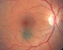
Choroidal Nevus in Macula 875 viewsNov 29, 2010
|
|

Radioactive Plaque Placement for Choroidal Melanoma Lead Patch after Surgery980 viewsNov 25, 2010
|
|

Radioactive Plaque Placement for Choroidal Melanoma Eye After Surgery with Conjunctiva Closed1098 viewsNov 25, 2010
|
|

Radioactive Plaque Placement for Choroidal Melanoma Radioactive Plaque Placement for Choroidal Melanoma 1125 viewsNov 25, 2010
|
|

Radioactive Plaque Placement for Choroidal Melanoma Plaque Sutured to Globe1113 viewsNov 25, 2010
|
|
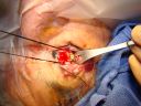
Radioactive Plaque Placement for Choroidal Melanoma Plaque Sutured to Globe987 viewsNov 25, 2010
|
|
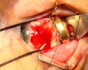
Radioactive Plaque Placement for Choroidal Melanoma Plaque Placed but not Sutured to Eye Yet1268 viewsNov 25, 2010
|
|
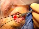
Radioactive Plaque Placement for Choroidal Melanoma Plaque Placed but not Sutured to Eye Ye1089 viewsNov 25, 2010
|
|

Radioactive Plaque Placement for Choroidal Melanoma Plaque Placed but not Sutured to Eye Ye1045 viewsNov 25, 2010
|
|

Radioactive Plaque Placement for Choroidal Melanoma Iodine Radioactive Seeds in Plaque1224 viewsNov 25, 2010
|
|

Radioactive Plaque Placement for Choroidal Melanoma Gold Plaque about the size of a Quarter988 viewsNov 25, 2010
|
|

Radioactive Plaque Placement for Choroidal Melanoma Transillumination of Globe to Localize Melanoma 974 viewsNov 25, 2010
|
|

Radioactive Plaque Placement for Choroidal Melanoma Transillumination of Globe to Localize Melanoma Arrow Points to Tumor Shadow898 viewsNov 25, 2010
|
|

Radioactive Plaque Placement for Choroidal Melanoma Transillumination of Globe to Localize Melanoma Arrow Points to Tumor Shadow1014 viewsNov 25, 2010
|
|
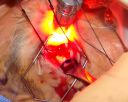
Radioactive Plaque Placement for Choroidal Melanoma Transillumination of Globe to Localize Melanoma 998 viewsNov 25, 2010
|
|

Radioactive Plaque Placement for Choroidal Melanoma Transillumination of Globe to Localize Melanoma 965 viewsNov 25, 2010
|
|

Radioactive Plaque Placement for Choroidal Melanoma Eye is Anesthetized Prior to Surgery1196 viewsNov 25, 2010
|
|
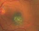
Macular Choroidal Nevus1713 views78-year-old woman has choroidal nevus in the macula of the left eye. You asked her to come in here for an evaluation and nevus was noted in the past about 7 years ago. She has not noticed any change in vision in that eye.
VISUAL ACUITY: OU 20/30. Nov 12, 2010
|
|

Retinoblastoma - Bilateral - Regressed - Chemotherapy, Thermal Therapy, Cryotherapy812 views10-year-old child was seen in the office on August 6, 2008. She was treated with a combination of chemotherapy at 10 months old as well as thermal therapy and cryotherapy for bilateral retinoblastoma tumors. She reportedly had nine tumors in the left eye and five tumors in the right eyeNov 09, 2010
|
|

Retinoblastoma - Bilateral - Regressed - Chemotherapy, Thermal Therapy, Cryotherapy688 views10-year-old child was seen in the office on August 6, 2008. She was treated with a combination of chemotherapy at 10 months old as well as thermal therapy and cryotherapy for bilateral retinoblastoma tumors. She reportedly had nine tumors in the left eye and five tumors in the right eyeNov 09, 2010
|
|

Retinoblastoma - Bilateral - Regressed - Chemotherapy, Thermal Therapy, Cryotherapy718 views10-year-old child was seen in the office on August 6, 2008. She was treated with a combination of chemotherapy at 10 months old as well as thermal therapy and cryotherapy for bilateral retinoblastoma tumors. She reportedly had nine tumors in the left eye and five tumors in the right eyeNov 09, 2010
|
|

Retinoblastoma - Bilateral - Regressed - Chemotherapy, Thermal Therapy, Cryotherapy755 views10-year-old child was seen in the office on August 6, 2008. She was treated with a combination of chemotherapy at 10 months old as well as thermal therapy and cryotherapy for bilateral retinoblastoma tumors. She reportedly had nine tumors in the left eye and five tumors in the right eyeNov 09, 2010
|
|
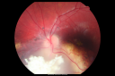
Retinoblastoma - Bilateral - Regressed - Chemotherapy, Thermal Therapy, Cryotherapy870 views10-year-old child was seen in the office on August 6, 2008. She was treated with a combination of chemotherapy at 10 months old as well as thermal therapy and cryotherapy for bilateral retinoblastoma tumors. She reportedly had nine tumors in the left eye and five tumors in the right eyeNov 09, 2010
|
|

Retinoblastoma - Bilateral - Regressed - Chemotherapy, Thermal Therapy, Cryotherapy728 views10-year-old child was seen in the office on August 6, 2008. She was treated with a combination of chemotherapy at 10 months old as well as thermal therapy and cryotherapy for bilateral retinoblastoma tumors. She reportedly had nine tumors in the left eye and five tumors in the right eyeNov 09, 2010
|
|

Retinoblastoma - Bilateral - Regressed - Chemotherapy, Thermal Therapy, Cryotherapy721 views10-year-old child was seen in the office on August 6, 2008. She was treated with a combination of chemotherapy at 10 months old as well as thermal therapy and cryotherapy for bilateral retinoblastoma tumors. She reportedly had nine tumors in the left eye and five tumors in the right eyeNov 09, 2010
|
|

Retinoblastoma - Bilateral - Regressed - Chemotherapy, Thermal Therapy, Cryotherapy784 views10-year-old child was seen in the office on August 6, 2008. She was treated with a combination of chemotherapy at 10 months old as well as thermal therapy and cryotherapy for bilateral retinoblastoma tumors. She reportedly had nine tumors in the left eye and five tumors in the right eyeNov 09, 2010
|
|

Retinoblastoma - Bilateral - Regressed - Chemotherapy, Thermal Therapy, Cryotherapy840 views10-year-old child was seen in the office on August 6, 2008. She was treated with a combination of chemotherapy at 10 months old as well as thermal therapy and cryotherapy for bilateral retinoblastoma tumors. She reportedly had nine tumors in the left eye and five tumors in the right eyeNov 09, 2010
|
|

Retinoblastoma - Bilateral - Regressed - Chemotherapy, Thermal Therapy, Cryotherapy868 views10-year-old child was seen in the office on August 6, 2008. She was treated with a combination of chemotherapy at 10 months old as well as thermal therapy and cryotherapy for bilateral retinoblastoma tumors. She reportedly had nine tumors in the left eye and five tumors in the right eyeNov 09, 2010
|
|

Retinoblastoma - Bilateral - Regressed - Chemotherapy, Thermal Therapy, Cryotherapy731 views10-year-old child was seen in the office on August 6, 2008. She was treated with a combination of chemotherapy at 10 months old as well as thermal therapy and cryotherapy for bilateral retinoblastoma tumors. She reportedly had nine tumors in the left eye and five tumors in the right eyeNov 09, 2010
|
|

Retinoblastoma - Bilateral - Regressed - Chemotherapy, Thermal Therapy, Cryotherapy797 views10-year-old child was seen in the office on August 6, 2008. She was treated with a combination of chemotherapy at 10 months old as well as thermal therapy and cryotherapy for bilateral retinoblastoma tumors. She reportedly had nine tumors in the left eye and five tumors in the right eyeNov 09, 2010
|
|

Retinoblastoma - Bilateral - Regressed - Chemotherapy, Thermal Therapy, Cryotherapy891 views10-year-old child was seen in the office on August 6, 2008. She was treated with a combination of chemotherapy at 10 months old as well as thermal therapy and cryotherapy for bilateral retinoblastoma tumors. She reportedly had nine tumors in the left eye and five tumors in the right eyeNov 09, 2010
|
|

Retinoblastoma - Bilateral - Regressed - Chemotherapy, Thermal Therapy, Cryotherapy1035 views10-year-old child was seen in the office on August 6, 2008. She was treated with a combination of chemotherapy at 10 months old as well as thermal therapy and cryotherapy for bilateral retinoblastoma tumors. She reportedly had nine tumors in the left eye and five tumors in the right eyeNov 09, 2010
|
|

Peripapillary Lesion Hemangioma or CNVM LATE FA546 views67-year-old man with a history of a peripapillary lesion in the right eye. VA 20/400. Worsening vision. Nov 08, 2010
|
|

Peripapillary Lesion Hemangioma or CNVM MID FA560 views67-year-old man with a history of a peripapillary lesion in the right eye. Nov 08, 2010
|
|

Peripapillary Lesion Hemangioma or CNVM Midphase FA455 views67-year-old man with a history of a peripapillary lesion in the right eye. Nov 08, 2010
|
|
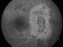
Peripapillary Lesion Hemangioma or CNVM Early FA491 views67-year-old man with a history of a peripapillary lesion in the right eye. Nov 08, 2010
|
|

Peripapillary Lesion Hemangioma or CNVM Early FA432 views67-year-old man with a history of a peripapillary lesion in the right eye. Nov 08, 2010
|
|

Peripapillary Lesion Hemangioma or CNVM Very Early FA463 views67-year-old man with a history of a peripapillary lesion in the right eye. Nov 08, 2010
|
|

Peripapillary Lesion Hemangioma or CNVM Red Free photo of normal left eye593 views67-year-old man with a history of a peripapillary lesion in the right eye. Nov 08, 2010
|
|

Peripapillary Lesion Hemangioma or CNVM Red Free photo of abnormal right eye496 views67-year-old man with a history of a peripapillary lesion in the right eye. Nov 08, 2010
|
|

Peripapillary Lesion Hemangioma or CNVM Line Scan shows Serous RD OD462 views67-year-old man with a history of a peripapillary lesion in the right eye. Nov 08, 2010
|
|

Peripapillary Lesion Hemangioma or CNVM Line Scan Shows Serous RD Right Eye622 views67-year-old man with a history of a peripapillary lesion in the right eye. Nov 08, 2010
|
|

Peripapillary Lesion Hemangioma or CNVM Color Fundus Photo Normal Left Eye592 views67-year-old man with a history of a peripapillary lesion in the right eye. Nov 08, 2010
|
|

Peripapillary Lesion Hemangioma or CNVM Color Fundus Photo Abnormal Right Eye764 views67-year-old man with a history of a peripapillary lesion in the right eye. Nov 08, 2010
|
|

Peripapillary Lesion Hemangioma or CNVM Fundus Photo Right Eye Shows Peripapillary Lesion672 views67-year-old man with a history of a peripapillary lesion in the right eye. Nov 08, 2010
|
|
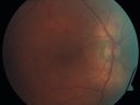
Peripapillary Lesion Hemangioma or CNVM Fundus Photo Right Eye693 views67-year-old man with a history of a peripapillary lesion in the right eye. Nov 08, 2010
|
|

Peripapillary Lesion Hemangioma or CNVM Late FA Shows Hyperfluorescent Lesion488 views67-year-old man with a history of a peripapillary lesion in the right eye. Nov 08, 2010
|
|

Choroidal Melanoma Medium590 views33-year-old man with a history of decreased in the right eye. He had blurring in the eye about a year ago. He reports that it improved spontaneously until it was “nearly resolvedâ€. He said he had some persistent distortion, but that over the past couple of weeks he has noticed this dark spot recurring in the right eye, the left eye is unaffected. He does not have any flashes, floaters or change in his peripheral visual acuity. 20/80 OD, 20/16 OS. No improvement with pinhole. IOP: 12 OD, 11 OS.
POSTERIOR SEGMENT EXAMINATION: Shows a clear view with no PVD in either eye. C/D ratio is 0.2 OU. Large retinal vessels are unremarkable. The macula of the right eye has a brown choroidal lesion with pigment overlying the surface and orange spots along its lateral surface. The left macula is normal.
B-SCAN: B-scan ecography of the ultrasound of the right eye shows a mass about 5 mm high with a moderate to low internal reflectivity on A-scan echogram. There is serous fluid inferiorly on the echogramNov 07, 2010
|
|

Choroidal Melanoma Medium732 views33-year-old man with a history of decreased in the right eye. He had blurring in the eye about a year ago. He reports that it improved spontaneously until it was “nearly resolvedâ€. He said he had some persistent distortion, but that over the past couple of weeks he has noticed this dark spot recurring in the right eye, the left eye is unaffected. He does not have any flashes, floaters or change in his peripheral visual acuity. 20/80 OD, 20/16 OS. No improvement with pinhole. IOP: 12 OD, 11 OS.
POSTERIOR SEGMENT EXAMINATION: Shows a clear view with no PVD in either eye. C/D ratio is 0.2 OU. Large retinal vessels are unremarkable. The macula of the right eye has a brown choroidal lesion with pigment overlying the surface and orange spots along its lateral surface. The left macula is normal.
B-SCAN: B-scan ecography of the ultrasound of the right eye shows a mass about 5 mm high with a moderate to low internal reflectivity on A-scan echogram. There is serous fluid inferiorly on the echogramNov 07, 2010
|
|
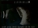
Choroidal Melanoma Medium672 views33-year-old man with a history of decreased in the right eye. He had blurring in the eye about a year ago. He reports that it improved spontaneously until it was “nearly resolvedâ€. He said he had some persistent distortion, but that over the past couple of weeks he has noticed this dark spot recurring in the right eye, the left eye is unaffected. He does not have any flashes, floaters or change in his peripheral visual acuity. 20/80 OD, 20/16 OS. No improvement with pinhole. IOP: 12 OD, 11 OS.
POSTERIOR SEGMENT EXAMINATION: Shows a clear view with no PVD in either eye. C/D ratio is 0.2 OU. Large retinal vessels are unremarkable. The macula of the right eye has a brown choroidal lesion with pigment overlying the surface and orange spots along its lateral surface. The left macula is normal.
B-SCAN: B-scan ecography of the ultrasound of the right eye shows a mass about 5 mm high with a moderate to low internal reflectivity on A-scan echogram. There is serous fluid inferiorly on the echogramNov 07, 2010
|
|

Choroidal Melanoma Medium766 views33-year-old man with a history of decreased in the right eye. He had blurring in the eye about a year ago. He reports that it improved spontaneously until it was “nearly resolvedâ€. He said he had some persistent distortion, but that over the past couple of weeks he has noticed this dark spot recurring in the right eye, the left eye is unaffected. He does not have any flashes, floaters or change in his peripheral visual acuity. 20/80 OD, 20/16 OS. No improvement with pinhole. IOP: 12 OD, 11 OS.
POSTERIOR SEGMENT EXAMINATION: Shows a clear view with no PVD in either eye. C/D ratio is 0.2 OU. Large retinal vessels are unremarkable. The macula of the right eye has a brown choroidal lesion with pigment overlying the surface and orange spots along its lateral surface. The left macula is normal.
B-SCAN: B-scan ecography of the ultrasound of the right eye shows a mass about 5 mm high with a moderate to low internal reflectivity on A-scan echogram. There is serous fluid inferiorly on the echogramNov 07, 2010
|
|

Choroidal Melanoma Medium500 views33-year-old man with a history of decreased in the right eye. He had blurring in the eye about a year ago. He reports that it improved spontaneously until it was “nearly resolvedâ€. He said he had some persistent distortion, but that over the past couple of weeks he has noticed this dark spot recurring in the right eye, the left eye is unaffected. He does not have any flashes, floaters or change in his peripheral visual acuity. 20/80 OD, 20/16 OS. No improvement with pinhole. IOP: 12 OD, 11 OS.
POSTERIOR SEGMENT EXAMINATION: Shows a clear view with no PVD in either eye. C/D ratio is 0.2 OU. Large retinal vessels are unremarkable. The macula of the right eye has a brown choroidal lesion with pigment overlying the surface and orange spots along its lateral surface. The left macula is normal.
B-SCAN: B-scan ecography of the ultrasound of the right eye shows a mass about 5 mm high with a moderate to low internal reflectivity on A-scan echogram. There is serous fluid inferiorly on the echogramNov 07, 2010
|
|
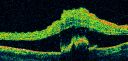
Choroidal Melanoma Medium588 views33-year-old man with a history of decreased in the right eye. He had blurring in the eye about a year ago. He reports that it improved spontaneously until it was “nearly resolvedâ€. He said he had some persistent distortion, but that over the past couple of weeks he has noticed this dark spot recurring in the right eye, the left eye is unaffected. He does not have any flashes, floaters or change in his peripheral visual acuity. 20/80 OD, 20/16 OS. No improvement with pinhole. IOP: 12 OD, 11 OS.
POSTERIOR SEGMENT EXAMINATION: Shows a clear view with no PVD in either eye. C/D ratio is 0.2 OU. Large retinal vessels are unremarkable. The macula of the right eye has a brown choroidal lesion with pigment overlying the surface and orange spots along its lateral surface. The left macula is normal.
B-SCAN: B-scan ecography of the ultrasound of the right eye shows a mass about 5 mm high with a moderate to low internal reflectivity on A-scan echogram. There is serous fluid inferiorly on the echogramNov 07, 2010
|
|

Choroidal Melanoma Medium500 views33-year-old man with a history of decreased in the right eye. He had blurring in the eye about a year ago. He reports that it improved spontaneously until it was “nearly resolvedâ€. He said he had some persistent distortion, but that over the past couple of weeks he has noticed this dark spot recurring in the right eye, the left eye is unaffected. He does not have any flashes, floaters or change in his peripheral visual acuity. 20/80 OD, 20/16 OS. No improvement with pinhole. IOP: 12 OD, 11 OS.
POSTERIOR SEGMENT EXAMINATION: Shows a clear view with no PVD in either eye. C/D ratio is 0.2 OU. Large retinal vessels are unremarkable. The macula of the right eye has a brown choroidal lesion with pigment overlying the surface and orange spots along its lateral surface. The left macula is normal.
B-SCAN: B-scan ecography of the ultrasound of the right eye shows a mass about 5 mm high with a moderate to low internal reflectivity on A-scan echogram. There is serous fluid inferiorly on the echogramNov 07, 2010
|
|

Choroidal Melanoma Medium482 views33-year-old man with a history of decreased in the right eye. He had blurring in the eye about a year ago. He reports that it improved spontaneously until it was “nearly resolvedâ€. He said he had some persistent distortion, but that over the past couple of weeks he has noticed this dark spot recurring in the right eye, the left eye is unaffected. He does not have any flashes, floaters or change in his peripheral visual acuity. 20/80 OD, 20/16 OS. No improvement with pinhole. IOP: 12 OD, 11 OS.
POSTERIOR SEGMENT EXAMINATION: Shows a clear view with no PVD in either eye. C/D ratio is 0.2 OU. Large retinal vessels are unremarkable. The macula of the right eye has a brown choroidal lesion with pigment overlying the surface and orange spots along its lateral surface. The left macula is normal.
B-SCAN: B-scan ecography of the ultrasound of the right eye shows a mass about 5 mm high with a moderate to low internal reflectivity on A-scan echogram. There is serous fluid inferiorly on the echogramNov 07, 2010
|
|

Choroidal Melanoma Medium513 views33-year-old man with a history of decreased in the right eye. He had blurring in the eye about a year ago. He reports that it improved spontaneously until it was “nearly resolvedâ€. He said he had some persistent distortion, but that over the past couple of weeks he has noticed this dark spot recurring in the right eye, the left eye is unaffected. He does not have any flashes, floaters or change in his peripheral visual acuity. 20/80 OD, 20/16 OS. No improvement with pinhole. IOP: 12 OD, 11 OS.
POSTERIOR SEGMENT EXAMINATION: Shows a clear view with no PVD in either eye. C/D ratio is 0.2 OU. Large retinal vessels are unremarkable. The macula of the right eye has a brown choroidal lesion with pigment overlying the surface and orange spots along its lateral surface. The left macula is normal.
B-SCAN: B-scan ecography of the ultrasound of the right eye shows a mass about 5 mm high with a moderate to low internal reflectivity on A-scan echogram. There is serous fluid inferiorly on the echogramNov 07, 2010
|
|

Choroidal Melanoma Medium498 views33-year-old man with a history of decreased in the right eye. He had blurring in the eye about a year ago. He reports that it improved spontaneously until it was “nearly resolvedâ€. He said he had some persistent distortion, but that over the past couple of weeks he has noticed this dark spot recurring in the right eye, the left eye is unaffected. He does not have any flashes, floaters or change in his peripheral visual acuity. 20/80 OD, 20/16 OS. No improvement with pinhole. IOP: 12 OD, 11 OS.
POSTERIOR SEGMENT EXAMINATION: Shows a clear view with no PVD in either eye. C/D ratio is 0.2 OU. Large retinal vessels are unremarkable. The macula of the right eye has a brown choroidal lesion with pigment overlying the surface and orange spots along its lateral surface. The left macula is normal.
B-SCAN: B-scan ecography of the ultrasound of the right eye shows a mass about 5 mm high with a moderate to low internal reflectivity on A-scan echogram. There is serous fluid inferiorly on the echogramNov 07, 2010
|
|

Choroidal Melanoma Medium470 views33-year-old man with a history of decreased in the right eye. He had blurring in the eye about a year ago. He reports that it improved spontaneously until it was “nearly resolvedâ€. He said he had some persistent distortion, but that over the past couple of weeks he has noticed this dark spot recurring in the right eye, the left eye is unaffected. He does not have any flashes, floaters or change in his peripheral visual acuity. 20/80 OD, 20/16 OS. No improvement with pinhole. IOP: 12 OD, 11 OS.
POSTERIOR SEGMENT EXAMINATION: Shows a clear view with no PVD in either eye. C/D ratio is 0.2 OU. Large retinal vessels are unremarkable. The macula of the right eye has a brown choroidal lesion with pigment overlying the surface and orange spots along its lateral surface. The left macula is normal.
B-SCAN: B-scan ecography of the ultrasound of the right eye shows a mass about 5 mm high with a moderate to low internal reflectivity on A-scan echogram. There is serous fluid inferiorly on the echogramNov 07, 2010
|
|

Choroidal Melanoma Medium485 views33-year-old man with a history of decreased in the right eye. He had blurring in the eye about a year ago. He reports that it improved spontaneously until it was “nearly resolvedâ€. He said he had some persistent distortion, but that over the past couple of weeks he has noticed this dark spot recurring in the right eye, the left eye is unaffected. He does not have any flashes, floaters or change in his peripheral visual acuity. 20/80 OD, 20/16 OS. No improvement with pinhole. IOP: 12 OD, 11 OS.
POSTERIOR SEGMENT EXAMINATION: Shows a clear view with no PVD in either eye. C/D ratio is 0.2 OU. Large retinal vessels are unremarkable. The macula of the right eye has a brown choroidal lesion with pigment overlying the surface and orange spots along its lateral surface. The left macula is normal.
B-SCAN: B-scan ecography of the ultrasound of the right eye shows a mass about 5 mm high with a moderate to low internal reflectivity on A-scan echogram. There is serous fluid inferiorly on the echogramNov 07, 2010
|
|

Choroidal Melanoma Medium494 views33-year-old man with a history of decreased in the right eye. He had blurring in the eye about a year ago. He reports that it improved spontaneously until it was “nearly resolvedâ€. He said he had some persistent distortion, but that over the past couple of weeks he has noticed this dark spot recurring in the right eye, the left eye is unaffected. He does not have any flashes, floaters or change in his peripheral visual acuity. 20/80 OD, 20/16 OS. No improvement with pinhole. IOP: 12 OD, 11 OS.
POSTERIOR SEGMENT EXAMINATION: Shows a clear view with no PVD in either eye. C/D ratio is 0.2 OU. Large retinal vessels are unremarkable. The macula of the right eye has a brown choroidal lesion with pigment overlying the surface and orange spots along its lateral surface. The left macula is normal.
B-SCAN: B-scan ecography of the ultrasound of the right eye shows a mass about 5 mm high with a moderate to low internal reflectivity on A-scan echogram. There is serous fluid inferiorly on the echogramNov 07, 2010
|
|

Choroidal Melanoma Medium498 views33-year-old man with a history of decreased in the right eye. He had blurring in the eye about a year ago. He reports that it improved spontaneously until it was “nearly resolvedâ€. He said he had some persistent distortion, but that over the past couple of weeks he has noticed this dark spot recurring in the right eye, the left eye is unaffected. He does not have any flashes, floaters or change in his peripheral visual acuity. 20/80 OD, 20/16 OS. No improvement with pinhole. IOP: 12 OD, 11 OS.
POSTERIOR SEGMENT EXAMINATION: Shows a clear view with no PVD in either eye. C/D ratio is 0.2 OU. Large retinal vessels are unremarkable. The macula of the right eye has a brown choroidal lesion with pigment overlying the surface and orange spots along its lateral surface. The left macula is normal.
B-SCAN: B-scan ecography of the ultrasound of the right eye shows a mass about 5 mm high with a moderate to low internal reflectivity on A-scan echogram. There is serous fluid inferiorly on the echogramNov 07, 2010
|
|

Choroidal Melanoma Medium505 views33-year-old man with a history of decreased in the right eye. He had blurring in the eye about a year ago. He reports that it improved spontaneously until it was “nearly resolvedâ€. He said he had some persistent distortion, but that over the past couple of weeks he has noticed this dark spot recurring in the right eye, the left eye is unaffected. He does not have any flashes, floaters or change in his peripheral visual acuity. 20/80 OD, 20/16 OS. No improvement with pinhole. IOP: 12 OD, 11 OS.
POSTERIOR SEGMENT EXAMINATION: Shows a clear view with no PVD in either eye. C/D ratio is 0.2 OU. Large retinal vessels are unremarkable. The macula of the right eye has a brown choroidal lesion with pigment overlying the surface and orange spots along its lateral surface. The left macula is normal.
B-SCAN: B-scan ecography of the ultrasound of the right eye shows a mass about 5 mm high with a moderate to low internal reflectivity on A-scan echogram. There is serous fluid inferiorly on the echogramNov 07, 2010
|
|

Choroidal Melanoma Medium498 views33-year-old man with a history of decreased in the right eye. He had blurring in the eye about a year ago. He reports that it improved spontaneously until it was “nearly resolvedâ€. He said he had some persistent distortion, but that over the past couple of weeks he has noticed this dark spot recurring in the right eye, the left eye is unaffected. He does not have any flashes, floaters or change in his peripheral visual acuity. 20/80 OD, 20/16 OS. No improvement with pinhole. IOP: 12 OD, 11 OS.
POSTERIOR SEGMENT EXAMINATION: Shows a clear view with no PVD in either eye. C/D ratio is 0.2 OU. Large retinal vessels are unremarkable. The macula of the right eye has a brown choroidal lesion with pigment overlying the surface and orange spots along its lateral surface. The left macula is normal.
B-SCAN: B-scan ecography of the ultrasound of the right eye shows a mass about 5 mm high with a moderate to low internal reflectivity on A-scan echogram. There is serous fluid inferiorly on the echogramNov 07, 2010
|
|

Choroidal Melanoma Medium544 views33-year-old man with a history of decreased in the right eye. He had blurring in the eye about a year ago. He reports that it improved spontaneously until it was “nearly resolvedâ€. He said he had some persistent distortion, but that over the past couple of weeks he has noticed this dark spot recurring in the right eye, the left eye is unaffected. He does not have any flashes, floaters or change in his peripheral visual acuity. 20/80 OD, 20/16 OS. No improvement with pinhole. IOP: 12 OD, 11 OS.
POSTERIOR SEGMENT EXAMINATION: Shows a clear view with no PVD in either eye. C/D ratio is 0.2 OU. Large retinal vessels are unremarkable. The macula of the right eye has a brown choroidal lesion with pigment overlying the surface and orange spots along its lateral surface. The left macula is normal.
B-SCAN: B-scan ecography of the ultrasound of the right eye shows a mass about 5 mm high with a moderate to low internal reflectivity on A-scan echogram. There is serous fluid inferiorly on the echogramNov 07, 2010
|
|

Choroidal Melanoma Medium568 views33-year-old man with a history of decreased in the right eye. He had blurring in the eye about a year ago. He reports that it improved spontaneously until it was “nearly resolvedâ€. He said he had some persistent distortion, but that over the past couple of weeks he has noticed this dark spot recurring in the right eye, the left eye is unaffected. He does not have any flashes, floaters or change in his peripheral visual acuity. 20/80 OD, 20/16 OS. No improvement with pinhole. IOP: 12 OD, 11 OS.
POSTERIOR SEGMENT EXAMINATION: Shows a clear view with no PVD in either eye. C/D ratio is 0.2 OU. Large retinal vessels are unremarkable. The macula of the right eye has a brown choroidal lesion with pigment overlying the surface and orange spots along its lateral surface. The left macula is normal.
B-SCAN: B-scan ecography of the ultrasound of the right eye shows a mass about 5 mm high with a moderate to low internal reflectivity on A-scan echogram. There is serous fluid inferiorly on the echogramNov 07, 2010
|
|

559 views33-year-old man with a history of decreased in the right eye. He had blurring in the eye about a year ago. He reports that it improved spontaneously until it was “nearly resolvedâ€. He said he had some persistent distortion, but that over the past couple of weeks he has noticed this dark spot recurring in the right eye, the left eye is unaffected. He does not have any flashes, floaters or change in his peripheral visual acuity. 20/80 OD, 20/16 OS. No improvement with pinhole. IOP: 12 OD, 11 OS.
POSTERIOR SEGMENT EXAMINATION: Shows a clear view with no PVD in either eye. C/D ratio is 0.2 OU. Large retinal vessels are unremarkable. The macula of the right eye has a brown choroidal lesion with pigment overlying the surface and orange spots along its lateral surface. The left macula is normal.
B-SCAN: B-scan ecography of the ultrasound of the right eye shows a mass about 5 mm high with a moderate to low internal reflectivity on A-scan echogram. There is serous fluid inferiorly on the echogramNov 07, 2010
|
|

Choroidal Melanoma Medium510 views33-year-old man with a history of decreased in the right eye. He had blurring in the eye about a year ago. He reports that it improved spontaneously until it was “nearly resolvedâ€. He said he had some persistent distortion, but that over the past couple of weeks he has noticed this dark spot recurring in the right eye, the left eye is unaffected. He does not have any flashes, floaters or change in his peripheral visual acuity. 20/80 OD, 20/16 OS. No improvement with pinhole. IOP: 12 OD, 11 OS.
POSTERIOR SEGMENT EXAMINATION: Shows a clear view with no PVD in either eye. C/D ratio is 0.2 OU. Large retinal vessels are unremarkable. The macula of the right eye has a brown choroidal lesion with pigment overlying the surface and orange spots along its lateral surface. The left macula is normal.
B-SCAN: B-scan ecography of the ultrasound of the right eye shows a mass about 5 mm high with a moderate to low internal reflectivity on A-scan echogram. There is serous fluid inferiorly on the echogramNov 07, 2010
|
|

Choroidal Melanoma Medium538 views33-year-old man with a history of decreased in the right eye. He had blurring in the eye about a year ago. He reports that it improved spontaneously until it was “nearly resolvedâ€. He said he had some persistent distortion, but that over the past couple of weeks he has noticed this dark spot recurring in the right eye, the left eye is unaffected. He does not have any flashes, floaters or change in his peripheral visual acuity. 20/80 OD, 20/16 OS. No improvement with pinhole. IOP: 12 OD, 11 OS.
POSTERIOR SEGMENT EXAMINATION: Shows a clear view with no PVD in either eye. C/D ratio is 0.2 OU. Large retinal vessels are unremarkable. The macula of the right eye has a brown choroidal lesion with pigment overlying the surface and orange spots along its lateral surface. The left macula is normal.
B-SCAN: B-scan ecography of the ultrasound of the right eye shows a mass about 5 mm high with a moderate to low internal reflectivity on A-scan echogram. There is serous fluid inferiorly on the echogramNov 07, 2010
|
|

Choroidal Melanoma Medium663 views33-year-old man with a history of decreased in the right eye. He had blurring in the eye about a year ago. He reports that it improved spontaneously until it was “nearly resolvedâ€. He said he had some persistent distortion, but that over the past couple of weeks he has noticed this dark spot recurring in the right eye, the left eye is unaffected. He does not have any flashes, floaters or change in his peripheral visual acuity. 20/80 OD, 20/16 OS. No improvement with pinhole. IOP: 12 OD, 11 OS.
POSTERIOR SEGMENT EXAMINATION: Shows a clear view with no PVD in either eye. C/D ratio is 0.2 OU. Large retinal vessels are unremarkable. The macula of the right eye has a brown choroidal lesion with pigment overlying the surface and orange spots along its lateral surface. The left macula is normal.
B-SCAN: B-scan ecography of the ultrasound of the right eye shows a mass about 5 mm high with a moderate to low internal reflectivity on A-scan echogram. There is serous fluid inferiorly on the echogramNov 07, 2010
|
|

Melanoma - Large Ciliary Body A scan shows medium to low internal reflectivity815 viewsOct 30, 2010
|
|
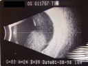
Melanoma - Large Ciliary Body B-Scan ultrasound shows tumore filling much of the eye959 viewsOct 30, 2010
|
|

Melanoma - Large Ciliary Body Photo shows tumor visible throught the pupil943 viewsOct 30, 2010
|
|

Melanoma - Large Ciliary Body Photo shows tumor visible through the pupil1117 viewsOct 30, 2010
|
|

Melanoma - Large Ciliary Body B Scan Ultrasound730 viewsOct 30, 2010
|
|
|
|
| 1098 files on 11 page(s) |
 |
|
|
|
|
|
|
|
11 |
|


