neueste Dateien - Tumors, Masses, Pigmented Lesions
|

Atypical High Risk Choroidal Nevus vs Macular Melanoma496 x angesehen70 year old female with vision loss in the left eye for a few weeks. She was referred to a tumor center and being managed as an "atypical high risk choroidal nevus". The size is 6 mm x 6.5 mm x 1.6 mm (elevated).
Vision 20/40 - having serial avastin injections.01. Dezember 2019
|
|
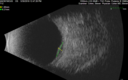
Atypical High Risk Choroidal Nevus vs Macular Melanoma396 x angesehen70 year old female with vision loss in the left eye for a few weeks. She was referred to a tumor center and being managed as an "atypical high risk choroidal nevus". The size is 6 mm x 6.5 mm x 1.6 mm (elevated).
Vision 20/40 - having serial avastin injections.01. Dezember 2019
|
|

Atypical High Risk Choroidal Nevus vs Macular Melanoma502 x angesehen70 year old female with vision loss in the left eye for a few weeks. She was referred to a tumor center and being managed as an "atypical high risk choroidal nevus". The size is 6 mm x 6.5 mm x 1.6 mm (elevated).
Vision 20/40 - having serial avastin injections.01. Dezember 2019
|
|

Atypical High Risk Choroidal Nevus vs Macular Melanoma468 x angesehen70 year old female with vision loss in the left eye for a few weeks. She was referred to a tumor center and being managed as an "atypical high risk choroidal nevus". The size is 6 mm x 6.5 mm x 1.6 mm (elevated).
Vision 20/40 - having serial avastin injections.01. Dezember 2019
|
|
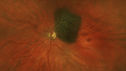
Atypical High Risk Choroidal Nevus vs Macular Melanoma452 x angesehen70 year old female with vision loss in the left eye for a few weeks. She was referred to a tumor center and being managed as an "atypical high risk choroidal nevus". The size is 6 mm x 6.5 mm x 1.6 mm (elevated).
Vision 20/40 - having serial avastin injections.01. Dezember 2019
|
|

Atypical High Risk Choroidal Nevus vs Macular Melanoma433 x angesehen70 year old female with vision loss in the left eye for a few weeks. She was referred to a tumor center and being managed as an "atypical high risk choroidal nevus". The size is 6 mm x 6.5 mm x 1.6 mm (elevated).
Vision 20/40 - having serial avastin injections.01. Dezember 2019
|
|

Atypical High Risk Choroidal Nevus vs Macular Melanoma376 x angesehen70 year old female with vision loss in the left eye for a few weeks. She was referred to a tumor center and being managed as an "atypical high risk choroidal nevus". The size is 6 mm x 6.5 mm x 1.6 mm (elevated).
Vision 20/40 - having serial avastin injections.01. Dezember 2019
|
|

Atypical High Risk Choroidal Nevus vs Macular Melanoma406 x angesehen70 year old female with vision loss in the left eye for a few weeks. She was referred to a tumor center and being managed as an "atypical high risk choroidal nevus". The size is 6 mm x 6.5 mm x 1.6 mm (elevated).
Vision 20/40 - having serial avastin injections.01. Dezember 2019
|
|

Atypical High Risk Choroidal Nevus vs Macular Melanoma468 x angesehen70 year old female with vision loss in the left eye for a few weeks. She was referred to a tumor center and being managed as an "atypical high risk choroidal nevus". The size is 6 mm x 6.5 mm x 1.6 mm (elevated).
Vision 20/40 - having serial avastin injections.01. Dezember 2019
|
|

Atypical High Risk Choroidal Nevus vs Macular Melanoma515 x angesehen70 year old female with vision loss in the left eye for a few weeks. She was referred to a tumor center and being managed as an "atypical high risk choroidal nevus". The size is 6 mm x 6.5 mm x 1.6 mm (elevated).
Vision 20/40 - having serial avastin injections.01. Dezember 2019
|
|

Atypical High Risk Choroidal Nevus vs Macular Melanoma492 x angesehen70 year old female with vision loss in the left eye for a few weeks. She was referred to a tumor center and being managed as an "atypical high risk choroidal nevus". The size is 6 mm x 6.5 mm x 1.6 mm (elevated).
Vision 20/40 - having serial avastin injections.01. Dezember 2019
|
|

Atypical High Risk Choroidal Nevus vs Macular Melanoma437 x angesehen70 year old female with vision loss in the left eye for a few weeks. She was referred to a tumor center and being managed as an "atypical high risk choroidal nevus". The size is 6 mm x 6.5 mm x 1.6 mm (elevated).
Vision 20/40 - having serial avastin injections.01. Dezember 2019
|
|

Atypical High Risk Choroidal Nevus vs Macular Melanoma444 x angesehen70 year old female with vision loss in the left eye for a few weeks. She was referred to a tumor center and being managed as an "atypical high risk choroidal nevus". The size is 6 mm x 6.5 mm x 1.6 mm (elevated).
Vision 20/40 - having serial avastin injections.01. Dezember 2019
|
|

Atypical High Risk Choroidal Nevus vs Macular Melanoma398 x angesehen70 year old female with vision loss in the left eye for a few weeks. She was referred to a tumor center and being managed as an "atypical high risk choroidal nevus". The size is 6 mm x 6.5 mm x 1.6 mm (elevated).
Vision 20/40 - having serial avastin injections.01. Dezember 2019
|
|

Atypical High Risk Choroidal Nevus vs Macular Melanoma457 x angesehen70 year old female with vision loss in the left eye for a few weeks. She was referred to a tumor center and being managed as an "atypical high risk choroidal nevus". The size is 6 mm x 6.5 mm x 1.6 mm (elevated).
Vision 20/40 - having serial avastin injections.01. Dezember 2019
|
|

Atypical High Risk Choroidal Nevus vs Macular Melanoma394 x angesehen70 year old female with vision loss in the left eye for a few weeks. She was referred to a tumor center and being managed as an "atypical high risk choroidal nevus". The size is 6 mm x 6.5 mm x 1.6 mm (elevated).
Vision 20/40 - having serial avastin injections.01. Dezember 2019
|
|

Melanosis Oculi and Choroidal Melanoma475 x angesehen88 year old female with 2.5 mm elevated choroidal melanoma and melanosis oculi. She also has geographic atrophy from AMD. The right eye is the better eye with 20/40 vision. After observation with growth, brachytherapy was done.01. Dezember 2019
|
|

Melanosis Oculi and Choroidal Melanoma425 x angesehen88 year old female with 2.5 mm elevated choroidal melanoma and melanosis oculi. She also has geographic atrophy from AMD. The right eye is the better eye with 20/40 vision. After observation with growth, brachytherapy was done.01. Dezember 2019
|
|
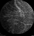
Melanosis Oculi and Choroidal Melanoma475 x angesehen88 year old female with 2.5 mm elevated choroidal melanoma and melanosis oculi. She also has geographic atrophy from AMD. The right eye is the better eye with 20/40 vision. After observation with growth, brachytherapy was done.01. Dezember 2019
|
|

Melanosis Oculi and Choroidal Melanoma428 x angesehen88 year old female with 2.5 mm elevated choroidal melanoma and melanosis oculi. She also has geographic atrophy from AMD. The right eye is the better eye with 20/40 vision. After observation with growth, brachytherapy was done.01. Dezember 2019
|
|

Melanosis Oculi and Choroidal Melanoma471 x angesehen88 year old female with 2.5 mm elevated choroidal melanoma and melanosis oculi. She also has geographic atrophy from AMD. The right eye is the better eye with 20/40 vision. After observation with growth, brachytherapy was done.01. Dezember 2019
|
|

Melanosis Oculi and Choroidal Melanoma442 x angesehen88 year old female with 2.5 mm elevated choroidal melanoma and melanosis oculi. She also has geographic atrophy from AMD. The right eye is the better eye with 20/40 vision. After observation with growth, brachytherapy was done.01. Dezember 2019
|
|

Melanosis Oculi and Choroidal Melanoma416 x angesehen88 year old female with 2.5 mm elevated choroidal melanoma and melanosis oculi. She also has geographic atrophy from AMD. The right eye is the better eye with 20/40 vision. After observation with growth, brachytherapy was done.01. Dezember 2019
|
|

Melanosis Oculi and Choroidal Melanoma453 x angesehen88 year old female with 2.5 mm elevated choroidal melanoma and melanosis oculi. She also has geographic atrophy from AMD. The right eye is the better eye with 20/40 vision. After observation with growth, brachytherapy was done.01. Dezember 2019
|
|
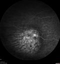
Melanosis Oculi and Choroidal Melanoma431 x angesehen88 year old female with 2.5 mm elevated choroidal melanoma and melanosis oculi. She also has geographic atrophy from AMD. The right eye is the better eye with 20/40 vision. After observation with growth, brachytherapy was done.01. Dezember 2019
|
|
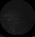
Melanosis Oculi and Choroidal Melanoma448 x angesehen88 year old female with 2.5 mm elevated choroidal melanoma and melanosis oculi. She also has geographic atrophy from AMD. The right eye is the better eye with 20/40 vision. After observation with growth, brachytherapy was done.01. Dezember 2019
|
|

Melanosis Oculi and Choroidal Melanoma425 x angesehen88 year old female with 2.5 mm elevated choroidal melanoma and melanosis oculi. She also has geographic atrophy from AMD. The right eye is the better eye with 20/40 vision. After observation with growth, brachytherapy was done.01. Dezember 2019
|
|

Melanosis Oculi and Choroidal Melanoma401 x angesehen88 year old female with 2.5 mm elevated choroidal melanoma and melanosis oculi. She also has geographic atrophy from AMD. The right eye is the better eye with 20/40 vision. After observation with growth, brachytherapy was done.01. Dezember 2019
|
|

Melanosis Oculi and Choroidal Melanoma565 x angesehen88 year old female with 2.5 mm elevated choroidal melanoma and melanosis oculi. She also has geographic atrophy from AMD. The right eye is the better eye with 20/40 vision. After observation with growth, brachytherapy was done.01. Dezember 2019
|
|

Melanosis Oculi and Choroidal Melanoma462 x angesehen88 year old female with 2.5 mm elevated choroidal melanoma and melanosis oculi. She also has geographic atrophy from AMD. The right eye is the better eye with 20/40 vision. After observation with growth, brachytherapy was done.01. Dezember 2019
|
|
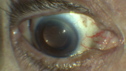
Melanosis Oculi and Choroidal Melanoma595 x angesehen88 year old female with 2.5 mm elevated choroidal melanoma and melanosis oculi. She also has geographic atrophy from AMD. The right eye is the better eye with 20/40 vision. After observation with growth, brachytherapy was done.01. Dezember 2019
|
|

Melanosis Oculi and Choroidal Melanoma489 x angesehen88 year old female with 2.5 mm elevated choroidal melanoma and melanosis oculi. She also has geographic atrophy from AMD. The right eye is the better eye with 20/40 vision. After observation with growth, brachytherapy was done.01. Dezember 2019
|
|

Melanosis Oculi and Choroidal Melanoma489 x angesehen88 year old female with 2.5 mm elevated choroidal melanoma and melanosis oculi. She also has geographic atrophy from AMD. The right eye is the better eye with 20/40 vision. After observation with growth, brachytherapy was done.01. Dezember 2019
|
|

Melanosis Oculi and Choroidal Melanoma570 x angesehen88 year old female with 2.5 mm elevated choroidal melanoma and melanosis oculi. She also has geographic atrophy from AMD. The right eye is the better eye with 20/40 vision. After observation with growth, brachytherapy was done.01. Dezember 2019
|
|

Melanosis Oculi and Choroidal Melanoma598 x angesehen88 year old female with 2.5 mm elevated choroidal melanoma and melanosis oculi. She also has geographic atrophy from AMD. The right eye is the better eye with 20/40 vision. After observation with growth, brachytherapy was done.01. Dezember 2019
|
|

Melanosis Oculi and Choroidal Melanoma867 x angesehen88 year old female with 2.5 mm elevated choroidal melanoma and melanosis oculi. She also has geographic atrophy from AMD. The right eye is the better eye with 20/40 vision. After observation with growth, brachytherapy was done.01. Dezember 2019
|
|

Melanosis Oculi and Choroidal Melanoma625 x angesehen88 year old female with 2.5 mm elevated choroidal melanoma and melanosis oculi. She also has geographic atrophy from AMD. The right eye is the better eye with 20/40 vision. After observation with growth, brachytherapy was done.01. Dezember 2019
|
|

Metastatic Non Small Cell Lung Cancer to Choroid - After External radiation631 x angesehen19. September 2019
|
|

Metastatic Non Small Cell Lung Cancer to Choroid - Before External radiation566 x angesehen19. September 2019
|
|

Lung Cancer Metastasis to Right Eye433 x angesehen63 year old female with vision loss for 6 weeks. She had been scheduled for cataract surgery and then a tumor was found. She was ultimately diagnosed with lung cancer. Vision is 20/10001. Juni 2019
|
|

Lung Cancer Metastasis to Right Eye428 x angesehen63 year old female with vision loss for 6 weeks. She had been scheduled for cataract surgery and then a tumor was found. She was ultimately diagnosed with lung cancer. Vision is 20/10001. Juni 2019
|
|

Lung Cancer Metastasis to Right Eye376 x angesehen63 year old female with vision loss for 6 weeks. She had been scheduled for cataract surgery and then a tumor was found. She was ultimately diagnosed with lung cancer. Vision is 20/10001. Juni 2019
|
|

Lung Cancer Metastasis to Right Eye412 x angesehen63 year old female with vision loss for 6 weeks. She had been scheduled for cataract surgery and then a tumor was found. She was ultimately diagnosed with lung cancer. Vision is 20/10001. Juni 2019
|
|
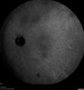
Lung Cancer Metastasis to Right Eye456 x angesehen63 year old female with vision loss for 6 weeks. She had been scheduled for cataract surgery and then a tumor was found. She was ultimately diagnosed with lung cancer. Vision is 20/10001. Juni 2019
|
|

Lung Cancer Metastasis to Right Eye404 x angesehen63 year old female with vision loss for 6 weeks. She had been scheduled for cataract surgery and then a tumor was found. She was ultimately diagnosed with lung cancer. Vision is 20/10001. Juni 2019
|
|

Lung Cancer Metastasis to Right Eye425 x angesehen63 year old female with vision loss for 6 weeks. She had been scheduled for cataract surgery and then a tumor was found. She was ultimately diagnosed with lung cancer. Vision is 20/10001. Juni 2019
|
|

Lung Cancer Metastasis to Right Eye404 x angesehen63 year old female with vision loss for 6 weeks. She had been scheduled for cataract surgery and then a tumor was found. She was ultimately diagnosed with lung cancer. Vision is 20/10001. Juni 2019
|
|

Lung Cancer Metastasis to Right Eye415 x angesehen63 year old female with vision loss for 6 weeks. She had been scheduled for cataract surgery and then a tumor was found. She was ultimately diagnosed with lung cancer. Vision is 20/10001. Juni 2019
|
|

Lung Cancer Metastasis to Right Eye449 x angesehen63 year old female with vision loss for 6 weeks. She had been scheduled for cataract surgery and then a tumor was found. She was ultimately diagnosed with lung cancer. Vision is 20/10001. Juni 2019
|
|
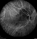
Lung Cancer Metastasis to Right Eye389 x angesehen63 year old female with vision loss for 6 weeks. She had been scheduled for cataract surgery and then a tumor was found. She was ultimately diagnosed with lung cancer. Vision is 20/10001. Juni 2019
|
|

Lung Cancer Metastasis to Right Eye465 x angesehen63 year old female with vision loss for 6 weeks. She had been scheduled for cataract surgery and then a tumor was found. She was ultimately diagnosed with lung cancer. Vision is 20/10001. Juni 2019
|
|

Lung Cancer Metastasis to Right Eye466 x angesehen63 year old female with vision loss for 6 weeks. She had been scheduled for cataract surgery and then a tumor was found. She was ultimately diagnosed with lung cancer. Vision is 20/10001. Juni 2019
|
|

Lung Cancer Metastasis to Right Eye430 x angesehen63 year old female with vision loss for 6 weeks. She had been scheduled for cataract surgery and then a tumor was found. She was ultimately diagnosed with lung cancer. Vision is 20/10001. Juni 2019
|
|
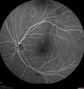
Lung Cancer Metastasis to Right Eye453 x angesehen63 year old female with vision loss for 6 weeks. She had been scheduled for cataract surgery and then a tumor was found. She was ultimately diagnosed with lung cancer. Vision is 20/10001. Juni 2019
|
|

Lung Cancer Metastasis to Right Eye402 x angesehen63 year old female with vision loss for 6 weeks. She had been scheduled for cataract surgery and then a tumor was found. She was ultimately diagnosed with lung cancer. Vision is 20/10001. Juni 2019
|
|

Lung Cancer Metastasis to Right Eye389 x angesehen63 year old female with vision loss for 6 weeks. She had been scheduled for cataract surgery and then a tumor was found. She was ultimately diagnosed with lung cancer. Vision is 20/10001. Juni 2019
|
|

Lung Cancer Metastasis to Right Eye415 x angesehen63 year old female with vision loss for 6 weeks. She had been scheduled for cataract surgery and then a tumor was found. She was ultimately diagnosed with lung cancer. Vision is 20/10001. Juni 2019
|
|
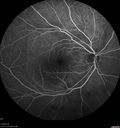
Lung Cancer Metastasis to Right Eye460 x angesehen63 year old female with vision loss for 6 weeks. She had been scheduled for cataract surgery and then a tumor was found. She was ultimately diagnosed with lung cancer. Vision is 20/10001. Juni 2019
|
|

Lung Cancer Metastasis to Right Eye402 x angesehen63 year old female with vision loss for 6 weeks. She had been scheduled for cataract surgery and then a tumor was found. She was ultimately diagnosed with lung cancer. Vision is 20/10001. Juni 2019
|
|

Lung Cancer Metastasis to Right Eye484 x angesehen63 year old female with vision loss for 6 weeks. She had been scheduled for cataract surgery and then a tumor was found. She was ultimately diagnosed with lung cancer. Vision is 20/10001. Juni 2019
|
|

Lung Cancer Metastasis to Right Eye482 x angesehen63 year old female with vision loss for 6 weeks. She had been scheduled for cataract surgery and then a tumor was found. She was ultimately diagnosed with lung cancer. Vision is 20/10001. Juni 2019
|
|
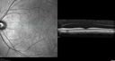
Lung Cancer Metastasis to Right Eye406 x angesehen63 year old female with vision loss for 6 weeks. She had been scheduled for cataract surgery and then a tumor was found. She was ultimately diagnosed with lung cancer. Vision is 20/10001. Juni 2019
|
|

Lung Cancer Metastasis to Right Eye442 x angesehen63 year old female with vision loss for 6 weeks. She had been scheduled for cataract surgery and then a tumor was found. She was ultimately diagnosed with lung cancer. Vision is 20/10001. Juni 2019
|
|

Lung Cancer Metastasis to Right Eye423 x angesehen63 year old female with vision loss for 6 weeks. She had been scheduled for cataract surgery and then a tumor was found. She was ultimately diagnosed with lung cancer. Vision is 20/10001. Juni 2019
|
|

Lung Cancer Metastasis to Right Eye411 x angesehen63 year old female with vision loss for 6 weeks. She had been scheduled for cataract surgery and then a tumor was found. She was ultimately diagnosed with lung cancer. Vision is 20/10001. Juni 2019
|
|

Lung Cancer Metastasis to Right Eye406 x angesehen63 year old female with vision loss for 6 weeks. She had been scheduled for cataract surgery and then a tumor was found. She was ultimately diagnosed with lung cancer. Vision is 20/10001. Juni 2019
|
|
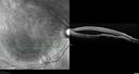
Lung Cancer Metastasis to Right Eye454 x angesehen63 year old female with vision loss for 6 weeks. She had been scheduled for cataract surgery and then a tumor was found. She was ultimately diagnosed with lung cancer. Vision is 20/10001. Juni 2019
|
|

Lung Cancer Metastasis to Right Eye437 x angesehen63 year old female with vision loss for 6 weeks. She had been scheduled for cataract surgery and then a tumor was found. She was ultimately diagnosed with lung cancer. Vision is 20/10001. Juni 2019
|
|

Lung Cancer Metastasis to Right Eye395 x angesehen63 year old female with vision loss for 6 weeks. She had been scheduled for cataract surgery and then a tumor was found. She was ultimately diagnosed with lung cancer. Vision is 20/10001. Juni 2019
|
|

Lung Cancer Metastasis to Right Eye406 x angesehen63 year old female with vision loss for 6 weeks. She had been scheduled for cataract surgery and then a tumor was found. She was ultimately diagnosed with lung cancer. Vision is 20/10001. Juni 2019
|
|
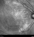
Lung Cancer Metastasis to Right Eye466 x angesehen63 year old female with vision loss for 6 weeks. She had been scheduled for cataract surgery and then a tumor was found. She was ultimately diagnosed with lung cancer. Vision is 20/10001. Juni 2019
|
|

Lung Cancer Metastasis to Right Eye439 x angesehen63 year old female with vision loss for 6 weeks. She had been scheduled for cataract surgery and then a tumor was found. She was ultimately diagnosed with lung cancer. Vision is 20/10001. Juni 2019
|
|

Lung Cancer Metastasis to Right Eye429 x angesehen63 year old female with vision loss for 6 weeks. She had been scheduled for cataract surgery and then a tumor was found. She was ultimately diagnosed with lung cancer. Vision is 20/10001. Juni 2019
|
|
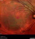
Lung Cancer Metastasis to Right Eye580 x angesehen63 year old female with vision loss for 6 weeks. She had been scheduled for cataract surgery and then a tumor was found. She was ultimately diagnosed with lung cancer. Vision is 20/10001. Juni 2019
|
|

Lung Cancer Metastasis to Right Eye506 x angesehen63 year old female with vision loss for 6 weeks. She had been scheduled for cataract surgery and then a tumor was found. She was ultimately diagnosed with lung cancer. Vision is 20/10001. Juni 2019
|
|
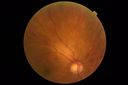
Lung Cancer Metastasis to Right Eye419 x angesehen63 year old female with vision loss for 6 weeks. She had been scheduled for cataract surgery and then a tumor was found. She was ultimately diagnosed with lung cancer. Vision is 20/10001. Juni 2019
|
|

Lung Cancer Metastasis to Right Eye460 x angesehen63 year old female with vision loss for 6 weeks. She had been scheduled for cataract surgery and then a tumor was found. She was ultimately diagnosed with lung cancer. Vision is 20/10001. Juni 2019
|
|

Lung Cancer Metastasis to Right Eye496 x angesehen63 year old female with vision loss for 6 weeks. She had been scheduled for cataract surgery and then a tumor was found. She was ultimately diagnosed with lung cancer. Vision is 20/10001. Juni 2019
|
|

Lung Cancer Metastasis to Right Eye423 x angesehen63 year old female with vision loss for 6 weeks. She had been scheduled for cataract surgery and then a tumor was found. She was ultimately diagnosed with lung cancer. Vision is 20/10001. Juni 2019
|
|

Lung Cancer Metastasis to Right Eye446 x angesehen63 year old female with vision loss for 6 weeks. She had been scheduled for cataract surgery and then a tumor was found. She was ultimately diagnosed with lung cancer. Vision is 20/10001. Juni 2019
|
|

Lung Cancer Metastasis to Right Eye414 x angesehen63 year old female with vision loss for 6 weeks. She had been scheduled for cataract surgery and then a tumor was found. She was ultimately diagnosed with lung cancer. Vision is 20/10001. Juni 2019
|
|
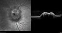
Melanocytoma - Lightly Pigmented489 x angesehen42 year old female with very mild visual field and vision loss in the left eye: 20/20 OU06. Mai 2019
|
|

Melanocytoma - Lightly Pigmented437 x angesehen42 year old female with very mild visual field and vision loss in the left eye: 20/20 OU06. Mai 2019
|
|
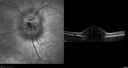
Melanocytoma - Lightly Pigmented421 x angesehen42 year old female with very mild visual field and vision loss in the left eye: 20/20 OU06. Mai 2019
|
|

Melanocytoma - Lightly Pigmented462 x angesehen42 year old female with very mild visual field and vision loss in the left eye: 20/20 OU06. Mai 2019
|
|

Melanocytoma - Lightly Pigmented452 x angesehen42 year old female with very mild visual field and vision loss in the left eye: 20/20 OU06. Mai 2019
|
|

Melanocytoma - Lightly Pigmented469 x angesehen42 year old female with very mild visual field and vision loss in the left eye: 20/20 OU06. Mai 2019
|
|

Melanocytoma - Lightly Pigmented519 x angesehen42 year old female with very mild visual field and vision loss in the left eye: 20/20 OU06. Mai 2019
|
|

Melanocytoma - Lightly Pigmented497 x angesehen42 year old female with very mild visual field and vision loss in the left eye: 20/20 OU06. Mai 2019
|
|

Melanocytoma - Lightly Pigmented445 x angesehen42 year old female with very mild visual field and vision loss in the left eye: 20/20 OU06. Mai 2019
|
|

Melanocytoma - Lightly Pigmented470 x angesehen42 year old female with very mild visual field and vision loss in the left eye: 20/20 OU06. Mai 2019
|
|

Melanocytoma - Lightly Pigmented419 x angesehen42 year old female with very mild visual field and vision loss in the left eye: 20/20 OU06. Mai 2019
|
|

Melanocytoma - Lightly Pigmented457 x angesehen42 year old female with very mild visual field and vision loss in the left eye: 20/20 OU06. Mai 2019
|
|

Melanocytoma - Lightly Pigmented429 x angesehen42 year old female with very mild visual field and vision loss in the left eye: 20/20 OU06. Mai 2019
|
|
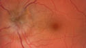
Melanocytoma - Lightly Pigmented532 x angesehen42 year old female with very mild visual field and vision loss in the left eye: 20/20 OU06. Mai 2019
|
|

Melanocytoma - Lightly Pigmented467 x angesehen42 year old female with very mild visual field and vision loss in the left eye: 20/20 OU06. Mai 2019
|
|

Melanocytoma - Lightly Pigmented385 x angesehen42 year old female with very mild visual field and vision loss in the left eye: 20/20 OU06. Mai 2019
|
|

Melanocytoma - Lightly Pigmented430 x angesehen42 year old female with very mild visual field and vision loss in the left eye: 20/20 OU06. Mai 2019
|
|

Melanocytoma - Lightly Pigmented442 x angesehen42 year old female with very mild visual field and vision loss in the left eye: 20/20 OU06. Mai 2019
|
|

Choroidal Nevus - Macula - Flat564 x angesehen37 year old female with 5 year stable nevus Left eye - OCT shows flat nevus (EDI scan)12. März 2018
|
|
| 1098 Dateien auf 11 Seite(n) |
 |
|
|
5 | |
|
|
|
|
|


