Last additions - Tumors, Masses, Pigmented Lesions
|

Metastatic Choroidal Lesion1110 viewsYoung female with choroidal lesion with subretinal fluid in the left eye. VA is finger count in the left eye. Sep 16, 2014
|
|

Choroidal Hemangioma698 viewsPatient comes in for eval on Choroidal Hemangioma in the left eye. VA is count fingers in the left eye. Patient will consider proton beam therapy treatment as Photo-dynamic Therapy and Avastin Injections did not help. Sep 16, 2014
|
|

Choroidal Melanoma with Retinal Detachment500 viewsChoroidal Melanoma centering off the optic nerve with retinal detachment inferiorly in the right eye. Sep 12, 2014
|
|

Choroidal Melanoma495 viewsEval for choroidal melanoma in the left eye. Fundus photo shows large choroidal mass centralized over the macula in the left eye. Aug 25, 2014
|
|

Treated Choroidal Melanoma387 viewsPatient that underwent proton therapy for choroidal melanoma. Fundus photo shows treated melanoma temporal inferior with scattered hemorrhages inferior of the retina. Jul 13, 2014
|
|

Treated Choroidal Melanoma1052 viewsFollow up patient for melanoma in the right eye that was treated with proton beam therapy. VA is 20/50 in the right eye. Fundus photo shows scattered hemorrhages inferiorly with tumor temporal inferior at 8-o'clock. Patient will be followed up in 6-monthsJul 10, 2014
|
|

Melanoma of Optic Nerve1107 viewsPatient comes in for evaluation on Optic Nerve Melanoma in the right eye. Patient's VA is 20/40 in the right eye. Fundus photograph reveals melanoma invading optic nerve and ultrasound reveals behind the optic nerve as well. Jul 09, 2014
|
|

Large Choroidal Melanoma with Retinal Detachment461 viewsPatient notices a decline in her vision for the past 3-weeks in her right eye. VA was light perception in the right eye. Fundus photo shows a very large melanoma temporal to the optic nerve and retinal detachment inferiorly. Patient will under go proton beam therapy. Jun 20, 2014
|
|
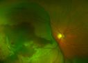
Large Choroidal Melanoma with Retinal Detachment991 viewsPatient notices a decline in her vision for the past 3-weeks in her right eye. VA was light perception in the right eye. Fundus photo shows a very large melanoma temporal to the optic nerve and retinal detachment inferiorly. Patient will under go proton beam therapy. Jun 20, 2014
|
|

Chorodial Melanoma1339 views80 year old male came in for retinal evaluation and presented with a melanoma.Jun 10, 2014
|
|

Retinoblastoma2016 viewsYoung male who has a retinal tumor inferior with a calcium core in the left eye. This is a regressed retinoblastoma. Apr 28, 2014
|
|

Treated Melanoma908 viewsTreated melanoma in the right eye. VA is light perception in the right eye. Mar 28, 2014
|
|

Choroidal Osteoma1433 viewsPatient with decreased vision within the past 2-years. VA 20/100 in the right eye. Fundus photo shows atrophic scar temporal to the macula in the right eye.Mar 27, 2014
|
|

Choroidal Nevus1063 viewsPatient comes in with pigmented spot in the right eye. VA is 20/25. Fundus photography shows elevated choroidal nevus inferiorily in the right eye. Will be reevaluated in 3-months... Feb 17, 2014
|
|

Choroidal Nevus499 viewsFundus exam shows choroidal nevus, inferior nasally in the right eye. VA is 20/40 in the right eye. BSCAN shows nevus is 2mm thick with no orange pigment. Will be reevaluated in 3-monthsFeb 17, 2014
|
|

Choroidal Melanoma1087 viewsFemale patient comes in with a visual defect in the right eye. VA is 20/40 in the right eye. Fundus photography shows choroidal melanoma with retinal detachment temporally at 3-o'clock in the right eye.Feb 17, 2014
|
|

Choroidal Melanoma with Sub Retinal Fluid479 viewsFundus photography shows choroidal melanoma superiorily between 11-12-o'clock in the left eye. VA is 20/150 in the left eye. Jan 23, 2014
|
|

Choroidal Melanoma with Sub Retinal Fluid1160 viewsFundus photography shows choroidal melanoma superiorily between 11-12-o'clock in the left eye. VA is 20/150 in the left eye. Jan 23, 2014
|
|

Optic Nerve Tumor475 viewsYoung male patient with a tumor present on the optic nerve in the right eye. VA is hand motion in the right eye.Dec 13, 2013
|
|

Optic Nerve Tumor1011 viewsYoung male patient with a tumor present on the optic nerve in the right eye. VA is hand motion in the right eyeDec 13, 2013
|
|

Choroidal Melanoma1139 viewsPatient comes in with spot in vision in the right eye. Fundus photography shows medium size melanoma adjacent to the optic nerve and measures 13mm across horizontally and 6mm diameter in thickness. Patient will undergo proton beam therapy. Sep 23, 2013
|
|

Choroidal-Melanoma with Retinal Hemorrhages573 viewsPatient comes in with blurry vision in his inferior visual field in his right eye. Fundus photography shows choroidal melanoma superiorly at 12-o'clock. Retinal hemorrhages scattered around the periphery. Sep 11, 2013
|
|

Choroidal Melanoma (Treated)578 viewsFundus photography shows choroidal melanoma at 2-o'clock superiorily. Patient had a radioactive implant back in August 2011. Sep 11, 2013
|
|

Large Choroidal Nevus452 viewsYoung male patient who comes in for follow up on choroidal nevus in the right eye. VA is 20/30 in the right eye. Fundus montage photo shows very large nevus branched out from the macula to the temporal aspect of the right eye. Patient will be followed up in 3-months.Jul 30, 2013
|
|

Cavernous Choroidal Hemangioma511 viewsPatient presents with Cavernous Choroidal Hemangioma in the right eye. VA is 20/50 after manifest refraction. Patient also notices decreased vision with shadow close to her central vision in the right eye. Patient will undergo photodynamic therapy in the right eye. Jul 30, 2013
|
|
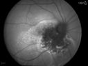
Cavernous Choroidal Hemangioma514 viewsPatient presents with Cavernous Choroidal Hemangioma in the right eye. VA is 20/50 after manifest refraction. Patient also notices decreased vision with shadow close to her central vision in the right eye. Patient will undergo photodynamic therapy in the right eye. Jul 30, 2013
|
|

Cavernous Choroidal Hemangioma519 viewsPatient presents with Cavernous Choroidal Hemangioma in the right eye. VA is 20/50 after manifest refraction. Patient also notices decreased vision with shadow close to her central vision in the right eye. Patient will undergo photodynamic therapy in the right eye. Jul 30, 2013
|
|
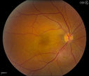
Cavernous Choroidal Hemangioma542 viewsPatient presents with Cavernous Choroidal Hemangioma in the right eye. VA is 20/50 after manifest refraction. Patient also notices decreased vision with shadow close to her central vision in the right eye. Patient will undergo photodynamic therapy in the right eye. Jul 30, 2013
|
|

Multiple Uveal Choroidal Nevi462 viewsPatient referred for CHRPE which in this case has unusual appearance. VA is 20/20 in the both eyes. Distinguishing if there was any relation to polyposis. This is not CHRPE. Jul 29, 2013
|
|

Multiple Uveal Choroidal Nevi450 viewsPatient referred for CHRPE which in this case has unusual appearance. VA is 20/20 in the both eyes. Distinguishing if there was any relation to polyposis. This is not CHRPE.Jul 29, 2013
|
|

Multiple Uveal Choroidal Nevi446 viewsPatient referred for CHRPE which in this case has unusual appearance. VA is 20/20 in the both eyes. Distinguishing if there was any relation to polyposis. This is not CHRPE.Jul 29, 2013
|
|

Multiple Uveal Choroidal Nevi473 viewsPatient referred for CHRPE which in this case has unusual appearance. VA is 20/20 in the both eyes. Distinguishing if there was any relation to polyposis. This is not CHRPE.Jul 29, 2013
|
|

Choroidal Metastasis Tumor 1067 viewsPatient that a history of breast cancer. She has a metastasis tumor superiority in the right eye. VA was 20/20, right eye. Patient underwent radiation and chemotherapy and mass has not changed in size for the past year. Jul 22, 2013
|
|

Ciliary Body Melanoma516 views71-year old male who was recently diagnosed with a large ciliary body melanoma that is pushing into the anterior chamber of the left eye. Patient is going to proceed with proton therapy. Jul 12, 2013
|
|

Ciliary Body Melanoma494 views71-year old male who was recently diagnosed with a large ciliary body melanoma that is pushing into the anterior chamber of the left eye. Patient is going to proceed with proton therapy. Jul 12, 2013
|
|

Ciliary Body Melanoma537 views71-year old male who was recently diagnosed with a large ciliary body melanoma that is pushing into the anterior chamber of the left eye. Patient is going to proceed with proton therapy. Jul 12, 2013
|
|

Ciliary Body Melanoma746 views71-year old male who was recently diagnosed with a large ciliary body melanoma that is pushing into the anterior chamber of the left eye. Patient is going to proceed with proton therapy. Jul 12, 2013
|
|

Ciliary Body Melanoma897 views71-year old male who was recently diagnosed with a large ciliary body melanoma that is pushing into the anterior chamber of the left eye. Patient is going to proceed with proton therapy. Jul 12, 2013
|
|

Choroidal Tumor consistent with Uveal Melanoma1152 viewsPatient comes in for second opinion for 2-melanomas in the right eye. Patient's VA is 20/20, both eyes. Fundus exam reveals melanoma superior to the macula which measured at 0.7-mm thick. Second melanoma is nasally which measured at 4.1-mm thick and has low internal reflectivity. Patient will be followed up with MRI and ocular protocol.May 20, 2013
|
|

503 viewsMay 06, 2013
|
|

472 viewsMay 06, 2013
|
|
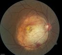
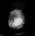
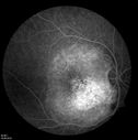
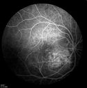
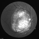
Choroidal Osteoma878 viewsYoung girl with vision loss from osteoma.May 06, 2013
|
|
394 viewsMay 06, 2013
|
|

364 viewsMay 06, 2013
|
|

366 viewsMay 06, 2013
|
|

450 viewsMay 06, 2013
|
|

435 viewsMay 06, 2013
|
|
461 viewsMay 06, 2013
|
|

538 viewsMay 06, 2013
|
|

443 viewsMay 06, 2013
|
|

436 viewsMay 06, 2013
|
|

453 viewsMay 06, 2013
|
|

382 viewsMay 06, 2013
|
|

385 viewsMay 06, 2013
|
|

428 viewsMay 06, 2013
|
|

404 viewsMay 06, 2013
|
|

425 viewsMay 06, 2013
|
|

380 viewsMay 06, 2013
|
|

397 viewsMay 06, 2013
|
|
442 viewsMay 06, 2013
|
|
505 viewsMay 06, 2013
|
|

472 viewsMay 06, 2013
|
|

595 viewsMay 06, 2013
|
|

634 viewsMay 06, 2013
|
|

Amelanotic Nevus Initial Visit1541 viewsMar 07, 2013
|
|

Amelanotic Nevus - 1 year Follow-up Visit - No Change1015 viewsMar 07, 2013
|
|

Medium Choroidal Melanoma Left Eye 677 viewsMar 04, 2013
|
|
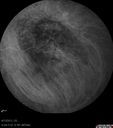
Medium Choroidal Melanoma Left Eye 613 viewsMar 04, 2013
|
|

Medium Choroidal Melanoma Left Eye 523 viewsMar 04, 2013
|
|

Medium Choroidal Melanoma Left Eye 566 viewsMar 04, 2013
|
|

Medium Choroidal Melanoma Left Eye 560 viewsMar 04, 2013
|
|

Medium Choroidal Melanoma Left Eye 499 viewsMar 04, 2013
|
|

Medium Choroidal Melanoma Left Eye 529 viewsMar 04, 2013
|
|

Medium Choroidal Melanoma Left Eye 497 viewsMar 04, 2013
|
|

Medium Choroidal Melanoma Left Eye 480 viewsMar 04, 2013
|
|

Medium Choroidal Melanoma Left Eye 474 viewsMar 04, 2013
|
|

Medium Choroidal Melanoma Left Eye 621 viewsMar 04, 2013
|
|

Medium Choroidal Melanoma Left Eye 523 viewsMar 04, 2013
|
|
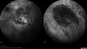
Medium Choroidal Melanoma Left Eye 548 viewsMar 04, 2013
|
|

Medium Choroidal Melanoma Left Eye 563 viewsMar 04, 2013
|
|
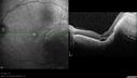
Medium Choroidal Melanoma Left Eye 471 viewsMar 04, 2013
|
|
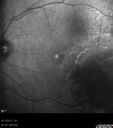
Medium Choroidal Melanoma Left Eye 486 viewsMar 04, 2013
|
|

Medium Choroidal Melanoma Left Eye 500 viewsMar 04, 2013
|
|

Medium Choroidal Melanoma Left Eye 521 viewsMar 04, 2013
|
|

Medium Choroidal Melanoma Left Eye 512 viewsMar 04, 2013
|
|

Medium Choroidal Melanoma Left Eye 597 viewsMar 04, 2013
|
|

Medium Choroidal Melanoma Left Eye 516 viewsMar 04, 2013
|
|

Medium Choroidal Melanoma Left Eye 534 viewsMar 04, 2013
|
|

Medium Choroidal Melanoma Left Eye 529 viewsMar 04, 2013
|
|

Medium Choroidal Melanoma Left Eye 642 viewsMar 04, 2013
|
|

Medium Choroidal Melanoma Left Eye 593 viewsMar 04, 2013
|
|

Uveal Choroidal Melanoma475 viewsPatient comes in for evaluation on a Choroidal Melanoma in the right eye. VA was 20/25 in both eyes. The melanoma is in the temporal aspect of the right eye. It measured at 0.7mm elevated after doing a BSCAN Ultrasound. Feb 26, 2013
|
|

Uveal Choroidal Melanoma477 viewsPatient comes in for evaluation on a Choroidal Melanoma in the right eye. VA was 20/25 in both eyes. The melanoma is in the temporal aspect of the right eye. It measured at 0.7mm elevated after doing a BSCAN Ultrasound. Feb 26, 2013
|
|

Uveal Choroidal Melanoma544 viewsPatient comes in for evaluation on a Choroidal Melanoma in the right eye. VA was 20/25 in both eyes. The melanoma is in the temporal aspect of the right eye. It measured at 0.7mm elevated after doing a BSCAN Ultrasound. Feb 26, 2013
|
|

Uveal Choroidal Melanoma452 viewsPatient comes in for evaluation on a Choroidal Melanoma in the right eye. VA was 20/25 in both eyes. The melanoma is in the temporal aspect of the right eye. It measured at 0.7mm elevated after doing a BSCAN Ultrasound. Feb 26, 2013
|
|

Uveal Choroidal Melanoma542 viewsPatient comes in for evaluation on a Choroidal Melanoma in the right eye. VA was 20/25 in both eyes. The melanoma is in the temporal aspect of the right eye. It measured at 0.7mm elevated after doing a BSCAN Ultrasound. Feb 26, 2013
|
|

Uveal Choroidal Melanoma463 viewsPatient comes in for evaluation on a Choroidal Melanoma in the right eye. VA was 20/25 in both eyes. The melanoma is in the temporal aspect of the right eye. It measured at 0.7mm elevated after doing a BSCAN Ultrasound. Feb 26, 2013
|
|

Uveal Choroidal Melanoma480 viewsPatient comes in for evaluation on a Choroidal Melanoma in the right eye. VA was 20/25 in both eyes. The melanoma is in the temporal aspect of the right eye. It measured at 0.7mm elevated after doing a BSCAN Ultrasound. Feb 26, 2013
|
|

Uveal Choroidal Melanoma506 viewsPatient comes in for evaluation on a Choroidal Melanoma in the right eye. VA was 20/25 in both eyes. The melanoma is in the temporal aspect of the right eye. It measured at 0.7mm elevated after doing a BSCAN Ultrasound. Feb 26, 2013
|
|

Uveal Choroidal Melanoma505 viewsPatient comes in for evaluation on a Choroidal Melanoma in the right eye. VA was 20/25 in both eyes. The melanoma is in the temporal aspect of the right eye. It measured at 0.7mm elevated after doing a BSCAN Ultrasound. Feb 26, 2013
|
|
| 1098 files on 11 page(s) |
 |
|
|
|
|
7 | |
|
|
|


