Last additions - Dystrophies
|

Retinitis Pigmentosa - Autosomal Dominant - Vision Loss at Young Age - Infrared553 views 54-year-old woman has autosomal dominant retinitis pigmentosa. Her mother and grandmother both had it and she has one son who has it and one daughter who doesn’t. She also has a sister with it. She lost vision since she was younger. She had cataract surgery done in January and February of this year. She has been seeing unusual flashing lights throughout the periphery and central vision for about the last year or two.
VISUAL ACUITY: OD 20/160, OS 20/160Nov 02, 2012
|
|
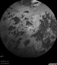
Retinitis Pigmentosa - Autosomal Dominant - Vision Loss at Young Age - Infrared604 views 54-year-old woman has autosomal dominant retinitis pigmentosa. Her mother and grandmother both had it and she has one son who has it and one daughter who doesn’t. She also has a sister with it. She lost vision since she was younger. She had cataract surgery done in January and February of this year. She has been seeing unusual flashing lights throughout the periphery and central vision for about the last year or two.
VISUAL ACUITY: OD 20/160, OS 20/160Nov 02, 2012
|
|

Retinitis Pigmentosa - Autosomal Dominant - Vision Loss at Young Age - Infrared628 views 54-year-old woman has autosomal dominant retinitis pigmentosa. Her mother and grandmother both had it and she has one son who has it and one daughter who doesn’t. She also has a sister with it. She lost vision since she was younger. She had cataract surgery done in January and February of this year. She has been seeing unusual flashing lights throughout the periphery and central vision for about the last year or two.
VISUAL ACUITY: OD 20/160, OS 20/160Nov 02, 2012
|
|

Retinitis Pigmentosa - Autosomal Dominant - Vision Loss at Young Age - Infrared569 views 54-year-old woman has autosomal dominant retinitis pigmentosa. Her mother and grandmother both had it and she has one son who has it and one daughter who doesn’t. She also has a sister with it. She lost vision since she was younger. She had cataract surgery done in January and February of this year. She has been seeing unusual flashing lights throughout the periphery and central vision for about the last year or two.
VISUAL ACUITY: OD 20/160, OS 20/160Nov 02, 2012
|
|

Retinitis Pigmentosa - Autosomal Dominant - Vision Loss at Young Age - Infrared634 views 54-year-old woman has autosomal dominant retinitis pigmentosa. Her mother and grandmother both had it and she has one son who has it and one daughter who doesn’t. She also has a sister with it. She lost vision since she was younger. She had cataract surgery done in January and February of this year. She has been seeing unusual flashing lights throughout the periphery and central vision for about the last year or two.
VISUAL ACUITY: OD 20/160, OS 20/160Nov 02, 2012
|
|

Retinitis Pigmentosa - Autosomal Dominant - Vision Loss at Young Age - Fundus Photo686 views 54-year-old woman has autosomal dominant retinitis pigmentosa. Her mother and grandmother both had it and she has one son who has it and one daughter who doesn’t. She also has a sister with it. She lost vision since she was younger. She had cataract surgery done in January and February of this year. She has been seeing unusual flashing lights throughout the periphery and central vision for about the last year or two.
VISUAL ACUITY: OD 20/160, OS 20/160Nov 02, 2012
|
|

Retinitis Pigmentosa - Autosomal Dominant - Vision Loss at Young Age - Fundus Photo723 views 54-year-old woman has autosomal dominant retinitis pigmentosa. Her mother and grandmother both had it and she has one son who has it and one daughter who doesn’t. She also has a sister with it. She lost vision since she was younger. She had cataract surgery done in January and February of this year. She has been seeing unusual flashing lights throughout the periphery and central vision for about the last year or two.
VISUAL ACUITY: OD 20/160, OS 20/160Nov 02, 2012
|
|

Retinitis Pigmentosa - Autosomal Dominant - Vision Loss at Young Age - Fundus Photo720 views 54-year-old woman has autosomal dominant retinitis pigmentosa. Her mother and grandmother both had it and she has one son who has it and one daughter who doesn’t. She also has a sister with it. She lost vision since she was younger. She had cataract surgery done in January and February of this year. She has been seeing unusual flashing lights throughout the periphery and central vision for about the last year or two.
VISUAL ACUITY: OD 20/160, OS 20/160Nov 02, 2012
|
|

Retinitis Pigmentosa - Autosomal Dominant - Vision Loss at Young Age - Fundus Photo712 views 54-year-old woman has autosomal dominant retinitis pigmentosa. Her mother and grandmother both had it and she has one son who has it and one daughter who doesn’t. She also has a sister with it. She lost vision since she was younger. She had cataract surgery done in January and February of this year. She has been seeing unusual flashing lights throughout the periphery and central vision for about the last year or two.
VISUAL ACUITY: OD 20/160, OS 20/160Nov 02, 2012
|
|

Retinitis Pigmentosa - Autosomal Dominant - Vision Loss at Young Age - Fundus Photo837 views 54-year-old woman has autosomal dominant retinitis pigmentosa. Her mother and grandmother both had it and she has one son who has it and one daughter who doesn’t. She also has a sister with it. She lost vision since she was younger. She had cataract surgery done in January and February of this year. She has been seeing unusual flashing lights throughout the periphery and central vision for about the last year or two.
VISUAL ACUITY: OD 20/160, OS 20/160Nov 02, 2012
|
|

Malattia leventinese2076 viewsBasal laminar drusen in a perifoveal radial distribution are noted in this rare condition. Inheritance is AD with variable expressivity but full penetrance. Sep 24, 2012
|
|

Retinitis Pigmentosa1150 viewsRetinitis PigmentosaSep 06, 2012
|
|

Choroideremia1241 views62-year old female who has extensive retinal damage in both eyes. VA 20/50 right eye, HM, left eye. Spot in vision, which is PSC, both eyes. Aug 14, 2012
|
|

Probable Measles Retinopathy776 views52-year old female wanted second opinion on retinal eval. Patient had PK and PDT in the left eye. VA was 20/30, right eye was count fingers. Pinhole was NI, both eyes. Patient's mother had measles. Notice pigment changes in both the color and FAF photo. Aug 14, 2012
|
|

Probable Measles Retinopathy1358 views52-year old female wanted second opinion on retinal eval. Patient had PK and PDT in the left eye. VA was 20/30, right eye was count fingers. Pinhole was NI, both eyes. Patient's mother had measles. Notice pigment changes in both the color and FAF photo. Aug 14, 2012
|
|

Bulls Eye Maculopathy548 viewsJul 06, 2012
|
|
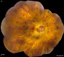
559 viewsJul 06, 2012
|
|

627 viewsJul 06, 2012
|
|
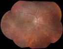
Choroidal Dystrophy571 viewsJul 03, 2012
|
|

Choroidal Dystrophy629 viewsJul 03, 2012
|
|

Alphamethlyacyl COA Racemase Deficiency1201 viewsMay 08, 2012
|
|

Alphamethlyacyl COA Racemase Deficiency594 viewsMay 08, 2012
|
|

Alphamethlyacyl COA Racemase Deficiency579 viewsMay 08, 2012
|
|

Alphamethlyacyl COA Racemase Deficiency593 viewsMay 08, 2012
|
|

Alphamethlyacyl COA Racemase Deficiency678 viewsMay 08, 2012
|
|

BESTS Disease721 viewsMay 07, 2012
|
|

BESTS Disease602 viewsMay 07, 2012
|
|

X-linked Juvenille Retinoschisis - Peripheral Retinal Vascular Anomalies - Vitreous Hemorrhage - 8 Year Old Boy630 views8-year-old child OD 20/40, OS 20/50
OD: Vertical C/D ratio is 0.2. There are foveal cysts. There is also a retinal elevation inferiorly and there are patchy peripheral retinal hemorrhages.
OS: Vertical C/D ratio is 0.2. There are foveal cysts. There is peripheral retinal issues with some hemorrhage in some areas of peripheral retinoschisis. May 03, 2012
|
|

Video - Takes a LONG TIME TO LOAD - X-linked Juvenille Retinoschisis - Peripheral Retinal Vascular Anomalies - Vitreous Hemorrhage - 8 Year Old Boy587 views8-year-old child OD 20/40, OS 20/50
OD: Vertical C/D ratio is 0.2. There are foveal cysts. There is also a retinal elevation inferiorly and there are patchy peripheral retinal hemorrhages.
OS: Vertical C/D ratio is 0.2. There are foveal cysts. There is peripheral retinal issues with some hemorrhage in some areas of peripheral retinoschisis. May 03, 2012
|
|

Video - TAKES A LONG TIME TO LOAD - X-linked Juvenille Retinoschisis - Peripheral Retinal Vascular Anomalies - Vitreous Hemorrhage - 8 Year Old Boy604 views8-year-old child OD 20/40, OS 20/50
OD: Vertical C/D ratio is 0.2. There are foveal cysts. There is also a retinal elevation inferiorly and there are patchy peripheral retinal hemorrhages.
OS: Vertical C/D ratio is 0.2. There are foveal cysts. There is peripheral retinal issues with some hemorrhage in some areas of peripheral retinoschisis. May 03, 2012
|
|

X-linked Juvenille Retinoschisis - Peripheral Retinal Vascular Anomalies - Vitreous Hemorrhage - 8 Year Old Boy563 views8-year-old child OD 20/40, OS 20/50
OD: Vertical C/D ratio is 0.2. There are foveal cysts. There is also a retinal elevation inferiorly and there are patchy peripheral retinal hemorrhages.
OS: Vertical C/D ratio is 0.2. There are foveal cysts. There is peripheral retinal issues with some hemorrhage in some areas of peripheral retinoschisis. May 03, 2012
|
|

X-linked Juvenille Retinoschisis - Peripheral Retinal Vascular Anomalies - Vitreous Hemorrhage - 8 Year Old Boy558 views8-year-old child OD 20/40, OS 20/50
OD: Vertical C/D ratio is 0.2. There are foveal cysts. There is also a retinal elevation inferiorly and there are patchy peripheral retinal hemorrhages.
OS: Vertical C/D ratio is 0.2. There are foveal cysts. There is peripheral retinal issues with some hemorrhage in some areas of peripheral retinoschisis. May 03, 2012
|
|

X-linked Juvenille Retinoschisis - Peripheral Retinal Vascular Anomalies - Vitreous Hemorrhage - 8 Year Old Boy532 views8-year-old child OD 20/40, OS 20/50
OD: Vertical C/D ratio is 0.2. There are foveal cysts. There is also a retinal elevation inferiorly and there are patchy peripheral retinal hemorrhages.
OS: Vertical C/D ratio is 0.2. There are foveal cysts. There is peripheral retinal issues with some hemorrhage in some areas of peripheral retinoschisis. May 03, 2012
|
|

X-linked Juvenille Retinoschisis - Peripheral Retinal Vascular Anomalies - Vitreous Hemorrhage - 8 Year Old Boy739 views8-year-old child OD 20/40, OS 20/50
OD: Vertical C/D ratio is 0.2. There are foveal cysts. There is also a retinal elevation inferiorly and there are patchy peripheral retinal hemorrhages.
OS: Vertical C/D ratio is 0.2. There are foveal cysts. There is peripheral retinal issues with some hemorrhage in some areas of peripheral retinoschisis. May 03, 2012
|
|

X-linked Juvenille Retinoschisis - Peripheral Retinal Vascular Anomalies - Vitreous Hemorrhage - 8 Year Old Boy463 views8-year-old child OD 20/40, OS 20/50
OD: Vertical C/D ratio is 0.2. There are foveal cysts. There is also a retinal elevation inferiorly and there are patchy peripheral retinal hemorrhages.
OS: Vertical C/D ratio is 0.2. There are foveal cysts. There is peripheral retinal issues with some hemorrhage in some areas of peripheral retinoschisis. May 03, 2012
|
|

X-linked Juvenille Retinoschisis - Peripheral Retinal Vascular Anomalies - Vitreous Hemorrhage - 8 Year Old Boy489 views8-year-old child OD 20/40, OS 20/50
OD: Vertical C/D ratio is 0.2. There are foveal cysts. There is also a retinal elevation inferiorly and there are patchy peripheral retinal hemorrhages.
OS: Vertical C/D ratio is 0.2. There are foveal cysts. There is peripheral retinal issues with some hemorrhage in some areas of peripheral retinoschisis. May 03, 2012
|
|

X-linked Juvenille Retinoschisis - Peripheral Retinal Vascular Anomalies - Vitreous Hemorrhage - 8 Year Old Boy448 views8-year-old child OD 20/40, OS 20/50
OD: Vertical C/D ratio is 0.2. There are foveal cysts. There is also a retinal elevation inferiorly and there are patchy peripheral retinal hemorrhages.
OS: Vertical C/D ratio is 0.2. There are foveal cysts. There is peripheral retinal issues with some hemorrhage in some areas of peripheral retinoschisis. May 03, 2012
|
|

X-linked Juvenille Retinoschisis - Peripheral Retinal Vascular Anomalies - Vitreous Hemorrhage - 8 Year Old Boy448 views8-year-old child OD 20/40, OS 20/50
OD: Vertical C/D ratio is 0.2. There are foveal cysts. There is also a retinal elevation inferiorly and there are patchy peripheral retinal hemorrhages.
OS: Vertical C/D ratio is 0.2. There are foveal cysts. There is peripheral retinal issues with some hemorrhage in some areas of peripheral retinoschisis. May 03, 2012
|
|

X-linked Juvenille Retinoschisis - Peripheral Retinal Vascular Anomalies - Vitreous Hemorrhage - 8 Year Old Boy504 views8-year-old child OD 20/40, OS 20/50
OD: Vertical C/D ratio is 0.2. There are foveal cysts. There is also a retinal elevation inferiorly and there are patchy peripheral retinal hemorrhages.
OS: Vertical C/D ratio is 0.2. There are foveal cysts. There is peripheral retinal issues with some hemorrhage in some areas of peripheral retinoschisis. May 03, 2012
|
|

X-linked Juvenille Retinoschisis - Peripheral Retinal Vascular Anomalies - Vitreous Hemorrhage - 8 Year Old Boy470 views8-year-old child OD 20/40, OS 20/50
OD: Vertical C/D ratio is 0.2. There are foveal cysts. There is also a retinal elevation inferiorly and there are patchy peripheral retinal hemorrhages.
OS: Vertical C/D ratio is 0.2. There are foveal cysts. There is peripheral retinal issues with some hemorrhage in some areas of peripheral retinoschisis. May 03, 2012
|
|

X-linked Juvenille Retinoschisis - Peripheral Retinal Vascular Anomalies - Vitreous Hemorrhage - 8 Year Old Boy433 views8-year-old child OD 20/40, OS 20/50
OD: Vertical C/D ratio is 0.2. There are foveal cysts. There is also a retinal elevation inferiorly and there are patchy peripheral retinal hemorrhages.
OS: Vertical C/D ratio is 0.2. There are foveal cysts. There is peripheral retinal issues with some hemorrhage in some areas of peripheral retinoschisis. May 03, 2012
|
|

X-linked Juvenille Retinoschisis - Peripheral Retinal Vascular Anomalies - Vitreous Hemorrhage - 8 Year Old Boy514 views8-year-old child OD 20/40, OS 20/50
OD: Vertical C/D ratio is 0.2. There are foveal cysts. There is also a retinal elevation inferiorly and there are patchy peripheral retinal hemorrhages.
OS: Vertical C/D ratio is 0.2. There are foveal cysts. There is peripheral retinal issues with some hemorrhage in some areas of peripheral retinoschisis. May 03, 2012
|
|

X-linked Juvenille Retinoschisis - Peripheral Retinal Vascular Anomalies - Vitreous Hemorrhage - 8 Year Old Boy547 views8-year-old child OD 20/40, OS 20/50
OD: Vertical C/D ratio is 0.2. There are foveal cysts. There is also a retinal elevation inferiorly and there are patchy peripheral retinal hemorrhages.
OS: Vertical C/D ratio is 0.2. There are foveal cysts. There is peripheral retinal issues with some hemorrhage in some areas of peripheral retinoschisis. May 03, 2012
|
|

X-linked Juvenille Retinoschisis - Peripheral Retinal Vascular Anomalies - Vitreous Hemorrhage - 8 Year Old Boy563 views8-year-old child OD 20/40, OS 20/50
OD: Vertical C/D ratio is 0.2. There are foveal cysts. There is also a retinal elevation inferiorly and there are patchy peripheral retinal hemorrhages.
OS: Vertical C/D ratio is 0.2. There are foveal cysts. There is peripheral retinal issues with some hemorrhage in some areas of peripheral retinoschisis. May 03, 2012
|
|

X-linked Juvenille Retinoschisis - Peripheral Retinal Vascular Anomalies - Vitreous Hemorrhage - 8 Year Old Boy622 views8-year-old child OD 20/40, OS 20/50
OD: Vertical C/D ratio is 0.2. There are foveal cysts. There is also a retinal elevation inferiorly and there are patchy peripheral retinal hemorrhages.
OS: Vertical C/D ratio is 0.2. There are foveal cysts. There is peripheral retinal issues with some hemorrhage in some areas of peripheral retinoschisis. May 03, 2012
|
|

X-linked Juvenille Retinoschisis - Peripheral Retinal Vascular Anomalies - Vitreous Hemorrhage - 8 Year Old Boy843 views8-year-old child OD 20/40, OS 20/50
OD: Vertical C/D ratio is 0.2. There are foveal cysts. There is also a retinal elevation inferiorly and there are patchy peripheral retinal hemorrhages.
OS: Vertical C/D ratio is 0.2. There are foveal cysts. There is peripheral retinal issues with some hemorrhage in some areas of peripheral retinoschisis. May 03, 2012
|
|

X-linked Juvenille Retinoschisis - Peripheral Retinal Vascular Anomalies - Vitreous Hemorrhage - 8 Year Old Boy629 views8-year-old child OD 20/40, OS 20/50
OD: Vertical C/D ratio is 0.2. There are foveal cysts. There is also a retinal elevation inferiorly and there are patchy peripheral retinal hemorrhages.
OS: Vertical C/D ratio is 0.2. There are foveal cysts. There is peripheral retinal issues with some hemorrhage in some areas of peripheral retinoschisis. May 03, 2012
|
|

X-linked Juvenille Retinoschisis - Peripheral Retinal Vascular Anomalies - Vitreous Hemorrhage - 8 Year Old Boy542 views8-year-old child OD 20/40, OS 20/50
OD: Vertical C/D ratio is 0.2. There are foveal cysts. There is also a retinal elevation inferiorly and there are patchy peripheral retinal hemorrhages.
OS: Vertical C/D ratio is 0.2. There are foveal cysts. There is peripheral retinal issues with some hemorrhage in some areas of peripheral retinoschisis. May 03, 2012
|
|

X-linked Juvenille Retinoschisis - Peripheral Retinal Vascular Anomalies - Vitreous Hemorrhage - 8 Year Old Boy535 views8-year-old child OD 20/40, OS 20/50
OD: Vertical C/D ratio is 0.2. There are foveal cysts. There is also a retinal elevation inferiorly and there are patchy peripheral retinal hemorrhages.
OS: Vertical C/D ratio is 0.2. There are foveal cysts. There is peripheral retinal issues with some hemorrhage in some areas of peripheral retinoschisis. May 03, 2012
|
|

X-linked Juvenille Retinoschisis - Peripheral Retinal Vascular Anomalies - Vitreous Hemorrhage - 8 Year Old Boy641 views8-year-old child OD 20/40, OS 20/50
OD: Vertical C/D ratio is 0.2. There are foveal cysts. There is also a retinal elevation inferiorly and there are patchy peripheral retinal hemorrhages.
OS: Vertical C/D ratio is 0.2. There are foveal cysts. There is peripheral retinal issues with some hemorrhage in some areas of peripheral retinoschisis. May 03, 2012
|
|

X-linked Juvenille Retinoschisis - Peripheral Retinal Vascular Anomalies - Vitreous Hemorrhage - 8 Year Old Boy513 views8-year-old child OD 20/40, OS 20/50
OD: Vertical C/D ratio is 0.2. There are foveal cysts. There is also a retinal elevation inferiorly and there are patchy peripheral retinal hemorrhages.
OS: Vertical C/D ratio is 0.2. There are foveal cysts. There is peripheral retinal issues with some hemorrhage in some areas of peripheral retinoschisis. May 03, 2012
|
|

X-linked Juvenille Retinoschisis - Peripheral Retinal Vascular Anomalies - Vitreous Hemorrhage - 8 Year Old Boy504 views8-year-old child OD 20/40, OS 20/50
OD: Vertical C/D ratio is 0.2. There are foveal cysts. There is also a retinal elevation inferiorly and there are patchy peripheral retinal hemorrhages.
OS: Vertical C/D ratio is 0.2. There are foveal cysts. There is peripheral retinal issues with some hemorrhage in some areas of peripheral retinoschisis. May 03, 2012
|
|

X-linked Juvenille Retinoschisis - Peripheral Retinal Vascular Anomalies - Vitreous Hemorrhage - 8 Year Old Boy583 views8-year-old child OD 20/40, OS 20/50
OD: Vertical C/D ratio is 0.2. There are foveal cysts. There is also a retinal elevation inferiorly and there are patchy peripheral retinal hemorrhages.
OS: Vertical C/D ratio is 0.2. There are foveal cysts. There is peripheral retinal issues with some hemorrhage in some areas of peripheral retinoschisis. May 03, 2012
|
|

X-linked Juvenille Retinoschisis - Peripheral Retinal Vascular Anomalies - Vitreous Hemorrhage - 8 Year Old Boy596 views8-year-old child OD 20/40, OS 20/50
OD: Vertical C/D ratio is 0.2. There are foveal cysts. There is also a retinal elevation inferiorly and there are patchy peripheral retinal hemorrhages.
OS: Vertical C/D ratio is 0.2. There are foveal cysts. There is peripheral retinal issues with some hemorrhage in some areas of peripheral retinoschisis. May 03, 2012
|
|

X-linked Juvenille Retinoschisis - Peripheral Retinal Vascular Anomalies - Vitreous Hemorrhage - 8 Year Old Boy654 views8-year-old child OD 20/40, OS 20/50
OD: Vertical C/D ratio is 0.2. There are foveal cysts. There is also a retinal elevation inferiorly and there are patchy peripheral retinal hemorrhages.
OS: Vertical C/D ratio is 0.2. There are foveal cysts. There is peripheral retinal issues with some hemorrhage in some areas of peripheral retinoschisis. May 03, 2012
|
|

X-linked Juvenille Retinoschisis - Peripheral Retinal Vascular Anomalies - Vitreous Hemorrhage - 8 Year Old Boy647 views8-year-old child OD 20/40, OS 20/50
OD: Vertical C/D ratio is 0.2. There are foveal cysts. There is also a retinal elevation inferiorly and there are patchy peripheral retinal hemorrhages.
OS: Vertical C/D ratio is 0.2. There are foveal cysts. There is peripheral retinal issues with some hemorrhage in some areas of peripheral retinoschisis. May 03, 2012
|
|

X-linked Juvenille Retinoschisis - Peripheral Retinal Vascular Anomalies - Vitreous Hemorrhage - 8 Year Old Boy682 views8-year-old child OD 20/40, OS 20/50
OD: Vertical C/D ratio is 0.2. There are foveal cysts. There is also a retinal elevation inferiorly and there are patchy peripheral retinal hemorrhages.
OS: Vertical C/D ratio is 0.2. There are foveal cysts. There is peripheral retinal issues with some hemorrhage in some areas of peripheral retinoschisis. May 03, 2012
|
|

X-linked Juvenille Retinoschisis - Peripheral Retinal Vascular Anomalies - Vitreous Hemorrhage - 8 Year Old Boy670 views8-year-old child OD 20/40, OS 20/50
OD: Vertical C/D ratio is 0.2. There are foveal cysts. There is also a retinal elevation inferiorly and there are patchy peripheral retinal hemorrhages.
OS: Vertical C/D ratio is 0.2. There are foveal cysts. There is peripheral retinal issues with some hemorrhage in some areas of peripheral retinoschisis. May 03, 2012
|
|

X-linked Juvenille Retinoschisis - Peripheral Retinal Vascular Anomalies - Vitreous Hemorrhage - 8 Year Old Boy553 views8-year-old child OD 20/40, OS 20/50
OD: Vertical C/D ratio is 0.2. There are foveal cysts. There is also a retinal elevation inferiorly and there are patchy peripheral retinal hemorrhages.
OS: Vertical C/D ratio is 0.2. There are foveal cysts. There is peripheral retinal issues with some hemorrhage in some areas of peripheral retinoschisis. May 03, 2012
|
|

X-linked Juvenille Retinoschisis - Peripheral Retinal Vascular Anomalies - Vitreous Hemorrhage - 8 Year Old Boy712 views8-year-old child OD 20/40, OS 20/50
OD: Vertical C/D ratio is 0.2. There are foveal cysts. There is also a retinal elevation inferiorly and there are patchy peripheral retinal hemorrhages.
OS: Vertical C/D ratio is 0.2. There are foveal cysts. There is peripheral retinal issues with some hemorrhage in some areas of peripheral retinoschisis. May 03, 2012
|
|

X-linked Juvenille Retinoschisis - Peripheral Retinal Vascular Anomalies - Vitreous Hemorrhage - 8 Year Old Boy608 views8-year-old child OD 20/40, OS 20/50
OD: Vertical C/D ratio is 0.2. There are foveal cysts. There is also a retinal elevation inferiorly and there are patchy peripheral retinal hemorrhages.
OS: Vertical C/D ratio is 0.2. There are foveal cysts. There is peripheral retinal issues with some hemorrhage in some areas of peripheral retinoschisis. May 03, 2012
|
|

X-linked Juvenille Retinoschisis - Peripheral Retinal Vascular Anomalies - Vitreous Hemorrhage - 8 Year Old Boy651 views8-year-old child OD 20/40, OS 20/50
OD: Vertical C/D ratio is 0.2. There are foveal cysts. There is also a retinal elevation inferiorly and there are patchy peripheral retinal hemorrhages.
OS: Vertical C/D ratio is 0.2. There are foveal cysts. There is peripheral retinal issues with some hemorrhage in some areas of peripheral retinoschisis. May 03, 2012
|
|

X-linked Juvenille Retinoschisis - Peripheral Retinal Vascular Anomalies - Vitreous Hemorrhage - 8 Year Old Boy873 views8-year-old child OD 20/40, OS 20/50
OD: Vertical C/D ratio is 0.2. There are foveal cysts. There is also a retinal elevation inferiorly and there are patchy peripheral retinal hemorrhages.
OS: Vertical C/D ratio is 0.2. There are foveal cysts. There is peripheral retinal issues with some hemorrhage in some areas of peripheral retinoschisis. May 03, 2012
|
|

Chorodial Sclerosis503 views74 year old female diagnosed with Chorodial Sclerosis OU and ARMD OU. Va 20/400 OD 2'200 OSApr 23, 2012
|
|
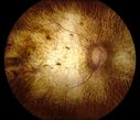
Chorodial Sclerosis1247 views74 year old female diagnosed with Chorodial Sclerosis OU and ARMD OU. Va 20/400 OD 2'200 OSApr 23, 2012
|
|
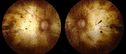
Chorodial Sclerosis563 views74 year old female diagnosed with Chorodial Sclerosis OU and ARMD OU. Va 20/400 OD 2'200 OSApr 23, 2012
|
|

Retinitis Pigmentosa Newly Diagnosed 55 Year Old734 views55-year-old noticed his vision declining recently, over the last few weeks, particularly in the right eye. He said previous to that the vision was not normal but was pretty good. OD is 20/100, OS is 20/50
OCT SCAN: There is macular thickening in both eyes with intraretinal cysts.Jan 20, 2012
|
|

Retinitis Pigmentosa Newly Diagnosed 55 Year Old646 views55-year-old noticed his vision declining recently, over the last few weeks, particularly in the right eye. He said previous to that the vision was not normal but was pretty good. OD is 20/100, OS is 20/50
OCT SCAN: There is macular thickening in both eyes with intraretinal cysts.Jan 20, 2012
|
|

Retinitis Pigmentosa Newly Diagnosed 55 Year Old623 views55-year-old noticed his vision declining recently, over the last few weeks, particularly in the right eye. He said previous to that the vision was not normal but was pretty good. OD is 20/100, OS is 20/50
OCT SCAN: There is macular thickening in both eyes with intraretinal cysts.Jan 20, 2012
|
|

Retinitis Pigmentosa Newly Diagnosed 55 Year Old541 views55-year-old noticed his vision declining recently, over the last few weeks, particularly in the right eye. He said previous to that the vision was not normal but was pretty good. OD is 20/100, OS is 20/50
OCT SCAN: There is macular thickening in both eyes with intraretinal cysts.Jan 20, 2012
|
|

Retinitis Pigmentosa Newly Diagnosed 55 Year Old583 views55-year-old noticed his vision declining recently, over the last few weeks, particularly in the right eye. He said previous to that the vision was not normal but was pretty good. OD is 20/100, OS is 20/50
OCT SCAN: There is macular thickening in both eyes with intraretinal cysts.Jan 20, 2012
|
|

Retinitis Pigmentosa Newly Diagnosed 55 Year Old609 views55-year-old noticed his vision declining recently, over the last few weeks, particularly in the right eye. He said previous to that the vision was not normal but was pretty good. OD is 20/100, OS is 20/50
OCT SCAN: There is macular thickening in both eyes with intraretinal cysts.Jan 20, 2012
|
|

Retinitis Pigmentosa Newly Diagnosed 55 Year Old642 views55-year-old noticed his vision declining recently, over the last few weeks, particularly in the right eye. He said previous to that the vision was not normal but was pretty good. OD is 20/100, OS is 20/50
OCT SCAN: There is macular thickening in both eyes with intraretinal cysts.Jan 20, 2012
|
|

Retinitis Pigmentosa Newly Diagnosed 55 Year Old570 views55-year-old noticed his vision declining recently, over the last few weeks, particularly in the right eye. He said previous to that the vision was not normal but was pretty good. OD is 20/100, OS is 20/50
OCT SCAN: There is macular thickening in both eyes with intraretinal cysts.Jan 20, 2012
|
|

Retinitis Pigmentosa Newly Diagnosed 55 Year Old603 views55-year-old noticed his vision declining recently, over the last few weeks, particularly in the right eye. He said previous to that the vision was not normal but was pretty good. OD is 20/100, OS is 20/50
OCT SCAN: There is macular thickening in both eyes with intraretinal cysts.Jan 20, 2012
|
|

Retinitis Pigmentosa Newly Diagnosed 55 Year Old579 views55-year-old noticed his vision declining recently, over the last few weeks, particularly in the right eye. He said previous to that the vision was not normal but was pretty good. OD is 20/100, OS is 20/50
OCT SCAN: There is macular thickening in both eyes with intraretinal cysts.Jan 20, 2012
|
|

Retinitis Pigmentosa Newly Diagnosed 55 Year Old525 views55-year-old noticed his vision declining recently, over the last few weeks, particularly in the right eye. He said previous to that the vision was not normal but was pretty good. OD is 20/100, OS is 20/50
OCT SCAN: There is macular thickening in both eyes with intraretinal cysts.Jan 20, 2012
|
|

Retinitis Pigmentosa Newly Diagnosed 55 Year Old548 views55-year-old noticed his vision declining recently, over the last few weeks, particularly in the right eye. He said previous to that the vision was not normal but was pretty good. OD is 20/100, OS is 20/50
OCT SCAN: There is macular thickening in both eyes with intraretinal cysts.Jan 20, 2012
|
|

Retinitis Pigmentosa Newly Diagnosed 55 Year Old500 views55-year-old noticed his vision declining recently, over the last few weeks, particularly in the right eye. He said previous to that the vision was not normal but was pretty good. OD is 20/100, OS is 20/50
OCT SCAN: There is macular thickening in both eyes with intraretinal cysts.Jan 20, 2012
|
|

Retinitis Pigmentosa Newly Diagnosed 55 Year Old529 views55-year-old noticed his vision declining recently, over the last few weeks, particularly in the right eye. He said previous to that the vision was not normal but was pretty good. OD is 20/100, OS is 20/50
OCT SCAN: There is macular thickening in both eyes with intraretinal cysts.Jan 20, 2012
|
|

Retinitis Pigmentosa Newly Diagnosed 55 Year Old556 views55-year-old noticed his vision declining recently, over the last few weeks, particularly in the right eye. He said previous to that the vision was not normal but was pretty good. OD is 20/100, OS is 20/50
OCT SCAN: There is macular thickening in both eyes with intraretinal cysts.Jan 20, 2012
|
|

Retinitis Pigmentosa Newly Diagnosed 55 Year Old656 views55-year-old noticed his vision declining recently, over the last few weeks, particularly in the right eye. He said previous to that the vision was not normal but was pretty good. OD is 20/100, OS is 20/50
OCT SCAN: There is macular thickening in both eyes with intraretinal cysts.Jan 20, 2012
|
|

Retinitis Pigmentosa Newly Diagnosed 55 Year Old659 views55-year-old noticed his vision declining recently, over the last few weeks, particularly in the right eye. He said previous to that the vision was not normal but was pretty good. OD is 20/100, OS is 20/50
OCT SCAN: There is macular thickening in both eyes with intraretinal cysts.Jan 20, 2012
|
|

Retinitis Pigmentosa Newly Diagnosed 55 Year Old636 views55-year-old noticed his vision declining recently, over the last few weeks, particularly in the right eye. He said previous to that the vision was not normal but was pretty good. OD is 20/100, OS is 20/50
OCT SCAN: There is macular thickening in both eyes with intraretinal cysts.Jan 20, 2012
|
|

Retinitis Pigmentosa Newly Diagnosed 55 Year Old669 views55-year-old noticed his vision declining recently, over the last few weeks, particularly in the right eye. He said previous to that the vision was not normal but was pretty good. OD is 20/100, OS is 20/50
OCT SCAN: There is macular thickening in both eyes with intraretinal cysts.Jan 20, 2012
|
|

Retinitis Pigmentosa Newly Diagnosed 55 Year Old535 views55-year-old noticed his vision declining recently, over the last few weeks, particularly in the right eye. He said previous to that the vision was not normal but was pretty good. OD is 20/100, OS is 20/50
OCT SCAN: There is macular thickening in both eyes with intraretinal cysts.Jan 20, 2012
|
|

Retinitis Pigmentosa Newly Diagnosed 55 Year Old665 views55-year-old noticed his vision declining recently, over the last few weeks, particularly in the right eye. He said previous to that the vision was not normal but was pretty good. OD is 20/100, OS is 20/50
OCT SCAN: There is macular thickening in both eyes with intraretinal cysts.Jan 20, 2012
|
|

Retinitis Pigmentosa Newly Diagnosed 55 Year Old602 views55-year-old noticed his vision declining recently, over the last few weeks, particularly in the right eye. He said previous to that the vision was not normal but was pretty good. OD is 20/100, OS is 20/50
OCT SCAN: There is macular thickening in both eyes with intraretinal cysts.Jan 20, 2012
|
|

Retinitis Pigmentosa Newly Diagnosed 55 Year Old597 views55-year-old noticed his vision declining recently, over the last few weeks, particularly in the right eye. He said previous to that the vision was not normal but was pretty good. OD is 20/100, OS is 20/50
OCT SCAN: There is macular thickening in both eyes with intraretinal cysts.Jan 20, 2012
|
|

Retinitis Pigmentosa Newly Diagnosed 55 Year Old663 views55-year-old noticed his vision declining recently, over the last few weeks, particularly in the right eye. He said previous to that the vision was not normal but was pretty good. OD is 20/100, OS is 20/50
OCT SCAN: There is macular thickening in both eyes with intraretinal cysts.Jan 20, 2012
|
|

Retinitis Pigmentosa Newly Diagnosed 55 Year Old608 views55-year-old noticed his vision declining recently, over the last few weeks, particularly in the right eye. He said previous to that the vision was not normal but was pretty good. OD is 20/100, OS is 20/50
OCT SCAN: There is macular thickening in both eyes with intraretinal cysts.Jan 20, 2012
|
|

Retinitis Pigmentosa Newly Diagnosed 55 Year Old573 views55-year-old noticed his vision declining recently, over the last few weeks, particularly in the right eye. He said previous to that the vision was not normal but was pretty good. OD is 20/100, OS is 20/50
OCT SCAN: There is macular thickening in both eyes with intraretinal cysts.Jan 20, 2012
|
|

Retinitis Pigmentosa Newly Diagnosed 55 Year Old620 views55-year-old noticed his vision declining recently, over the last few weeks, particularly in the right eye. He said previous to that the vision was not normal but was pretty good. OD is 20/100, OS is 20/50
OCT SCAN: There is macular thickening in both eyes with intraretinal cysts.Jan 20, 2012
|
|

Retinitis Pigmentosa Newly Diagnosed 55 Year Old623 views55-year-old noticed his vision declining recently, over the last few weeks, particularly in the right eye. He said previous to that the vision was not normal but was pretty good. OD is 20/100, OS is 20/50
OCT SCAN: There is macular thickening in both eyes with intraretinal cysts.Jan 20, 2012
|
|
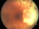
Retinitis Pigmentosa Newly Diagnosed 55 Year Old818 views55-year-old noticed his vision declining recently, over the last few weeks, particularly in the right eye. He said previous to that the vision was not normal but was pretty good. OD is 20/100, OS is 20/50
OCT SCAN: There is macular thickening in both eyes with intraretinal cysts.Jan 20, 2012
|
|

Congenital Achromatopsia 763 views26-year-old man was diagnosed with congenital achromatopsia by ERG as a child. He has family members who also have this disorder. OD 20/200, OS 20/200.Jan 17, 2012
|
|

Congenital Achromatopsia 632 views26-year-old man was diagnosed with congenital achromatopsia by ERG as a child. He has family members who also have this disorder. OD 20/200, OS 20/200.Jan 17, 2012
|
|

Congenital Achromatopsia 681 views26-year-old man was diagnosed with congenital achromatopsia by ERG as a child. He has family members who also have this disorder. OD 20/200, OS 20/200.Jan 17, 2012
|
|

Congenital Achromatopsia 624 views26-year-old man was diagnosed with congenital achromatopsia by ERG as a child. He has family members who also have this disorder. OD 20/200, OS 20/200.Jan 17, 2012
|
|

Congenital Achromatopsia 585 views26-year-old man was diagnosed with congenital achromatopsia by ERG as a child. He has family members who also have this disorder. OD 20/200, OS 20/200.Jan 17, 2012
|
|
| 1242 files on 13 page(s) |
 |
|
|
|
|
|
|
10 | |
|
|


