Last additions - Dystrophies
|

Stargardt's Macular Dystrophy ABCA4 positive534 views Fundus Autofluorescence shows central atrophy with Hyper FAF pisciform triradiate lesions. Images show some progression over 3 years of macular dystrophy.Feb 16, 2016
|
|

Stargardt's Macular Dystrophy ABCA4 positive595 views Fundus Autofluorescence shows central atrophy with Hyper FAF pisciform triradiate lesions. Images show some progression over 3 years of macular dystrophy.Feb 16, 2016
|
|

Stargardt's Macular Dystrophy ABCA4 positive501 viewsImages show some progression over 3 years of macular dystrophy.Feb 16, 2016
|
|

Stargardt's Macular Dystrophy ABCA4 positive469 viewsImages show some progression over 3 years of macular dystrophy.Feb 16, 2016
|
|

Stargardt's Macular Dystrophy ABCA4 positive494 views Fundus Autofluorescence shows central atrophy with Hyper FAF pisciform triradiate lesions. Images show some progression over 3 years of macular dystrophy.Feb 16, 2016
|
|

Stargardt's Macular Dystrophy ABCA4 positive582 views Fundus Autofluorescence shows central atrophy with Hyper FAF pisciform triradiate lesions. Images show some progression over 3 years of macular dystrophy.Feb 16, 2016
|
|

Stargardt's Macular Dystrophy ABCA4 positive611 views Fundus Autofluorescence shows central atrophy with Hyper FAF pisciform triradiate lesions. Images show some progression over 3 years of macular dystrophy.Feb 16, 2016
|
|

Stargardt's Macular Dystrophy ABCA4 positive567 viewsImages show some progression over 3 years of macular dystrophy.Feb 16, 2016
|
|

Stargardt's Macular Dystrophy ABCA4 positive677 viewsImages show some progression over 3 years of macular dystrophy.Feb 16, 2016
|
|

Stargardt's Macular Dystrophy ABCA4 positive568 viewsImages show some progression over 3 years of macular dystrophy.Feb 16, 2016
|
|

Stargardt's Macular Dystrophy ABCA4 positive619 viewsImages show some progression over 3 years of macular dystrophy.Feb 16, 2016
|
|

Stargardt's Macular Dystrophy ABCA4 positive653 viewsImages show some progression over 3 years of macular dystrophy.Feb 16, 2016
|
|

Stargardt's Macular Dystrophy ABCA4 positive549 viewsImages show some progression over 3 years of macular dystrophy.Feb 16, 2016
|
|

Stargardt's Macular Dystrophy ABCA4 positive607 viewsImages show some progression over 3 years of macular dystrophy.Feb 16, 2016
|
|

Choroideremia - Female Carrier - Patchy Atrophy of Choroid and RPE - Lyonization744 viewsMar 06, 2015
|
|

Choroideremia - Female Carrier - Patchy Atrophy of Choroid and RPE - Lyonization697 viewsMar 06, 2015
|
|

Choroideremia - Female Carrier - Patchy Atrophy of Choroid and RPE - Lyonization609 viewsMar 06, 2015
|
|

Choroideremia - Female Carrier - Patchy Atrophy of Choroid and RPE - Lyonization617 viewsMar 06, 2015
|
|

Choroideremia - Female Carrier - Patchy Atrophy of Choroid and RPE - Lyonization667 viewsMar 06, 2015
|
|

Choroideremia - Female Carrier - Patchy Atrophy of Choroid and RPE - Lyonization629 viewsMar 06, 2015
|
|

Choroideremia - Female Carrier - Patchy Atrophy of Choroid and RPE - Lyonization571 viewsMar 06, 2015
|
|

Choroideremia - Female Carrier - Patchy Atrophy of Choroid and RPE - Lyonization647 viewsMar 06, 2015
|
|

Choroideremia - Female Carrier - Patchy Atrophy of Choroid and RPE - Lyonization551 viewsMar 06, 2015
|
|

Choroideremia - Female Carrier - Patchy Atrophy of Choroid and RPE - Lyonization618 viewsMar 06, 2015
|
|

Choroideremia - Female Carrier - Patchy Atrophy of Choroid and RPE - Lyonization587 viewsMar 06, 2015
|
|

Choroideremia - Female Carrier - Patchy Atrophy of Choroid and RPE - Lyonization651 viewsMar 06, 2015
|
|

Choroideremia - Female Carrier - Patchy Atrophy of Choroid and RPE - Lyonization653 viewsMar 06, 2015
|
|

Choroideremia - Female Carrier - Patchy Atrophy of Choroid and RPE - Lyonization626 viewsMar 06, 2015
|
|

Choroideremia - Female Carrier - Patchy Atrophy of Choroid and RPE - Lyonization651 viewsMar 06, 2015
|
|

Choroideremia - Female Carrier - Patchy Atrophy of Choroid and RPE - Lyonization606 viewsMar 06, 2015
|
|

Choroideremia - Female Carrier - Patchy Atrophy of Choroid and RPE - Lyonization581 viewsMar 06, 2015
|
|

Choroideremia - Female Carrier - Patchy Atrophy of Choroid and RPE - Lyonization564 viewsMar 06, 2015
|
|

Choroideremia - Female Carrier - Patchy Atrophy of Choroid and RPE - Lyonization546 viewsMar 06, 2015
|
|

Choroideremia - Female Carrier - Patchy Atrophy of Choroid and RPE - Lyonization564 viewsMar 06, 2015
|
|

Choroideremia - Female Carrier - Patchy Atrophy of Choroid and RPE - Lyonization591 viewsMar 06, 2015
|
|

Choroideremia - Female Carrier - Patchy Atrophy of Choroid and RPE - Lyonization584 viewsMar 06, 2015
|
|

Choroideremia - Female Carrier - Patchy Atrophy of Choroid and RPE - Lyonization566 viewsMar 06, 2015
|
|

Choroideremia - Female Carrier - Patchy Atrophy of Choroid and RPE - Lyonization679 viewsMar 06, 2015
|
|

Choroideremia - Female Carrier - Patchy Atrophy of Choroid and RPE - Lyonization638 viewsMar 06, 2015
|
|

Choroideremia - Female Carrier - Patchy Atrophy of Choroid and RPE - Lyonization819 viewsMar 06, 2015
|
|

Choroideremia - Female Carrier - Patchy Atrophy of Choroid and RPE - Lyonization667 viewsMar 06, 2015
|
|

Choroideremia - Female Carrier - Patchy Atrophy of Choroid and RPE - Lyonization677 viewsMar 06, 2015
|
|

Choroideremia - Female Carrier - Patchy Atrophy of Choroid and RPE - Lyonization637 viewsMar 06, 2015
|
|

Choroideremia - Female Carrier - Patchy Atrophy of Choroid and RPE - Lyonization542 viewsMar 06, 2015
|
|

Choroideremia - Female Carrier - Patchy Atrophy of Choroid and RPE - Lyonization562 viewsMar 06, 2015
|
|

Choroideremia - Female Carrier - Patchy Atrophy of Choroid and RPE - Lyonization651 viewsMar 06, 2015
|
|

Choroideremia - Female Carrier - Patchy Atrophy of Choroid and RPE - Lyonization628 viewsMar 06, 2015
|
|

Choroideremia - Female Carrier - Patchy Atrophy of Choroid and RPE - Lyonization622 viewsMar 06, 2015
|
|

Choroideremia - Female Carrier - Patchy Atrophy of Choroid and RPE - Lyonization618 viewsMar 06, 2015
|
|

Choroideremia - Female Carrier - Patchy Atrophy of Choroid and RPE - Lyonization715 viewsMar 06, 2015
|
|

Choroideremia - Female Carrier - Patchy Atrophy of Choroid and RPE - Lyonization761 viewsMar 06, 2015
|
|

Retinitis Pigmentosa - Autosomal Recessive - 82 year old man with Good Vision572 viewsVA 20/25, 20/32 - Bone spicules in periphery - FAF is very symmetricMar 06, 2015
|
|
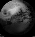
Retinitis Pigmentosa - Autosomal Recessive - 82 year old man with Good Vision497 viewsVA 20/25, 20/32 - Bone spicules in periphery - FAF is very symmetricMar 06, 2015
|
|

Retinitis Pigmentosa - Autosomal Recessive - 82 year old man with Good Vision511 viewsVA 20/25, 20/32 - Bone spicules in periphery - FAF is very symmetricMar 06, 2015
|
|

Retinitis Pigmentosa - Autosomal Recessive - 82 year old man with Good Vision518 viewsVA 20/25, 20/32 - Bone spicules in periphery - FAF is very symmetricMar 06, 2015
|
|

Retinitis Pigmentosa - Autosomal Recessive - 82 year old man with Good Vision483 viewsVA 20/25, 20/32 - Bone spicules in periphery - FAF is very symmetricMar 06, 2015
|
|

Retinitis Pigmentosa - Autosomal Recessive - 82 year old man with Good Vision511 viewsVA 20/25, 20/32 - Bone spicules in periphery - FAF is very symmetricMar 06, 2015
|
|

Retinitis Pigmentosa - Autosomal Recessive - 82 year old man with Good Vision483 viewsVA 20/25, 20/32 - Bone spicules in periphery - FAF is very symmetricMar 06, 2015
|
|
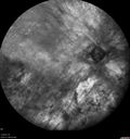
Retinitis Pigmentosa - Autosomal Recessive - 82 year old man with Good Vision545 viewsVA 20/25, 20/32 - Bone spicules in periphery - FAF is very symmetricMar 06, 2015
|
|

Retinitis Pigmentosa - Autosomal Recessive - 82 year old man with Good Vision500 viewsVA 20/25, 20/32 - Bone spicules in periphery - FAF is very symmetricMar 06, 2015
|
|

Retinitis Pigmentosa - Autosomal Recessive - 82 year old man with Good Vision488 viewsVA 20/25, 20/32 - Bone spicules in periphery - FAF is very symmetricMar 06, 2015
|
|

Retinitis Pigmentosa - Autosomal Recessive - 82 year old man with Good Vision499 viewsVA 20/25, 20/32 - Bone spicules in periphery - FAF is very symmetricMar 06, 2015
|
|

Retinitis Pigmentosa - Autosomal Recessive - 82 year old man with Good Vision554 viewsVA 20/25, 20/32 - Bone spicules in periphery - FAF is very symmetricMar 06, 2015
|
|

Retinitis Pigmentosa - Autosomal Recessive - 82 year old man with Good Vision579 viewsVA 20/25, 20/32 - Bone spicules in periphery - FAF is very symmetricMar 06, 2015
|
|

Retinitis Pigmentosa - Autosomal Recessive - 82 year old man with Good Vision559 viewsVA 20/25, 20/32 - Bone spicules in periphery - FAF is very symmetricMar 06, 2015
|
|
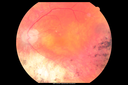
Retinitis Pigmentosa - Autosomal Recessive - 82 year old man with Good Vision562 viewsVA 20/25, 20/32 - Bone spicules in periphery - FAF is very symmetricMar 06, 2015
|
|

Retinitis Pigmentosa - Autosomal Recessive - 82 year old man with Good Vision506 viewsVA 20/25, 20/32 - Bone spicules in periphery - FAF is very symmetricMar 06, 2015
|
|

Retinitis Pigmentosa - Autosomal Recessive - 82 year old man with Good Vision562 viewsVA 20/25, 20/32 - Bone spicules in periphery - FAF is very symmetricMar 06, 2015
|
|

Retinitis Pigmentosa Sine Pigmento 665 viewsSep 06, 2014
|
|

Retinitis Pigmentosa Sine Pigmento 600 viewsSep 06, 2014
|
|

Retinitis Pigmentosa Sine Pigmento 638 viewsSep 06, 2014
|
|

Retinitis Pigmentosa Sine Pigmento 792 viewsSep 06, 2014
|
|

Retinitis Pigmentosa Sine Pigmento 657 viewsSep 06, 2014
|
|

Retinitis Pigmentosa Sine Pigmento 599 viewsSep 06, 2014
|
|

Retinitis Pigmentosa Sine Pigmento 579 viewsSep 06, 2014
|
|

Retinitis Pigmentosa Sine Pigmento 662 viewsSep 06, 2014
|
|

Retinitis Pigmentosa Sine Pigmento 777 viewsSep 06, 2014
|
|

Retinitis Pigmentosa Sine Pigmento 1001 viewsSep 06, 2014
|
|

Retinitis Pigmentosa Sine Pigmento 1456 viewsSep 06, 2014
|
|

Retinitis Pigmentosa Sine Pigmento 1303 viewsSep 06, 2014
|
|

Retinitis Pigmentosa Sine Pigmento 968 viewsSep 06, 2014
|
|

Retinitis Pigmentosa Sine Pigmento 943 viewsSep 06, 2014
|
|

Retinitis Pigmentosa591 viewsPatient comes in with cloudy vision. VA is 20/200 in both eyes. Patient has had retinal pigmentosa since 1986. Patient to consider cataract surgery to help her visual symptoms...Jul 23, 2014
|
|

Retinitis Pigmentosa1827 viewsPatient comes in with cloudy vision. VA is 20/200 in both eyes. Patient has had retinal pigmentosa since 1986. Patient to consider cataract surgery to help her visual symptoms...Jul 23, 2014
|
|

Fundus Flavimaculatus - Asymptomatic - Color Photo708 views45 year old man was told to come in because of ‘white spots on their retina’.
Mar 17, 2014
|
|

Fundus Flavimaculatus - Asymptomatic - Spectral Domain OCT Line Scan819 views45 year old man was told to come in because of ‘white spots on their retina’.
Mar 17, 2014
|
|

Fundus Flavimaculatus - Asymptomatic - Spectral Domain OCT Line Scan726 views45 year old man was told to come in because of ‘white spots on their retina’.
Mar 17, 2014
|
|

Fundus Flavimaculatus - Asymptomatic660 views45 year old man was told to come in because of ‘white spots on their retina’.
Mar 17, 2014
|
|

Fundus Flavimaculatus - Asymptomatic - Spectral Domain OCT Line Scan823 views45 year old man was told to come in because of ‘white spots on their retina’.
Mar 17, 2014
|
|

Fundus Flavimaculatus - Asymptomatic - Spectral Domain OCT Line Scan1070 views45 year old man was told to come in because of ‘white spots on their retina’.
Mar 17, 2014
|
|

Fundus Flavimaculatus - Asymptomatic - Spectral Domain OCT Line Scan752 views45 year old man was told to come in because of ‘white spots on their retina’.
Mar 17, 2014
|
|

Fundus Flavimaculatus - Asymptomatic - Spectral Domain OCT Line Scan759 views45 year old man was told to come in because of ‘white spots on their retina’.
Mar 17, 2014
|
|

Fundus Flavimaculatus - Asymptomatic - Spectral Domain OCT Line Scan770 views45 year old man was told to come in because of ‘white spots on their retina’.
Mar 17, 2014
|
|

Fundus Flavimaculatus - Asymptomatic - Spectral Domain OCT Line Scan799 views45 year old man was told to come in because of ‘white spots on their retina’.
Mar 17, 2014
|
|

Fundus Flavimaculatus - Asymptomatic668 views45 year old man was told to come in because of ‘white spots on their retina’.
Mar 17, 2014
|
|

Fundus Flavimaculatus - Asymptomatic - InfraRed Reflectance752 views45 year old man was told to come in because of ‘white spots on their retina’.
Mar 17, 2014
|
|

Fundus Flavimaculatus - Asymptomatic - InfraRed Reflectance753 views45 year old man was told to come in because of ‘white spots on their retina’.
Mar 17, 2014
|
|

Fundus Flavimaculatus - Asymptomatic - Fundus Autofluorescence745 views45 year old man was told to come in because of ‘white spots on their retina’. - FAF image shows that spots are strongly autofluorescent
Mar 17, 2014
|
|

Fundus Flavimaculatus - Asymptomatic - Fundus Autofluorescence778 views45 year old man was told to come in because of ‘white spots on their retina’. - FAF image shows that spots are strongly autofluorescent
Mar 17, 2014
|
|

Fundus Flavimaculatus - Asymptomatic - Fundus Autofluorescence1108 views45 year old man was told to come in because of ‘white spots on their retina’. - FAF image shows that spots are strongly autofluorescent
Mar 17, 2014
|
|
| 1242 files on 13 page(s) |
 |
|
|
|
|
7 | |
|
|
|
|


