Bilder-Suchergebnisse - "classic"
|
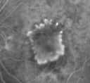
Subfoveal CNVM wet AMD early fa (contrast enhanced with photoshop)895 x angesehenShe has noticed decreased vision in the left eye. It is hard to say for how long, maybe a few months. She had her last eye exam about a year ago. She thoughts perhaps it was cataracts, but you saw a problem with the macula and suggested she come here for evaluation.
VISUAL ACUITY: Vision OD is 20/25, OS is 2/200
|
|

Classic Subfoveal Choroidal Neovascular Membrane - 20/400 Vision - 6 - 12 months old lesion885 x angesehen78-year-old woman six months or a year ago decreasing vision in the right eye. OD 20/400. Fluorescein angiography shows a classic subfoveal choroidal neovascular membrane in the right eye 3 disc-diameters across, which has brisk leakage. The left eye shows staining drusen.
|
|

Classic Subfoveal Choroidal Neovascular Membrane - 20/400 Vision - 6 - 12 months old lesion857 x angesehen78-year-old woman six months or a year ago decreasing vision in the right eye. OD 20/400. Fluorescein angiography shows a classic subfoveal choroidal neovascular membrane in the right eye 3 disc-diameters across, which has brisk leakage. The left eye shows staining drusen.
|
|

Classic Subfoveal Choroidal Neovascular Membrane - 20/400 Vision - 6 - 12 months old lesion864 x angesehen78-year-old woman six months or a year ago decreasing vision in the right eye. OD 20/400. Fluorescein angiography shows a classic subfoveal choroidal neovascular membrane in the right eye 3 disc-diameters across, which has brisk leakage. The left eye shows staining drusen.
|
|

Classic Subfoveal Choroidal Neovascular Membrane - 20/400 Vision - 6 - 12 months old lesion784 x angesehen78-year-old woman six months or a year ago decreasing vision in the right eye. OD 20/400. Fluorescein angiography shows a classic subfoveal choroidal neovascular membrane in the right eye 3 disc-diameters across, which has brisk leakage. The left eye shows staining drusen.
|
|

Classic Subfoveal Choroidal Neovascular Membrane - 20/400 Vision - 6 - 12 months old lesion731 x angesehen78-year-old woman six months or a year ago decreasing vision in the right eye. OD 20/400. Fluorescein angiography shows a classic subfoveal choroidal neovascular membrane in the right eye 3 disc-diameters across, which has brisk leakage. The left eye shows staining drusen.
|
|

Classic Subfoveal Choroidal Neovascular Membrane - 20/400 Vision - 6 - 12 months old lesion734 x angesehen78-year-old woman six months or a year ago decreasing vision in the right eye. OD 20/400. Fluorescein angiography shows a classic subfoveal choroidal neovascular membrane in the right eye 3 disc-diameters across, which has brisk leakage. The left eye shows staining drusen.
|
|

Classic Subfoveal Choroidal Neovascular Membrane - 20/400 Vision - 6 - 12 months old lesion765 x angesehen78-year-old woman six months or a year ago decreasing vision in the right eye. OD 20/400. Fluorescein angiography shows a classic subfoveal choroidal neovascular membrane in the right eye 3 disc-diameters across, which has brisk leakage. The left eye shows staining drusen.
|
|

Subfoveal CNVM wet AMD fellow eye841 x angesehenShe has noticed decreased vision in the left eye. It is hard to say for how long, maybe a few months. She had her last eye exam about a year ago. She thoughts perhaps it was cataracts, but you saw a problem with the macula and suggested she come here for evaluation.
VISUAL ACUITY: Vision OD is 20/25, OS is 2/200
|
|

Subfoveal CNVM wet AMD fellow eye oct line scan718 x angesehenShe has noticed decreased vision in the left eye. It is hard to say for how long, maybe a few months. She had her last eye exam about a year ago. She thoughts perhaps it was cataracts, but you saw a problem with the macula and suggested she come here for evaluation.
VISUAL ACUITY: Vision OD is 20/25, OS is 2/200
|
|

Subfoveal CNVM wet AMD color photo1006 x angesehenShe has noticed decreased vision in the left eye. It is hard to say for how long, maybe a few months. She had her last eye exam about a year ago. She thoughts perhaps it was cataracts, but you saw a problem with the macula and suggested she come here for evaluation.
VISUAL ACUITY: Vision OD is 20/25, OS is 2/200
|
|
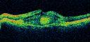
Subfoveal CNVM wet AMD oct line scan1058 x angesehenShe has noticed decreased vision in the left eye. It is hard to say for how long, maybe a few months. She had her last eye exam about a year ago. She thoughts perhaps it was cataracts, but you saw a problem with the macula and suggested she come here for evaluation.
VISUAL ACUITY: Vision OD is 20/25, OS is 2/200
|
|
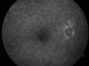
Subfoveal CNVM wet AMD fellow eye610 x angesehenShe has noticed decreased vision in the left eye. It is hard to say for how long, maybe a few months. She had her last eye exam about a year ago. She thoughts perhaps it was cataracts, but you saw a problem with the macula and suggested she come here for evaluation.
VISUAL ACUITY: Vision OD is 20/25, OS is 2/200
|
|

Subfoveal CNVM wet AMD early fa full image832 x angesehenShe has noticed decreased vision in the left eye. It is hard to say for how long, maybe a few months. She had her last eye exam about a year ago. She thoughts perhaps it was cataracts, but you saw a problem with the macula and suggested she come here for evaluation.
VISUAL ACUITY: Vision OD is 20/25, OS is 2/200
|
|

Subfoveal CNVM wet AMD early fa632 x angesehenShe has noticed decreased vision in the left eye. It is hard to say for how long, maybe a few months. She had her last eye exam about a year ago. She thoughts perhaps it was cataracts, but you saw a problem with the macula and suggested she come here for evaluation.
VISUAL ACUITY: Vision OD is 20/25, OS is 2/200
|
|

Subfoveal CNVM wet AMD late fa full image843 x angesehenShe has noticed decreased vision in the left eye. It is hard to say for how long, maybe a few months. She had her last eye exam about a year ago. She thoughts perhaps it was cataracts, but you saw a problem with the macula and suggested she come here for evaluation.
VISUAL ACUITY: Vision OD is 20/25, OS is 2/200
|
|

Subfoveal CNVM wet AMD late fa646 x angesehen85-year-old woman was seen in the office on 11/8/07. She has noticed decreased vision in the left eye. It is hard to say for how long, maybe a few months. She had her last eye exam about a year ago. She thoughts perhaps it was cataracts, but you saw a problem with the macula and suggested she come here for evaluation.
VISUAL ACUITY: Vision OD is 20/25, OS is 2/200
|
|
|
|
|
| 17 Dateien auf 1 Seite(n) |
|


