Resultado de la búsqueda de archivos - "detachment"
|

Pseudophakic Macula-Off Retinal Detachment with Larger Retinal Tearvista 1150 veces68-year-old woman with veil over her vision just the last few days. She had cataract surgery a year ago. OD 20/200 -- One year later vision 20/30!
|
|

Pseudophakic Macula-Off Retinal Detachment with Larger Retinal Tearvista 673 veces68-year-old woman with veil over her vision just the last few days. She had cataract surgery a year ago. OD 20/200 -- One year later vision 20/30!
|
|

Pseudophakic Macula-Off Retinal Detachment with Larger Retinal Tearvista 878 veces68-year-old woman with veil over her vision just the last few days. She had cataract surgery a year ago. OD 20/200 -- One year later vision 20/30!
|
|

Pseudophakic Macula-Off Retinal Detachment with Larger Retinal Tearvista 652 veces68-year-old woman with veil over her vision just the last few days. She had cataract surgery a year ago. OD 20/200 -- One year later vision 20/30!
|
|
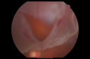
Pseudophakic Macula-Off Retinal Detachment with Larger Retinal Tearvista 863 veces68-year-old woman with veil over her vision just the last few days. She had cataract surgery a year ago. OD 20/200 -- One year later vision 20/30!
|
|

Pseudophakic Macula-Off Retinal Detachment with Larger Retinal Tearvista 698 veces68-year-old woman with veil over her vision just the last few days. She had cataract surgery a year ago. OD 20/200 -- One year later vision 20/30!
|
|
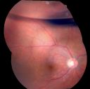
Pseudophakic Macula-Off Retinal Detachment with Larger Retinal Tearvista 752 veces68-year-old woman with veil over her vision just the last few days. She had cataract surgery a year ago. OD 20/200 -- One year later vision 20/30!
|
|

Hypertensive Retinopathy with Choroidal Ischemia Right Eye and Vision 8/200 - Blood Pressure 240/120 mmHg IgA Nephropathyvista 1018 veces39-year-old man two days ago he noticed poor vision in his right eye OD: 8/200; OS: 20/20. BP is 240/120
|
|

Hypertensive Retinopathy with Choroidal Ischemia Right Eye and Vision 8/200 - Blood Pressure 240/120 mmHg IgA Nephropathyvista 835 veces39-year-old man two days ago he noticed poor vision in his right eye OD: 8/200; OS: 20/20. BP is 240/120
|
|
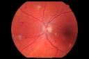
Hypertensive Retinopathy with Choroidal Ischemia Right Eye and Vision 8/200 - Blood Pressure 240/120 mmHg IgA Nephropathyvista 782 veces39-year-old man two days ago he noticed poor vision in his right eye OD: 8/200; OS: 20/20. BP is 240/120
|
|

Hypertensive Retinopathy with Choroidal Ischemia Right Eye and Vision 8/200 - Blood Pressure 240/120 mmHg IgA Nephropathyvista 764 veces39-year-old man two days ago he noticed poor vision in his right eye OD: 8/200; OS: 20/20. BP is 240/120
|
|

Hypertensive Retinopathy with Choroidal Ischemia Right Eye and Vision 8/200 - Blood Pressure 240/120 mmHg IgA Nephropathyvista 829 veces39-year-old man two days ago he noticed poor vision in his right eye OD: 8/200; OS: 20/20. BP is 240/120
|
|

Hypertensive Retinopathy with Choroidal Ischemia Right Eye and Vision 8/200 - Blood Pressure 240/120 mmHg IgA Nephropathyvista 744 veces39-year-old man two days ago he noticed poor vision in his right eye OD: 8/200; OS: 20/20. BP is 240/120
|
|

Hypertensive Retinopathy with Choroidal Ischemia Right Eye and Vision 8/200 - Blood Pressure 240/120 mmHg IgA Nephropathyvista 698 veces39-year-old man two days ago he noticed poor vision in his right eye OD: 8/200; OS: 20/20. BP is 240/120
|
|

Hypertensive Retinopathy with Choroidal Ischemia Right Eye and Vision 8/200 - Blood Pressure 240/120 mmHg IgA Nephropathyvista 709 veces39-year-old man two days ago he noticed poor vision in his right eye OD: 8/200; OS: 20/20. BP is 240/120
|
|
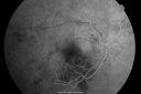
Hypertensive Retinopathy with Choroidal Ischemia Right Eye and Vision 8/200 - Blood Pressure 240/120 mmHg IgA Nephropathyvista 770 veces39-year-old man two days ago he noticed poor vision in his right eye OD: 8/200; OS: 20/20. BP is 240/120
|
|
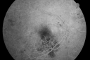
Hypertensive Retinopathy with Choroidal Ischemia Right Eye and Vision 8/200 - Blood Pressure 240/120 mmHg IgA Nephropathyvista 668 veces39-year-old man two days ago he noticed poor vision in his right eye OD: 8/200; OS: 20/20. BP is 240/120
|
|

Hypertensive Retinopathy with Choroidal Ischemia Right Eye and Vision 8/200 - Blood Pressure 240/120 mmHg IgA Nephropathyvista 629 veces39-year-old man two days ago he noticed poor vision in his right eye OD: 8/200; OS: 20/20. BP is 240/120
|
|
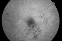
Hypertensive Retinopathy with Choroidal Ischemia Right Eye and Vision 8/200 - Blood Pressure 240/120 mmHg IgA Nephropathyvista 518 veces39-year-old man two days ago he noticed poor vision in his right eye OD: 8/200; OS: 20/20. BP is 240/120
|
|

Hypertensive Retinopathy with Choroidal Ischemia Right Eye and Vision 8/200 - Blood Pressure 240/120 mmHg IgA Nephropathyvista 619 veces39-year-old man two days ago he noticed poor vision in his right eye OD: 8/200; OS: 20/20. BP is 240/120
|
|

Hypertensive Retinopathy with Choroidal Ischemia Right Eye and Vision 8/200 - Blood Pressure 240/120 mmHg IgA Nephropathyvista 738 veces39-year-old man two days ago he noticed poor vision in his right eye OD: 8/200; OS: 20/20. BP is 240/120
|
|

Hypertensive Retinopathy with Choroidal Ischemia Right Eye and Vision 8/200 - Blood Pressure 240/120 mmHg IgA Nephropathyvista 644 veces39-year-old man two days ago he noticed poor vision in his right eye OD: 8/200; OS: 20/20. BP is 240/120
|
|

Hypertensive Retinopathy with Choroidal Ischemia Right Eye and Vision 8/200 - Blood Pressure 240/120 mmHg IgA Nephropathyvista 571 veces39-year-old man two days ago he noticed poor vision in his right eye OD: 8/200; OS: 20/20. BP is 240/120
|
|

Hypertensive Retinopathy with Choroidal Ischemia Right Eye and Vision 8/200 - Blood Pressure 240/120 mmHg IgA Nephropathyvista 557 veces39-year-old man two days ago he noticed poor vision in his right eye OD: 8/200; OS: 20/20. BP is 240/120
|
|

Serous Choroidal Effusionsvista 2484 vecesSerous choroidal effusions following anterior segment surgery. Final visual outcome was 20/20.
|
|
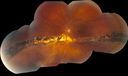
Chronic Retinal Detachmentvista 1405 veces51 year old female, with a chronic retinal detachment. The patient has been stable since 1999.
|
|
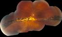
Chronic Retinal Detachmentvista 1070 veces51 year old female, with a chronic retinal detachment. The patient has been stable since 1999.
|
|

Retinal Tear with Laservista 901 veces
|
|

Horseshoe Retinal Tear with bridging blood vesselvista 1449 veces
|
|

Horseshoe Retinal Tear with bridging blood vesselvista 2630 veces
|
|

Horseshoe Retinal Tear with bridging blood vesselvista 880 veces
|
|

Giant Retinal Tearvista 1716 veces
|
|

vista 683 veces
|
|
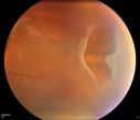
Horseshoe Retinal Tearvista 789 veces
|
|

Retinal Tearvista 581 veces
|
|

Retinal Tear treated with Laser vista 580 veces
|
|

Retinal Tear treated with Laser vista 551 veces
|
|

Retinal Tear vista 564 veces
|
|

Retinal Tear, the retina tear is flipped overvista 648 veces
|
|

Retinal Tearvista 623 veces
|
|

Combined schisis-rhegmatogenous RDvista 2260 vecesMultiple outer layer and inner layer holes
|
|

Macular fold after PPV for RDvista 3049 vecesA 53-year-old man with a macula-off RD underwent left eye pars plana vitrectomy with air-fluid exchange, laser retinopexy, and injection of 14% SF6 gas. There were 4 retinal breaks between 10 o’clock and 12 o’clock including one large tear. He was compliant with facedown positioning. On the 8th post-operative day, a retinal fold along the 4-10 o’clock meridian was seen coursing through his central macula. He underwent repeat PPV but multiple attempts to lift and flatten the retina were unsuccessful.
|
|
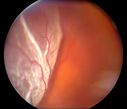
Retinal Detachmentvista 537 veces
|
|

Retinal Detachmentvista 598 veces
|
|

Retinal Detachmentvista 520 veces
|
|

Retinal Detachmentvista 576 veces
|
|

Retinal Detachmentvista 594 veces
|
|

Retinal Detachmentvista 528 veces
|
|

Retinal Detachmentvista 467 veces
|
|

C3F8 Gas Bubble for retinal tear/ Pneumatic Retinopexyvista 825 veces
|
|

Retinal Tearvista 525 veces
|
|

Retinal Tearvista 531 veces
|
|

C3F8 Gas Bubble for retinal tear/ Pneumatic Retinopexyvista 743 veces
|
|

2 - Horseshoe Tearsvista 1309 veces
|
|

SF6 Gas Bubble vista 942 veces
|
|

Giant Retinal Tear 6 weeks s/p PPV, SB, and GFEvista 604 vecesPt. s/p SB/PPV/PFO/AFX/C3F8 gas. All flat with superior residual gas bubble.
|
|

Giant Retinal Tear Initial Presentationvista 564 veces61 yo phakic male with giant retinal tear OD
|
|

Retinal Detachment Secondary to Irregular Retinal Tearvista 985 vecesRetinal Detachment with Irregular Retinal Tear
|
|

Displaced Scleral Bucklevista 1358 vecesDisplaced Scleral Buckle in OD of a 75 yr old male
|
|
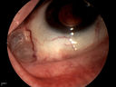
Displaced Scleral Bucklevista 766 vecesDisplaced Scleral Buckle in OD of a 75 yr old male
|
|

PDR w/TRDvista 450 vecesPDR w/heme and traction retinal detachment
|
|

Proliferative Diabetic Retinopathy with Tractional Retinal Detachmentvista 1033 vecesProliferative Diabetic Retinopathy with Tractional Retinal Detachment
|
|

Large serous PEDvista 1136 veces63-year-old female with acute onset large retinal pigment epithelium detachment from age-related macular degeneration
|
|

Ocular Trauma with Retinal Detachmentvista 897 vecesPatient suffered blunt force trauma to the eye resulting total retinal detachment. The retina is visible in the pupillary space.
|
|
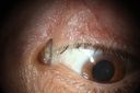
Silicon implant is seen inserted in to the superior lid skinvista 475 vecesanother view of the implant intruded in the lid skin
|
|

Superior Retinal Detachmentvista 401 veces
|
|

Retinal Detachmentvista 382 veces
|
|

Diabetic Tractional Detachmentvista 460 veces
|
|

Retinal Detachmentvista 771 veces
|
|
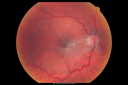
Macular Pucker in eye following giant retinal tear RD repair pre-op pucker photovista 628 vecesnow that the bubble is gone, but he is starting to notice distortion of central vision. He had a giant retinal tear retinal detachment repair emergently done on November 8, 2007, and subsequent laser on November 15th.
VISUAL ACUITY: Vision OD 20/400.
6 weeks post-op VA 20/40
|
|

Chronic Retinal Detachment and Macular hole Pre-Opvista 747 veces51-year-old woman about 3 or 4 months ago she noticed some flashing lights. Her vision became hazy and dark, especially when she was covering her left eye. OD 2/200, OS 20/20. Post op vision 20/200.
|
|

Juxtapapillary CNVM and Serous Macular Detachment Wet AMD Rx Laservista 849 veces85-year-old man OD is 20/25, OS is 20/100. IOP: OD 21, OS 22.
OS: Vertical C/D ratio is 0.1. There is a posterior vitreous separation. There is a hemorrhagic pigment epithelial detachment on the superior pole of the optic nerve, extending a disc-and-a-half diameter off the nerve, with adjacent exudates. There is a serous macular detachment involving the center of the fovea.
VA improved from 20/100 to 20/50 in four months from laser.
|
|
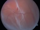
Retinal Detachmentvista 644 veces
|
|

Macular Pucker in eye following giant retinal tear RD repair pre-op Macular Photo Puckervista 731 vecesnow that the bubble is gone, but he is starting to notice distortion of central vision. He had a giant retinal tear retinal detachment repair emergently done on November 8, 2007, and subsequent laser on November 15th.
VISUAL ACUITY: Vision OD 20/400.
6 weeks post-op VA 20/40
|
|

Chronic Retinal Detachment and Macular hole Pre-Op Post-Opvista 722 veces51-year-old woman about 3 or 4 months ago she noticed some flashing lights. Her vision became hazy and dark, especially when she was covering her left eye. OD 2/200, OS 20/20. Post op vision 20/200.
|
|

Juxtapapillary CNVM and Serous Macular Detachment Wet AMD Rx Laservista 653 veces85-year-old man OD is 20/25, OS is 20/100. IOP: OD 21, OS 22.
OS: Vertical C/D ratio is 0.1. There is a posterior vitreous separation. There is a hemorrhagic pigment epithelial detachment on the superior pole of the optic nerve, extending a disc-and-a-half diameter off the nerve, with adjacent exudates. There is a serous macular detachment involving the center of the fovea.
VA improved from 20/100 to 20/50 in four months from laser.
|
|

Retinal Detachmentvista 683 veces
|
|

Macular Pucker in eye following giant retinal tear RD repair photo of tearvista 608 vecesnow that the bubble is gone, but he is starting to notice distortion of central vision. He had a giant retinal tear retinal detachment repair emergently done on November 8, 2007, and subsequent laser on November 15th.
VISUAL ACUITY: Vision OD 20/400.
6 weeks post-op VA 20/40
|
|

Chronic Retinal Detachment and Macular hole Pre-Op Montagevista 860 veces51-year-old woman about 3 or 4 months ago she noticed some flashing lights. Her vision became hazy and dark, especially when she was covering her left eye. OD 2/200, OS 20/20. Post op vision 20/200.
|
|

Juxtapapillary CNVM and Serous Macular Detachment Wet AMD Rx Laservista 636 veces85-year-old man OD is 20/25, OS is 20/100. IOP: OD 21, OS 22.
OS: Vertical C/D ratio is 0.1. There is a posterior vitreous separation. There is a hemorrhagic pigment epithelial detachment on the superior pole of the optic nerve, extending a disc-and-a-half diameter off the nerve, with adjacent exudates. There is a serous macular detachment involving the center of the fovea.
VA improved from 20/100 to 20/50 in four months from laser.
|
|

Retinal Detachmentvista 824 veces
|
|

Macular Pucker in eye following giant retinal tear RD repair - photo of tearvista 750 veces 42-year old man has noticed his vision is changing now that the bubble is gone, but he is starting to notice distortion of central vision. He had a giant retinal tear retinal detachment repair emergently done on November 8, 2007, and subsequent laser on November 15th.
VISUAL ACUITY: Vision OD 20/400.
6 weeks post-op VA 20/40
|
|
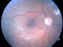
Chronic Retinal Detachment and Macular hole Post-Opvista 852 veces51-year-old woman about 3 or 4 months ago she noticed some flashing lights. Her vision became hazy and dark, especially when she was covering her left eye. OD 2/200, OS 20/20. Post op vision 20/200.
|
|

Juxtapapillary CNVM and Serous Macular Detachment Wet AMD Rx Laservista 618 veces85-year-old man OD is 20/25, OS is 20/100. IOP: OD 21, OS 22.
OS: Vertical C/D ratio is 0.1. There is a posterior vitreous separation. There is a hemorrhagic pigment epithelial detachment on the superior pole of the optic nerve, extending a disc-and-a-half diameter off the nerve, with adjacent exudates. There is a serous macular detachment involving the center of the fovea.
VA improved from 20/100 to 20/50 in four months from laser.
|
|

post retinal detachment repairvista 736 veces72-year-old with a history of retinal detachment in the right eye. He had pneumatic retinopexy followed by panretinal laser. He returns today reporting that he is seeing fairly well but the central vision in the right eye is not as clear as it used to be.
OPHTHALMIC EXAM: Visual acuity with correction of 20/100 OD, 20/20 OS; PH 20/80 OD. IOP: 18 OD.
|
|

Macular Pucker in eye following giant retinal tear RD repair pre-op OCT Mapvista 604 vecesnow that the bubble is gone, but he is starting to notice distortion of central vision. He had a giant retinal tear retinal detachment repair emergently done on November 8, 2007, and subsequent laser on November 15th.
VISUAL ACUITY: Vision OD 20/400.
6 weeks post-op VA 20/40
|
|

Chronic Retinal Detachment and Macular hole Post-Opvista 791 veces51-year-old woman about 3 or 4 months ago she noticed some flashing lights. Her vision became hazy and dark, especially when she was covering her left eye. OD 2/200, OS 20/20. Post op vision 20/200.
|
|

Juxtapapillary CNVM and Serous Macular Detachment Wet AMD Rx Laservista 876 veces85-year-old man OD is 20/25, OS is 20/100. IOP: OD 21, OS 22.
OS: Vertical C/D ratio is 0.1. There is a posterior vitreous separation. There is a hemorrhagic pigment epithelial detachment on the superior pole of the optic nerve, extending a disc-and-a-half diameter off the nerve, with adjacent exudates. There is a serous macular detachment involving the center of the fovea.
VA improved from 20/100 to 20/50 in four months from laser.
|
|

Macular Pucker in eye following giant retinal tear RD repair pre-op OCTvista 559 vecesnow that the bubble is gone, but he is starting to notice distortion of central vision. He had a giant retinal tear retinal detachment repair emergently done on November 8, 2007, and subsequent laser on November 15th.
VISUAL ACUITY: Vision OD 20/400.
6 weeks post-op VA 20/40
|
|

Chronic Retinal Detachment and Macular hole Pre-Opvista 1076 veces51-year-old woman about 3 or 4 months ago she noticed some flashing lights. Her vision became hazy and dark, especially when she was covering her left eye. OD 2/200, OS 20/20. Post op vision 20/200.
|
|

Juxtapapillary CNVM and Serous Macular Detachment Wet AMD Rx Laservista 629 veces85-year-old man OD is 20/25, OS is 20/100. IOP: OD 21, OS 22.
OS: Vertical C/D ratio is 0.1. There is a posterior vitreous separation. There is a hemorrhagic pigment epithelial detachment on the superior pole of the optic nerve, extending a disc-and-a-half diameter off the nerve, with adjacent exudates. There is a serous macular detachment involving the center of the fovea.
VA improved from 20/100 to 20/50 in four months from laser.
|
|

Macular Pucker in eye following giant retinal tear RD repair pre-op OCTvista 648 vecesnow that the bubble is gone, but he is starting to notice distortion of central vision. He had a giant retinal tear retinal detachment repair emergently done on November 8, 2007, and subsequent laser on November 15th.
VISUAL ACUITY: Vision OD 20/400.
6 weeks post-op VA 20/40
|
|

Juxtapapillary CNVM and Serous Macular Detachment Wet AMD Rx Laservista 651 veces85-year-old man OD is 20/25, OS is 20/100. IOP: OD 21, OS 22.
OS: Vertical C/D ratio is 0.1. There is a posterior vitreous separation. There is a hemorrhagic pigment epithelial detachment on the superior pole of the optic nerve, extending a disc-and-a-half diameter off the nerve, with adjacent exudates. There is a serous macular detachment involving the center of the fovea.
VA improved from 20/100 to 20/50 in four months from laser.
|
|
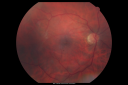
Macular Pucker in eye following giant retinal tear RD repair post-op Fundus Photovista 621 vecesnow that the bubble is gone, but he is starting to notice distortion of central vision. He had a giant retinal tear retinal detachment repair emergently done on November 8, 2007, and subsequent laser on November 15th.
VISUAL ACUITY: Vision OD 20/400.
6 weeks post-op VA 20/40
|
|
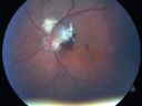
Juxtapapillary CNVM and Serous Macular Detachment Wet AMD Rx Laservista 662 veces85-year-old man OD is 20/25, OS is 20/100. IOP: OD 21, OS 22.
OS: Vertical C/D ratio is 0.1. There is a posterior vitreous separation. There is a hemorrhagic pigment epithelial detachment on the superior pole of the optic nerve, extending a disc-and-a-half diameter off the nerve, with adjacent exudates. There is a serous macular detachment involving the center of the fovea.
VA improved from 20/100 to 20/50 in four months from laser.
|
|

Macular Pucker in eye following giant retinal tear RD repair post-op macular photovista 621 vecesnow that the bubble is gone, but he is starting to notice distortion of central vision. He had a giant retinal tear retinal detachment repair emergently done on November 8, 2007, and subsequent laser on November 15th.
VISUAL ACUITY: Vision OD 20/400.
6 weeks post-op VA 20/40
|
|

Juxtapapillary CNVM and Serous Macular Detachment Wet AMD Rx Laservista 555 veces85-year-old man OD is 20/25, OS is 20/100. IOP: OD 21, OS 22.
OS: Vertical C/D ratio is 0.1. There is a posterior vitreous separation. There is a hemorrhagic pigment epithelial detachment on the superior pole of the optic nerve, extending a disc-and-a-half diameter off the nerve, with adjacent exudates. There is a serous macular detachment involving the center of the fovea.
VA improved from 20/100 to 20/50 in four months from laser.
|
|

Macular Pucker in eye following giant retinal tear RD repair post-op OCTvista 598 vecesnow that the bubble is gone, but he is starting to notice distortion of central vision. He had a giant retinal tear retinal detachment repair emergently done on November 8, 2007, and subsequent laser on November 15th.
VISUAL ACUITY: Vision OD 20/400.
6 weeks post-op VA 20/40
|
|

Juxtapapillary CNVM and Serous Macular Detachment Wet AMD Rx Laservista 617 veces85-year-old man OD is 20/25, OS is 20/100. IOP: OD 21, OS 22.
OS: Vertical C/D ratio is 0.1. There is a posterior vitreous separation. There is a hemorrhagic pigment epithelial detachment on the superior pole of the optic nerve, extending a disc-and-a-half diameter off the nerve, with adjacent exudates. There is a serous macular detachment involving the center of the fovea.
VA improved from 20/100 to 20/50 in four months from laser.
|
|
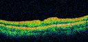
Macular Pucker in eye following giant retinal tear RD repair post-op OCTvista 542 vecesnow that the bubble is gone, but he is starting to notice distortion of central vision. He had a giant retinal tear retinal detachment repair emergently done on November 8, 2007, and subsequent laser on November 15th.
VISUAL ACUITY: Vision OD 20/400.
6 weeks post-op VA 20/40
|
|
| 101 archivos en 2 página(s) |
1 |
|


