Image search results - "february"
|

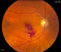
Choroidal Melanoma with Radiation Retinopathy986 viewsPatient comes with follow up on Choroidal Melanoma, Right Eye that was treated back in June of 2009 with Radioactive Implant. Vein Occlusion is also present with VA - Hand Motion. Hemorrhages visible with hard exudates from the Radiation Retinopathy.
|
|

Nonproliferative Diabetic Retinopathy7340 views65-year old female with diabetes. Has had cataract surgery in the left eye with VA 20/25. She has had laser in the past. Fundus examination shows microaneurysms with retinal hemorrahages and exudates in the left eye.
|
|

RD640 viewsInferior RD
|
|

RD with PVR1172 viewsRD with Proliferative Vitreoretinopathy
|
|

Hemangioma801 viewsFP Composite of Hemangioma in OD
|
|

Pigmented Peripheral Retinal Degeneration1447 views42-year old male comes in for routine eye exam and to follow up on peripheral retinal degeneration in both eyes. VA is 20/20, right eye and 20/25, left eye. Patient is asymptomatic with no visual complaints.
|
|

Uveal Choroidal Melanoma727 viewsPatient comes in for evaluation on a Choroidal Melanoma in the right eye. VA was 20/25 in both eyes. The melanoma is in the temporal aspect of the right eye. It measured at 0.7mm elevated after doing a BSCAN Ultrasound.
|
|

Gonioscopy, Blood in the Anterior Chamber from Hyphema683 viewsPatient comes in with blunt trauma to the right eye due to a BB gun incident. Patient was present with a hyphema at 8-o'clock about 1mm thick. Gonioscopy photos were then taken to show blood from the hyphema entered into the anterior chamber. Patient had no angle recession in the right eye.
|
|

Corneal Abrasion with Foreign Body Present 670 viewsER patient comes in with corneal abrasion in the left eye which was getting worst. VA 20/25. Slit lamp exam showed corneal abrasion superiorly at 2-o'clock. Flipped eyelid and foreign body appeared on the upper tarsal plate. Foreign body was removed.
|
|

Corneal Abrasion with Foreign Body Present 786 viewsER patient comes in with corneal abrasion in the left eye which was getting worst. VA 20/25. Slit lamp exam showed corneal abrasion superiorly at 2-o'clock. Flipped eyelid and foreign body appeared on the upper tarsal plate. Foreign body was removed.
|
|

Proliferative Diabetic Retinopathy1000 viewsFundus photography shows severe fibrosis and arterial narrowing. Peripheral laser scars in both eyes. VA is 20/40, right eye and 20/50, left eye.
|
|

Choroidal Melanoma758 viewsFemale patient comes in with a visual defect in the right eye. VA is 20/40 in the right eye. Fundus photography shows choroidal melanoma with retinal detachment temporally at 3-o'clock in the right eye.
|
|

Choroidal Nevus777 viewsPatient comes in with pigmented spot in the right eye. VA is 20/25. Fundus photography shows elevated choroidal nevus inferiorily in the right eye. Will be reevaluated in 3-months...
|
|

Retinal Detachment with Macula Detached749 viewsPatient had sudden loss of vision in the left eye. VA is 20/60 in the left eye. Fundus exam reveals retinal detachment inferiorly from 3-9 o'clock with tear at 3:30. Patient underwent scleral buckle in the left eye.
|
|

Branch Retinal Vein Occlusion772 viewsPatient comes in for eval on retinal hemorrhage in the right eye. VA is 20/30 in the right eye. Fundus photography shows branch retinal vein occlusion inferior of the macula with retinal hemorrhage present. Will reevaluate in 2-months.
|
|

Choroidal Rupture Secondary to Ocular Trauma665 views
|
|

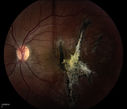
Traumatic Chorioretinal Scar466 views
|
|

Intraocular Self injection697 viewsThe treatment of wet macular degeneration was revolutionized in late 2013 when Geneneron introduced its innovative and inexpensive take-home kit. Now, millions of patients around the world treat themselves on a PRN basis, using instructions written in 10 different languages.
April Fool!
|
|

Pseudoexfoliation Material Peeling.596 viewsPatient comes in for regular eye exam. Last exam was 8-years ago. Complains of decrease in VA. VA was 20/30, right eye and 20/40, left eye. Slit lamp photography shows pseudoexfoliation peeling of the lens within 360-degrees. Pseudoexfoliation ring is present centrally. IOP was normal. Will revisit in 3-months for follow up.
|
|

Optic Nerve Edema with Retinal Hemorrhages.707 viewsFemale patient diagnosed with lupus a year ago, comes in with spot and decreased vision in the right eye. VA is 20/400 in the right eye and 20/40, in the left eye. Fundus photography shows multiple retinal hemorrhages and possible bilateral papilledema.
|
|

ARMD: Macular Degeneration with Subretinal Neovascular Membrane336 viewsARMD: Macular Degeneration with Subretinal Neovascular Membrane.
Upon presentation, this patient was found to have blots of subretinal hemorrhage extending into her FAZ, secondary to a subretinal neovascular membrane.
|
|
|
|
|
|
|