Retina Gallery ~ Full Sized Retina Images

Library of Free, Non-Copyrighted Retina Images and Videos

Vali keel:

Enimvaadatud - Bilateral Branch Retinal Artery Occlusion with Hollenhorst Plaques

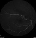
67 year old female smoker. Both eyes are developing shadows in the vision. The vision was normal starting recently. May 25th she noticed in the left eye a shadow that developed in the vision that she could not see through that did not move. The shadow is nasal in the bottom half of the vision. Mushroom shaped. This is not getting any better. Then yesterday she got a new shadow in the right eye this time. This is the same type of problem. The shadow in the right eye is more in the bottom half of the vision. The right eye started out worse and has diminshed a little. The right eye shadow is bigger than the shadow in the left eye. Vision 20/25 OD 20/32 OS. Photos show acute bilateral branch retinal artery occlusions.

bao_kasp1_060616_01.png
Bilateral Branch Retinal Artery Occlusion - Intraarterial (hollenhorst) plaques visiblevaatamisi: 63567 year old with acute visual field loss in each eye. Vision is 20/25 right eye 20/32 left eye. Images show intra-arterial plaques in each eye. The right eye on the optic nerve and the left eye in a branch retinal artery.
bao_kasp1_060616_02.png
Bilateral Branch Retinal Artery Occlusion - Intraarterial (hollenhorst) plaques visiblevaatamisi: 57867 year old with acute visual field loss in each eye. Vision is 20/25 right eye 20/32 left eye. Images show intra-arterial plaques in each eye. The right eye on the optic nerve and the left eye in a branch retinal artery.
bao_kasp1_060616_26.jpg
Bilateral Branch Retinal Artery Occlusion - Intraarterial (hollenhorst) plaques visiblevaatamisi: 57067 year old with acute visual field loss in each eye. Vision is 20/25 right eye 20/32 left eye. Images show intra-arterial plaques in each eye. The right eye on the optic nerve and the left eye in a branch retinal artery.
bao_kasp1_060616_03.png
Bilateral Branch Retinal Artery Occlusion - Intraarterial (hollenhorst) plaques visiblevaatamisi: 54467 year old with acute visual field loss in each eye. Vision is 20/25 right eye 20/32 left eye. Images show intra-arterial plaques in each eye. The right eye on the optic nerve and the left eye in a branch retinal artery.
bao_kasp1_060616_14.jpg
Bilateral Branch Retinal Artery Occlusion - Intraarterial (hollenhorst) plaques visiblevaatamisi: 52967 year old with acute visual field loss in each eye. Vision is 20/25 right eye 20/32 left eye. Images show intra-arterial plaques in each eye. The right eye on the optic nerve and the left eye in a branch retinal artery.
bao_kasp1_060616_18.jpg
Bilateral Branch Retinal Artery Occlusion - Intraarterial (hollenhorst) plaques visiblevaatamisi: 52667 year old with acute visual field loss in each eye. Vision is 20/25 right eye 20/32 left eye. Images show intra-arterial plaques in each eye. The right eye on the optic nerve and the left eye in a branch retinal artery.
bao_kasp1_060616_04.png
Bilateral Branch Retinal Artery Occlusion - Intraarterial (hollenhorst) plaques visiblevaatamisi: 51567 year old with acute visual field loss in each eye. Vision is 20/25 right eye 20/32 left eye. Images show intra-arterial plaques in each eye. The right eye on the optic nerve and the left eye in a branch retinal artery.
bao_kasp1_060616_23.jpg
Bilateral Branch Retinal Artery Occlusion - Intraarterial (hollenhorst) plaques visiblevaatamisi: 51367 year old with acute visual field loss in each eye. Vision is 20/25 right eye 20/32 left eye. Images show intra-arterial plaques in each eye. The right eye on the optic nerve and the left eye in a branch retinal artery.
bao_kasp1_060616_10.jpg
Bilateral Branch Retinal Artery Occlusion - Intraarterial (hollenhorst) plaques visiblevaatamisi: 51267 year old with acute visual field loss in each eye. Vision is 20/25 right eye 20/32 left eye. Images show intra-arterial plaques in each eye. The right eye on the optic nerve and the left eye in a branch retinal artery.
bao_kasp1_060616_28.jpg
Bilateral Branch Retinal Artery Occlusion - Intraarterial (hollenhorst) plaques visiblevaatamisi: 51067 year old with acute visual field loss in each eye. Vision is 20/25 right eye 20/32 left eye. Images show intra-arterial plaques in each eye. The right eye on the optic nerve and the left eye in a branch retinal artery.
bao_kasp1_060616_21.jpg
Bilateral Branch Retinal Artery Occlusion - Intraarterial (hollenhorst) plaques visiblevaatamisi: 49967 year old with acute visual field loss in each eye. Vision is 20/25 right eye 20/32 left eye. Images show intra-arterial plaques in each eye. The right eye on the optic nerve and the left eye in a branch retinal artery.
bao_kasp1_060616_19.jpg
Bilateral Branch Retinal Artery Occlusion - Intraarterial (hollenhorst) plaques visiblevaatamisi: 49867 year old with acute visual field loss in each eye. Vision is 20/25 right eye 20/32 left eye. Images show intra-arterial plaques in each eye. The right eye on the optic nerve and the left eye in a branch retinal artery.
bao_kasp1_060616_25.jpg
Bilateral Branch Retinal Artery Occlusion - Intraarterial (hollenhorst) plaques visiblevaatamisi: 49467 year old with acute visual field loss in each eye. Vision is 20/25 right eye 20/32 left eye. Images show intra-arterial plaques in each eye. The right eye on the optic nerve and the left eye in a branch retinal artery.
bao_kasp1_060616_12.jpg
Bilateral Branch Retinal Artery Occlusion - Intraarterial (hollenhorst) plaques visiblevaatamisi: 49367 year old with acute visual field loss in each eye. Vision is 20/25 right eye 20/32 left eye. Images show intra-arterial plaques in each eye. The right eye on the optic nerve and the left eye in a branch retinal artery.
bao_kasp1_060616_17.jpg
Bilateral Branch Retinal Artery Occlusion - Intraarterial (hollenhorst) plaques visiblevaatamisi: 49367 year old with acute visual field loss in each eye. Vision is 20/25 right eye 20/32 left eye. Images show intra-arterial plaques in each eye. The right eye on the optic nerve and the left eye in a branch retinal artery.
bao_kasp1_060616_07.jpg
Bilateral Branch Retinal Artery Occlusion - Intraarterial (hollenhorst) plaques visiblevaatamisi: 49067 year old with acute visual field loss in each eye. Vision is 20/25 right eye 20/32 left eye. Images show intra-arterial plaques in each eye. The right eye on the optic nerve and the left eye in a branch retinal artery.
bao_kasp1_060616_15.jpg
Bilateral Branch Retinal Artery Occlusion - Intraarterial (hollenhorst) plaques visiblevaatamisi: 48467 year old with acute visual field loss in each eye. Vision is 20/25 right eye 20/32 left eye. Images show intra-arterial plaques in each eye. The right eye on the optic nerve and the left eye in a branch retinal artery.
bao_kasp1_060616_11.jpg
Bilateral Branch Retinal Artery Occlusion - Intraarterial (hollenhorst) plaques visiblevaatamisi: 48367 year old with acute visual field loss in each eye. Vision is 20/25 right eye 20/32 left eye. Images show intra-arterial plaques in each eye. The right eye on the optic nerve and the left eye in a branch retinal artery.
bao_kasp1_060616_06.jpg
Bilateral Branch Retinal Artery Occlusion - Intraarterial (hollenhorst) plaques visiblevaatamisi: 48067 year old with acute visual field loss in each eye. Vision is 20/25 right eye 20/32 left eye. Images show intra-arterial plaques in each eye. The right eye on the optic nerve and the left eye in a branch retinal artery.
bao_kasp1_060616_29.jpg
Bilateral Branch Retinal Artery Occlusion - Intraarterial (hollenhorst) plaques visiblevaatamisi: 47967 year old with acute visual field loss in each eye. Vision is 20/25 right eye 20/32 left eye. Images show intra-arterial plaques in each eye. The right eye on the optic nerve and the left eye in a branch retinal artery.
bao_kasp1_060616_13.jpg
Bilateral Branch Retinal Artery Occlusion - Intraarterial (hollenhorst) plaques visiblevaatamisi: 47767 year old with acute visual field loss in each eye. Vision is 20/25 right eye 20/32 left eye. Images show intra-arterial plaques in each eye. The right eye on the optic nerve and the left eye in a branch retinal artery.
bao_kasp1_060616_16.jpg
Bilateral Branch Retinal Artery Occlusion - Intraarterial (hollenhorst) plaques visiblevaatamisi: 47467 year old with acute visual field loss in each eye. Vision is 20/25 right eye 20/32 left eye. Images show intra-arterial plaques in each eye. The right eye on the optic nerve and the left eye in a branch retinal artery.
bao_kasp1_060616_05.jpg
Bilateral Branch Retinal Artery Occlusion - Intraarterial (hollenhorst) plaques visiblevaatamisi: 47367 year old with acute visual field loss in each eye. Vision is 20/25 right eye 20/32 left eye. Images show intra-arterial plaques in each eye. The right eye on the optic nerve and the left eye in a branch retinal artery.
bao_kasp1_060616_09.jpg
Bilateral Branch Retinal Artery Occlusion - Intraarterial (hollenhorst) plaques visiblevaatamisi: 46767 year old with acute visual field loss in each eye. Vision is 20/25 right eye 20/32 left eye. Images show intra-arterial plaques in each eye. The right eye on the optic nerve and the left eye in a branch retinal artery.
bao_kasp1_060616_08.jpg
Bilateral Branch Retinal Artery Occlusion - Intraarterial (hollenhorst) plaques visiblevaatamisi: 46667 year old with acute visual field loss in each eye. Vision is 20/25 right eye 20/32 left eye. Images show intra-arterial plaques in each eye. The right eye on the optic nerve and the left eye in a branch retinal artery.
bao_kasp1_060616_24.jpg
Bilateral Branch Retinal Artery Occlusion - Intraarterial (hollenhorst) plaques visiblevaatamisi: 46267 year old with acute visual field loss in each eye. Vision is 20/25 right eye 20/32 left eye. Images show intra-arterial plaques in each eye. The right eye on the optic nerve and the left eye in a branch retinal artery.
bao_kasp1_060616_20.jpg
Bilateral Branch Retinal Artery Occlusion - Intraarterial (hollenhorst) plaques visiblevaatamisi: 44267 year old with acute visual field loss in each eye. Vision is 20/25 right eye 20/32 left eye. Images show intra-arterial plaques in each eye. The right eye on the optic nerve and the left eye in a branch retinal artery.
bao_kasp1_060616_27.jpg
Bilateral Branch Retinal Artery Occlusion - Intraarterial (hollenhorst) plaques visiblevaatamisi: 43767 year old with acute visual field loss in each eye. Vision is 20/25 right eye 20/32 left eye. Images show intra-arterial plaques in each eye. The right eye on the optic nerve and the left eye in a branch retinal artery.
bao_kasp1_060616_22.jpg
Bilateral Branch Retinal Artery Occlusion - Intraarterial (hollenhorst) plaques visiblevaatamisi: 42567 year old with acute visual field loss in each eye. Vision is 20/25 right eye 20/32 left eye. Images show intra-arterial plaques in each eye. The right eye on the optic nerve and the left eye in a branch retinal artery.
     
29 faili 1 lehel

67 year old female smoker. Both eyes are developing shadows in the vision. The vision was normal starting recently. May 25th she noticed in the left eye a shadow that developed in the vision that she could not see through that did not move. The shadow is nasal in the bottom half of the vision. Mushroom shaped. This is not getting any better. Then yesterday she got a new shadow in the right eye this time. This is the same type of problem. The shadow in the right eye is more in the bottom half of the vision. The right eye started out worse and has diminshed a little. The right eye shadow is bigger than the shadow in the left eye. Vision 20/25 OD 20/32 OS. Photos show acute bilateral branch retinal artery occlusions.