Most viewed - Multifocal Choroiditis Panuveitis - 32-year-old woman CNVM OS Recurrs 4 years after initial therapy (avastin) 32-year-old woman in the office on February 21, 2008. I appreciate you sending along her old notes from 1999 when she had an episode of vision loss in the right eye associated with macular scarring and multifocal choroiditis. At that time she was not seen to have any active lesions in her macula and comes in now with the vision complaints lasting about two weeks of blurred vision in the left and good eye, obviously causing her concern.
She is not having any health problems. She does have a history of migraine and she also takes a multivitamin. She does not have any fevers, rashes, or arthritis. She has no evidence of any oral or genital sores, nor does she have any breathing problems.
VISUAL ACUITY: OD 20/400, OS 20/50. IOP: OD 15, OS 16.
SLIT LAMP EXAM: Normal clear lenses.
EXTENDED OPHTHALMOSCOPY:
OD: Vertical C/D ratio is 0.1. There are multifocal chorioretinal scars with a foveal scar.
OS: Vertical C/D ratio is 0.1. There are multifocal chorioretinal scars. There is a focus of choroiditis just inferonasal to the fovea about 500 microns short of the fovea.
OCT SCAN: The right eye shows diffuse retinal atrophy. The left eye shows thickening of the retinal pigment epithelium with overlying retinal edema just nasal to the fovea. Photos confirm clinical findings.
FLUORESCEIN ANGIOGRAM: Shows increasing hyperfluorescence in the lesion of the macula of the left eye. There is, however, no definite vascularity to it. The right eye has staining macular scars.
IMPRESSION:
1. PIC – BOTH EYES
2. NEW LESION OF ACTIVE CHOROIDITIS THREATENING THE FOVEA – LEFT EYE
3. POSSIBLE CHOROIDAL NEOVASCULAR MEMBRANE – LEFT EYE
DISCUSSION: I explained to the patient given her history of multifocal scarring and the new lesion which appears to have no vascularity, no hemorrhage, very little overlying fluid, and is not associated with an old macular scar, I think this is most likely a focus of active inflammation and not an area of neovascularization. Patients with multifocal choroiditis rarely develop choroidal neovascular membranes, whereas patients with punctate inner choroidopathy do, but she does not look to have punctate inner choroidopathy. Because the lesion looks inflammatory, I am starting her on 60 mg of oral steroids daily. I asked her to return for a check in one week, sooner should she notice any problem. If we have no response at that time, I will probably try her with intravitreal Kenalog. If we do have response, we will need to do something about keeping her on anti-inflammatory medicines to prevent central vision loss in the left eye. If the lesion starts to look more vascular, I will treat her with intravitreal blood vessel inhibitors.
When she comes next time, I will also obtain blood tests from Dr. Miller’s office and see what testing needs to be obtained in her to rule-out possible pathologies for possible causes of multifocal choroiditis.
TWO MONTHS LATER:
Her vision in the left eye is doing a little bit better. Interestingly, the right eye is doing substantially better since I first encountered her. She since has had treatment in the left eye with a posterior sub-Tenon Kenalog in February and intravitreal Avastin in March.
VISUAL ACUITY: Vision OD is 20/30, OS is 20/40.
FU: 4 years later:
June 7, 2012. This pleasant 35-year-old woman noticed decreasing vision in the left eye recently. She returns now noticing the vision in the left eye is declining.
VISUAL ACUITY: OD 20/50, OS 20/80. IOP: OD 14, OS 17. There is 1+ nuclear sclerosis in both eyes.
EXTENDED OPHTHALMOSCOPY:
OD: Vertical C/D ratio is 0.3. There is multifocal chorioretinal scars in the macula as well as nasal to the optic nerve. There is some hemorrhage nasal to the optic nerve.
OS: Vertical C/D ratio is 0.3. There is a scar just inferonasal to the fovea, which has grown some towards the fovea with a little blood just under the fovea.
SPECTRALIS-SD-OCT SCAN: The OCT scan in the right eye shows the macula to be dry. The left eye shows macular edema. The photos confirm the retinal hemorrhage in the left eye.
FLUORESCEIN ANGIOGRAPHY: Fluorescein angiography of the right eye shows multiple hyperfluorescence spots in the macula where the scarring is. The left eye shows an increasing hyperfluorescencent area under the fovea, which does look to be vascular, likely from a neovascular complex.
INDOCYANINE GREEN ANGIOGRAPHY: An indocyanine green angiogram was obtained in the both eyes, because of the multifocal chorioditis. The indocyanine green does show hypofluorescence centrally where the neovascular complex is and in the later frames there is a small hypofluorescence lesion. The right eye shows multifocal hypofluorescence spots corresponding to the macular scar.
IMPRESSION:
1. MULTIFOCAL CHOROIDITIS SUBRETINAL FIBROSIS – BOTH EYES
2. NEW FOVEAL HEMORRHAGE WITH A NEOVASCULAR MEMBRANE – LEFT EYE
3. ECCENTRIC HEMORRHAGE – RIGHT EYE
DISCUSSION: I explained to the patient the left macula does appear to have an area of scarring that is reactivated and there is a new blood vessel growth extending towards the fovea, which looks like it may at the moment be subfoveal. I treated the left eye with intravitreal injection of Avastin (1.25 mg/0.05 ml) without any difficulty today.
Vision returned to 20/40 after 3 treatments. Decision was made to treat every 3 months.
|
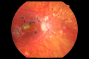
Multifocal Choroiditis and Subretinal Fibrosis - 32 yo Female Multifocal and macular scar OD1311 views32-year-old woman vision loss in the right eye associated with macular scarring and multifocal choroiditis in 1999 with new vision loss in left eye: OD 20/400, OS 20/50.
|
|

Multifocal Choroiditis and Subretinal Fibrosis - 32 yo Female Macula OS 2 months post rx1221 views32-year-old woman vision loss in the right eye associated with macular scarring and multifocal choroiditis in 1999 with new vision loss in left eye: OD 20/400, OS 20/50.
2 months post-rx with posterior subtenons kenalog and intravitreal avastin - va os 20/30 and lesion has retracted and organized. It never subsequently grew over 2 years follow-up
|
|

Multifocal Choroiditis and Subretinal Fibrosis - 32 yo Female Old Scar OD1137 views32-year-old woman vision loss in the right eye associated with macular scarring and multifocal choroiditis in 1999 with new vision loss in left eye: OD 20/400, OS 20/50.
|
|

Multifocal Choroiditis and Subretinal Fibrosis - 32 yo Female Old Macular Scar OD1093 views32-year-old woman vision loss in the right eye associated with macular scarring and multifocal choroiditis in 1999 with new vision loss in left eye: OD 20/400, OS 20/50.
|
|
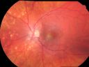
Multifocal Choroiditis and Subretinal Fibrosis - 32 yo Female Montage Initial Visit1018 views32-year-old woman vision loss in the right eye associated with macular scarring and multifocal choroiditis in 1999 with new vision loss in left eye: OD 20/400, OS 20/50.
2 months post-rx with posterior subtenons kenalog and intravitreal avastin - va os 20/30 and lesion has retracted and organized. It never subsequently grew over 2 years follow-up
|
|

Multifocal Choroiditis and Subretinal Fibrosis - 32 yo Female OCT map967 views32-year-old woman vision loss in the right eye associated with macular scarring and multifocal choroiditis in 1999 with new vision loss in left eye: OD 20/400, OS 20/50.
|
|

Punctate Inner Choroidopathy - Recurrent CNVM 4 years after initial treatment Left Eye 958 viewsJune 7, 2012. This pleasant 35-year-old woman noticed decreasing vision in the left eye recently. She returns now noticing the vision in the left eye is declining. OD 20/50, OS 20/80. OS: Vertical C/D ratio is 0.3. There is a scar just inferonasal to the fovea, which has grown some towards the fovea with a little blood just under the fovea.
|
|

Punctate Inner Choroidopathy - Recurrent CNVM 4 years after initial treatment Left Eye 952 viewsJune 7, 2012. This pleasant 35-year-old woman noticed decreasing vision in the left eye recently. She returns now noticing the vision in the left eye is declining. OD 20/50, OS 20/80. OS: Vertical C/D ratio is 0.3. There is a scar just inferonasal to the fovea, which has grown some towards the fovea with a little blood just under the fovea.
|
|

Multifocal Choroiditis and Subretinal Fibrosis - 32 yo Female New Lesion OS899 views32-year-old woman vision loss in the right eye associated with macular scarring and multifocal choroiditis in 1999 with new vision loss in left eye: OD 20/400, OS 20/50.
|
|

Punctate Inner Choroidopathy - Recurrent CNVM 4 years after initial treatment Left Eye - SD OCT893 viewsJune 7, 2012. This pleasant 35-year-old woman noticed decreasing vision in the left eye recently. She returns now noticing the vision in the left eye is declining. OD 20/50, OS 20/80. OS: Vertical C/D ratio is 0.3. There is a scar just inferonasal to the fovea, which has grown some towards the fovea with a little blood just under the fovea.
|
|
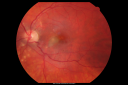
Multifocal Choroiditis and Subretinal Fibrosis - 32 yo Female Macula OS855 views32-year-old woman vision loss in the right eye associated with macular scarring and multifocal choroiditis in 1999 with new vision loss in left eye: OD 20/400, OS 20/50.
|
|

Punctate Inner Choroidopathy - Recurrent CNVM 4 years after initial treatment Left Eye - Fluorescein Angiogram843 viewsJune 7, 2012. This pleasant 35-year-old woman noticed decreasing vision in the left eye recently. She returns now noticing the vision in the left eye is declining. OD 20/50, OS 20/80. OS: Vertical C/D ratio is 0.3. There is a scar just inferonasal to the fovea, which has grown some towards the fovea with a little blood just under the fovea.
|
|

Multifocal Choroiditis and Subretinal Fibrosis - 32 yo Female Line Scan New Lesion OS841 views32-year-old woman vision loss in the right eye associated with macular scarring and multifocal choroiditis in 1999 with new vision loss in left eye: OD 20/400, OS 20/50.
|
|

Multifocal Choroiditis and Subretinal Fibrosis - 32 yo Female Multifocal Scars - PIC Like827 views32-year-old woman vision loss in the right eye associated with macular scarring and multifocal choroiditis in 1999 with new vision loss in left eye: OD 20/400, OS 20/50.
|
|

Multifocal Choroiditis and Subretinal Fibrosis - 32 yo Female FA Old Scar OS798 views32-year-old woman vision loss in the right eye associated with macular scarring and multifocal choroiditis in 1999 with new vision loss in left eye: OD 20/400, OS 20/50.
|
|
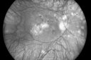
Multifocal Choroiditis and Subretinal Fibrosis - 32 yo Female Old Scar OD794 views32-year-old woman vision loss in the right eye associated with macular scarring and multifocal choroiditis in 1999 with new vision loss in left eye: OD 20/400, OS 20/50.
|
|
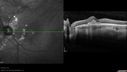
Punctate Inner Choroidopathy - Recurrent CNVM 4 years after initial treatment Left Eye - SD OCT784 viewsJune 7, 2012. This pleasant 35-year-old woman noticed decreasing vision in the left eye recently. She returns now noticing the vision in the left eye is declining. OD 20/50, OS 20/80. OS: Vertical C/D ratio is 0.3. There is a scar just inferonasal to the fovea, which has grown some towards the fovea with a little blood just under the fovea.
|
|

Multifocal Choroiditis and Subretinal Fibrosis - 32 yo Female New Lesion OS766 views32-year-old woman vision loss in the right eye associated with macular scarring and multifocal choroiditis in 1999 with new vision loss in left eye: OD 20/400, OS 20/50.
|
|

Multifocal Choroiditis and Subretinal Fibrosis - 32 yo Female FA New Lesion OS765 views32-year-old woman vision loss in the right eye associated with macular scarring and multifocal choroiditis in 1999 with new vision loss in left eye: OD 20/400, OS 20/50.
|
|

Punctate Inner Choroidopathy - Recurrent CNVM 4 years after initial treatment Left Eye - Fluorescein Angiogram758 viewsJune 7, 2012. This pleasant 35-year-old woman noticed decreasing vision in the left eye recently. She returns now noticing the vision in the left eye is declining. OD 20/50, OS 20/80. OS: Vertical C/D ratio is 0.3. There is a scar just inferonasal to the fovea, which has grown some towards the fovea with a little blood just under the fovea.
|
|

Punctate Inner Choroidopathy - Recurrent CNVM 4 years after initial treatment Left Eye 739 viewsJune 7, 2012. This pleasant 35-year-old woman noticed decreasing vision in the left eye recently. She returns now noticing the vision in the left eye is declining. OD 20/50, OS 20/80. OS: Vertical C/D ratio is 0.3. There is a scar just inferonasal to the fovea, which has grown some towards the fovea with a little blood just under the fovea.
|
|

Punctate Inner Choroidopathy - Recurrent CNVM 4 years after initial treatment Left Eye 736 viewsJune 7, 2012. This pleasant 35-year-old woman noticed decreasing vision in the left eye recently. She returns now noticing the vision in the left eye is declining. OD 20/50, OS 20/80. OS: Vertical C/D ratio is 0.3. There is a scar just inferonasal to the fovea, which has grown some towards the fovea with a little blood just under the fovea.
|
|
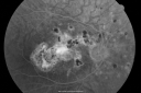
Multifocal Choroiditis and Subretinal Fibrosis - 32 yo Female Old Scar OD731 views32-year-old woman vision loss in the right eye associated with macular scarring and multifocal choroiditis in 1999 with new vision loss in left eye: OD 20/400, OS 20/50.
|
|

Punctate Inner Choroidopathy - Recurrent CNVM 4 years after initial treatment Left Eye 722 viewsJune 7, 2012. This pleasant 35-year-old woman noticed decreasing vision in the left eye recently. She returns now noticing the vision in the left eye is declining. OD 20/50, OS 20/80. OS: Vertical C/D ratio is 0.3. There is a scar just inferonasal to the fovea, which has grown some towards the fovea with a little blood just under the fovea.
|
|

Multifocal Choroiditis and Subretinal Fibrosis - 32 yo Female New Lesion OS716 views32-year-old woman vision loss in the right eye associated with macular scarring and multifocal choroiditis in 1999 with new vision loss in left eye: OD 20/400, OS 20/50.
|
|

Punctate Inner Choroidopathy - Recurrent CNVM 4 years after initial treatment Left Eye - Fluorescein Angiogram716 viewsJune 7, 2012. This pleasant 35-year-old woman noticed decreasing vision in the left eye recently. She returns now noticing the vision in the left eye is declining. OD 20/50, OS 20/80. OS: Vertical C/D ratio is 0.3. There is a scar just inferonasal to the fovea, which has grown some towards the fovea with a little blood just under the fovea.
|
|

Punctate Inner Choroidopathy - Recurrent CNVM 4 years after initial treatment Left Eye 715 viewsJune 7, 2012. This pleasant 35-year-old woman noticed decreasing vision in the left eye recently. She returns now noticing the vision in the left eye is declining. OD 20/50, OS 20/80. OS: Vertical C/D ratio is 0.3. There is a scar just inferonasal to the fovea, which has grown some towards the fovea with a little blood just under the fovea.
|
|

Punctate Inner Choroidopathy - Recurrent CNVM 4 years after initial treatment Left Eye - FAF713 viewsJune 7, 2012. This pleasant 35-year-old woman noticed decreasing vision in the left eye recently. She returns now noticing the vision in the left eye is declining. OD 20/50, OS 20/80. OS: Vertical C/D ratio is 0.3. There is a scar just inferonasal to the fovea, which has grown some towards the fovea with a little blood just under the fovea.
|
|

Multifocal Choroiditis and Subretinal Fibrosis - 32 yo Female New Lesion OS705 views32-year-old woman vision loss in the right eye associated with macular scarring and multifocal choroiditis in 1999 with new vision loss in left eye: OD 20/400, OS 20/50.
|
|
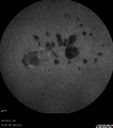
Punctate Inner Choroidopathy - Recurrent CNVM 4 years after initial treatment Left Eye - Indocyanine Green Angiogram703 viewsJune 7, 2012. This pleasant 35-year-old woman noticed decreasing vision in the left eye recently. She returns now noticing the vision in the left eye is declining. OD 20/50, OS 20/80. OS: Vertical C/D ratio is 0.3. There is a scar just inferonasal to the fovea, which has grown some towards the fovea with a little blood just under the fovea.
|
|

Punctate Inner Choroidopathy - Recurrent CNVM 4 years after initial treatment Left Eye - Indocyanine Green Angiogram700 viewsJune 7, 2012. This pleasant 35-year-old woman noticed decreasing vision in the left eye recently. She returns now noticing the vision in the left eye is declining. OD 20/50, OS 20/80. OS: Vertical C/D ratio is 0.3. There is a scar just inferonasal to the fovea, which has grown some towards the fovea with a little blood just under the fovea.
|
|

Punctate Inner Choroidopathy - Recurrent CNVM 4 years after initial treatment Left Eye - Fluorescein Angiogram698 viewsJune 7, 2012. This pleasant 35-year-old woman noticed decreasing vision in the left eye recently. She returns now noticing the vision in the left eye is declining. OD 20/50, OS 20/80. OS: Vertical C/D ratio is 0.3. There is a scar just inferonasal to the fovea, which has grown some towards the fovea with a little blood just under the fovea.
|
|
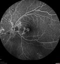
Punctate Inner Choroidopathy - Recurrent CNVM 4 years after initial treatment Left Eye - Fluorescein Angiogram693 viewsJune 7, 2012. This pleasant 35-year-old woman noticed decreasing vision in the left eye recently. She returns now noticing the vision in the left eye is declining. OD 20/50, OS 20/80. OS: Vertical C/D ratio is 0.3. There is a scar just inferonasal to the fovea, which has grown some towards the fovea with a little blood just under the fovea.
|
|

Punctate Inner Choroidopathy - Recurrent CNVM 4 years after initial treatment Left Eye - FAF680 viewsJune 7, 2012. This pleasant 35-year-old woman noticed decreasing vision in the left eye recently. She returns now noticing the vision in the left eye is declining. OD 20/50, OS 20/80. OS: Vertical C/D ratio is 0.3. There is a scar just inferonasal to the fovea, which has grown some towards the fovea with a little blood just under the fovea.
|
|
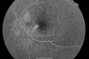
Multifocal Choroiditis and Subretinal Fibrosis - 32 yo Female New Lesion OS678 views32-year-old woman vision loss in the right eye associated with macular scarring and multifocal choroiditis in 1999 with new vision loss in left eye: OD 20/400, OS 20/50.
|
|

Punctate Inner Choroidopathy - Recurrent CNVM 4 years after initial treatment Left Eye - SDOCT678 viewsJune 7, 2012. This pleasant 35-year-old woman noticed decreasing vision in the left eye recently. She returns now noticing the vision in the left eye is declining. OD 20/50, OS 20/80. OS: Vertical C/D ratio is 0.3. There is a scar just inferonasal to the fovea, which has grown some towards the fovea with a little blood just under the fovea.
|
|
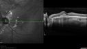
Punctate Inner Choroidopathy - Recurrent CNVM 4 years after initial treatment Left Eye - SD OCT671 viewsJune 7, 2012. This pleasant 35-year-old woman noticed decreasing vision in the left eye recently. She returns now noticing the vision in the left eye is declining. OD 20/50, OS 20/80. OS: Vertical C/D ratio is 0.3. There is a scar just inferonasal to the fovea, which has grown some towards the fovea with a little blood just under the fovea.
|
|

Punctate Inner Choroidopathy - Recurrent CNVM 4 years after initial treatment Left Eye - Fluorescein Angiogram667 viewsJune 7, 2012. This pleasant 35-year-old woman noticed decreasing vision in the left eye recently. She returns now noticing the vision in the left eye is declining. OD 20/50, OS 20/80. OS: Vertical C/D ratio is 0.3. There is a scar just inferonasal to the fovea, which has grown some towards the fovea with a little blood just under the fovea.
|
|

Punctate Inner Choroidopathy - Recurrent CNVM 4 years after initial treatment Left Eye - Fluorescein Angiogram666 viewsJune 7, 2012. This pleasant 35-year-old woman noticed decreasing vision in the left eye recently. She returns now noticing the vision in the left eye is declining. OD 20/50, OS 20/80. OS: Vertical C/D ratio is 0.3. There is a scar just inferonasal to the fovea, which has grown some towards the fovea with a little blood just under the fovea.
|
|

Punctate Inner Choroidopathy - Recurrent CNVM 4 years after initial treatment Left Eye - Indocyanine Green Angiogram662 viewsJune 7, 2012. This pleasant 35-year-old woman noticed decreasing vision in the left eye recently. She returns now noticing the vision in the left eye is declining. OD 20/50, OS 20/80. OS: Vertical C/D ratio is 0.3. There is a scar just inferonasal to the fovea, which has grown some towards the fovea with a little blood just under the fovea.
|
|

Multifocal Choroiditis and Subretinal Fibrosis - 32 yo Female New Lesion OS653 views32-year-old woman vision loss in the right eye associated with macular scarring and multifocal choroiditis in 1999 with new vision loss in left eye: OD 20/400, OS 20/50.
|
|

Punctate Inner Choroidopathy - Recurrent CNVM 4 years after initial treatment Left Eye 651 viewsJune 7, 2012. This pleasant 35-year-old woman noticed decreasing vision in the left eye recently. She returns now noticing the vision in the left eye is declining. OD 20/50, OS 20/80. OS: Vertical C/D ratio is 0.3. There is a scar just inferonasal to the fovea, which has grown some towards the fovea with a little blood just under the fovea.
|
|

Punctate Inner Choroidopathy - Recurrent CNVM 4 years after initial treatment Left Eye - Fluorescein Angiogram651 viewsJune 7, 2012. This pleasant 35-year-old woman noticed decreasing vision in the left eye recently. She returns now noticing the vision in the left eye is declining. OD 20/50, OS 20/80. OS: Vertical C/D ratio is 0.3. There is a scar just inferonasal to the fovea, which has grown some towards the fovea with a little blood just under the fovea.
|
|
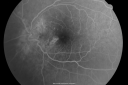
Multifocal Choroiditis and Subretinal Fibrosis - 32 yo Female New Lesion OS649 views32-year-old woman vision loss in the right eye associated with macular scarring and multifocal choroiditis in 1999 with new vision loss in left eye: OD 20/400, OS 20/50.
|
|

Punctate Inner Choroidopathy - Recurrent CNVM 4 years after initial treatment Left Eye - Fluorescein Angiogram647 viewsJune 7, 2012. This pleasant 35-year-old woman noticed decreasing vision in the left eye recently. She returns now noticing the vision in the left eye is declining. OD 20/50, OS 20/80. OS: Vertical C/D ratio is 0.3. There is a scar just inferonasal to the fovea, which has grown some towards the fovea with a little blood just under the fovea.
|
|

Punctate Inner Choroidopathy - Recurrent CNVM 4 years after initial treatment Left Eye - Fluorescein Angiogram645 viewsJune 7, 2012. This pleasant 35-year-old woman noticed decreasing vision in the left eye recently. She returns now noticing the vision in the left eye is declining. OD 20/50, OS 20/80. OS: Vertical C/D ratio is 0.3. There is a scar just inferonasal to the fovea, which has grown some towards the fovea with a little blood just under the fovea.
|
|

Punctate Inner Choroidopathy - Recurrent CNVM 4 years after initial treatment Left Eye - Indocyanine Green Angiogram622 viewsJune 7, 2012. This pleasant 35-year-old woman noticed decreasing vision in the left eye recently. She returns now noticing the vision in the left eye is declining. OD 20/50, OS 20/80. OS: Vertical C/D ratio is 0.3. There is a scar just inferonasal to the fovea, which has grown some towards the fovea with a little blood just under the fovea.
|
|

Punctate Inner Choroidopathy - Recurrent CNVM 4 years after initial treatment Left Eye - Fluorescein Angiogram621 viewsJune 7, 2012. This pleasant 35-year-old woman noticed decreasing vision in the left eye recently. She returns now noticing the vision in the left eye is declining. OD 20/50, OS 20/80. OS: Vertical C/D ratio is 0.3. There is a scar just inferonasal to the fovea, which has grown some towards the fovea with a little blood just under the fovea.
|
|

Punctate Inner Choroidopathy - Recurrent CNVM 4 years after initial treatment Left Eye - Indocyanine Green Angiogram619 viewsJune 7, 2012. This pleasant 35-year-old woman noticed decreasing vision in the left eye recently. She returns now noticing the vision in the left eye is declining. OD 20/50, OS 20/80. OS: Vertical C/D ratio is 0.3. There is a scar just inferonasal to the fovea, which has grown some towards the fovea with a little blood just under the fovea.
|
|

Punctate Inner Choroidopathy - Recurrent CNVM 4 years after initial treatment Left Eye - Fluorescein Angiogram614 viewsJune 7, 2012. This pleasant 35-year-old woman noticed decreasing vision in the left eye recently. She returns now noticing the vision in the left eye is declining. OD 20/50, OS 20/80. OS: Vertical C/D ratio is 0.3. There is a scar just inferonasal to the fovea, which has grown some towards the fovea with a little blood just under the fovea.
|
|

Punctate Inner Choroidopathy - Recurrent CNVM 4 years after initial treatment Left Eye - Indocyanine Green Angiogram612 viewsJune 7, 2012. This pleasant 35-year-old woman noticed decreasing vision in the left eye recently. She returns now noticing the vision in the left eye is declining. OD 20/50, OS 20/80. OS: Vertical C/D ratio is 0.3. There is a scar just inferonasal to the fovea, which has grown some towards the fovea with a little blood just under the fovea.
|
|

Punctate Inner Choroidopathy - Recurrent CNVM 4 years after initial treatment Left Eye 606 viewsJune 7, 2012. This pleasant 35-year-old woman noticed decreasing vision in the left eye recently. She returns now noticing the vision in the left eye is declining. OD 20/50, OS 20/80. OS: Vertical C/D ratio is 0.3. There is a scar just inferonasal to the fovea, which has grown some towards the fovea with a little blood just under the fovea.
|
|

Punctate Inner Choroidopathy - Recurrent CNVM 4 years after initial treatment Left Eye - Fluorescein Angiogram605 viewsJune 7, 2012. This pleasant 35-year-old woman noticed decreasing vision in the left eye recently. She returns now noticing the vision in the left eye is declining. OD 20/50, OS 20/80. OS: Vertical C/D ratio is 0.3. There is a scar just inferonasal to the fovea, which has grown some towards the fovea with a little blood just under the fovea.
|
|

Punctate Inner Choroidopathy - Recurrent CNVM 4 years after initial treatment Left Eye - Indocyanine Green Angiogram592 viewsJune 7, 2012. This pleasant 35-year-old woman noticed decreasing vision in the left eye recently. She returns now noticing the vision in the left eye is declining. OD 20/50, OS 20/80. OS: Vertical C/D ratio is 0.3. There is a scar just inferonasal to the fovea, which has grown some towards the fovea with a little blood just under the fovea.
|
|
|
|
|
|
32-year-old woman in the office on February 21, 2008. I appreciate you sending along her old notes from 1999 when she had an episode of vision loss in the right eye associated with macular scarring and multifocal choroiditis. At that time she was not seen to have any active lesions in her macula and comes in now with the vision complaints lasting about two weeks of blurred vision in the left and good eye, obviously causing her concern.
She is not having any health problems. She does have a history of migraine and she also takes a multivitamin. She does not have any fevers, rashes, or arthritis. She has no evidence of any oral or genital sores, nor does she have any breathing problems.
VISUAL ACUITY: OD 20/400, OS 20/50. IOP: OD 15, OS 16.
SLIT LAMP EXAM: Normal clear lenses.
EXTENDED OPHTHALMOSCOPY:
OD: Vertical C/D ratio is 0.1. There are multifocal chorioretinal scars with a foveal scar.
OS: Vertical C/D ratio is 0.1. There are multifocal chorioretinal scars. There is a focus of choroiditis just inferonasal to the fovea about 500 microns short of the fovea.
OCT SCAN: The right eye shows diffuse retinal atrophy. The left eye shows thickening of the retinal pigment epithelium with overlying retinal edema just nasal to the fovea. Photos confirm clinical findings.
FLUORESCEIN ANGIOGRAM: Shows increasing hyperfluorescence in the lesion of the macula of the left eye. There is, however, no definite vascularity to it. The right eye has staining macular scars.
IMPRESSION:
1. PIC – BOTH EYES
2. NEW LESION OF ACTIVE CHOROIDITIS THREATENING THE FOVEA – LEFT EYE
3. POSSIBLE CHOROIDAL NEOVASCULAR MEMBRANE – LEFT EYE
DISCUSSION: I explained to the patient given her history of multifocal scarring and the new lesion which appears to have no vascularity, no hemorrhage, very little overlying fluid, and is not associated with an old macular scar, I think this is most likely a focus of active inflammation and not an area of neovascularization. Patients with multifocal choroiditis rarely develop choroidal neovascular membranes, whereas patients with punctate inner choroidopathy do, but she does not look to have punctate inner choroidopathy. Because the lesion looks inflammatory, I am starting her on 60 mg of oral steroids daily. I asked her to return for a check in one week, sooner should she notice any problem. If we have no response at that time, I will probably try her with intravitreal Kenalog. If we do have response, we will need to do something about keeping her on anti-inflammatory medicines to prevent central vision loss in the left eye. If the lesion starts to look more vascular, I will treat her with intravitreal blood vessel inhibitors.
When she comes next time, I will also obtain blood tests from Dr. Miller’s office and see what testing needs to be obtained in her to rule-out possible pathologies for possible causes of multifocal choroiditis.
TWO MONTHS LATER:
Her vision in the left eye is doing a little bit better. Interestingly, the right eye is doing substantially better since I first encountered her. She since has had treatment in the left eye with a posterior sub-Tenon Kenalog in February and intravitreal Avastin in March.
VISUAL ACUITY: Vision OD is 20/30, OS is 20/40.
FU: 4 years later:
June 7, 2012. This pleasant 35-year-old woman noticed decreasing vision in the left eye recently. She returns now noticing the vision in the left eye is declining.
VISUAL ACUITY: OD 20/50, OS 20/80. IOP: OD 14, OS 17. There is 1+ nuclear sclerosis in both eyes.
EXTENDED OPHTHALMOSCOPY:
OD: Vertical C/D ratio is 0.3. There is multifocal chorioretinal scars in the macula as well as nasal to the optic nerve. There is some hemorrhage nasal to the optic nerve.
OS: Vertical C/D ratio is 0.3. There is a scar just inferonasal to the fovea, which has grown some towards the fovea with a little blood just under the fovea.
SPECTRALIS-SD-OCT SCAN: The OCT scan in the right eye shows the macula to be dry. The left eye shows macular edema. The photos confirm the retinal hemorrhage in the left eye.
FLUORESCEIN ANGIOGRAPHY: Fluorescein angiography of the right eye shows multiple hyperfluorescence spots in the macula where the scarring is. The left eye shows an increasing hyperfluorescencent area under the fovea, which does look to be vascular, likely from a neovascular complex.
INDOCYANINE GREEN ANGIOGRAPHY: An indocyanine green angiogram was obtained in the both eyes, because of the multifocal chorioditis. The indocyanine green does show hypofluorescence centrally where the neovascular complex is and in the later frames there is a small hypofluorescence lesion. The right eye shows multifocal hypofluorescence spots corresponding to the macular scar.
IMPRESSION:
1. MULTIFOCAL CHOROIDITIS SUBRETINAL FIBROSIS – BOTH EYES
2. NEW FOVEAL HEMORRHAGE WITH A NEOVASCULAR MEMBRANE – LEFT EYE
3. ECCENTRIC HEMORRHAGE – RIGHT EYE
DISCUSSION: I explained to the patient the left macula does appear to have an area of scarring that is reactivated and there is a new blood vessel growth extending towards the fovea, which looks like it may at the moment be subfoveal. I treated the left eye with intravitreal injection of Avastin (1.25 mg/0.05 ml) without any difficulty today.
Vision returned to 20/40 after 3 treatments. Decision was made to treat every 3 months.


