Most viewed - Diabetic Macular Edema - SD-OCT - Clinically Significant Macular Edema with Normal Central Vision 63-year-old man was seen in the office on May 10, 2012. He has been diabetic for twenty two years. He did have a stroke in September. He lost his right peripheral vision. His central vision is fine.
VISUAL ACUITY: OD 20/25, OS 20/25. IOP: OD 11, OS 14.
SLIT EXAMINATION: There is 2+ nuclear sclerosis in both eyes.
EXTENDED OPHTHALMOSCOPY:
OD: Vertical C/D ratio is 0.6. There is no posterior vitreous separation. There is exudate and microaneurysms clustered particularly inferior to the fovea.
OS: Vertical C/D ratio is 0.6. There is no posterior vitreous separation. There is exudate and microaneurysms clustered superior to the fovea. In both eyes there does appear to be edema within 500 microns of the center of the fovea.
SPECTRALIS-SD-OCT SCAN: The OCT scan in the right eye shows an average central foveal thickness of 270 microns, which is normal. The left eye shows an average central foveal thickness of 258 microns, which is normal. Both eyes though do have edema within less than a disc-diameter in the center.
FLUORESCEIN ANGIOGRAPHY: Fluorescein angiography shows microaneurysms leaking inferior to the fovea in the right eye and superior to the fovea in the left eye.
FUNDUS PHOTOGRAPHY - INFRA RED: The infra red and auto fluorescence images show a cluster of microaneurysms inferior to the fovea with some hemorrhage in that area. The infra red and auto fluorescence images in the left eye show microaneurysms clustered superior to the fovea with some hemorrhage in that area.
IMPRESSION:
1. BACKGROUND DIABETIC RETINOPATHY – BOTH EYES
2. CLINICALLY SIGNIFICANT MACULAR EDEMA – BOTH EYES
DISCUSSION: I explained to the patient he does have diabetic macular edema in both eyes. The ETDRS study shows that laser treatment is beneficial in this situation, however, the sub group analysis show that people with good vision there didn’t seem to be any difference between treated and non treated patients in two years. Therefore I think it is reasonable with today’s technology, to follow him closely and treat him should the edema worsen and not treat him should the edema stabilize or improve. To that I asked him to return for a check in three to four months or sooner should he notice a problem.
|

912 views63-year-old man diabetic for twenty two years. His central vision is fine.
VISUAL ACUITY: OD 20/25, OS 20/25
|
|

Diabetic Edema Better - 3 Months Follow-UP - No treatment - SD OCT781 viewsPatient remains asymptomatic and OCT shows less edema
|
|

769 views63-year-old man diabetic for twenty two years. His central vision is fine.
VISUAL ACUITY: OD 20/25, OS 20/25
|
|

702 views63-year-old man diabetic for twenty two years. His central vision is fine.
VISUAL ACUITY: OD 20/25, OS 20/25
|
|
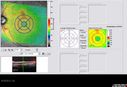
686 views63-year-old man diabetic for twenty two years. His central vision is fine.
VISUAL ACUITY: OD 20/25, OS 20/25
|
|

674 views63-year-old man diabetic for twenty two years. His central vision is fine.
VISUAL ACUITY: OD 20/25, OS 20/25
|
|

671 views63-year-old man diabetic for twenty two years. His central vision is fine.
VISUAL ACUITY: OD 20/25, OS 20/25
|
|

Diabetic Edema Better - 3 Months Follow-UP - No treatment - SD OCT658 viewsPatient remains asymptomatic and OCT shows less edema
|
|

647 views63-year-old man diabetic for twenty two years. His central vision is fine.
VISUAL ACUITY: OD 20/25, OS 20/25
|
|

609 views63-year-old man diabetic for twenty two years. His central vision is fine.
VISUAL ACUITY: OD 20/25, OS 20/25
|
|

579 views63-year-old man diabetic for twenty two years. His central vision is fine.
VISUAL ACUITY: OD 20/25, OS 20/25
|
|

570 views63-year-old man diabetic for twenty two years. His central vision is fine.
VISUAL ACUITY: OD 20/25, OS 20/25
|
|

569 views63-year-old man diabetic for twenty two years. His central vision is fine.
VISUAL ACUITY: OD 20/25, OS 20/25
|
|

556 views63-year-old man diabetic for twenty two years. His central vision is fine.
VISUAL ACUITY: OD 20/25, OS 20/25
|
|

553 views63-year-old man diabetic for twenty two years. His central vision is fine.
VISUAL ACUITY: OD 20/25, OS 20/25
|
|

550 views63-year-old man diabetic for twenty two years. His central vision is fine.
VISUAL ACUITY: OD 20/25, OS 20/25
|
|

549 views63-year-old man diabetic for twenty two years. His central vision is fine.
VISUAL ACUITY: OD 20/25, OS 20/25
|
|

548 views63-year-old man diabetic for twenty two years. His central vision is fine.
VISUAL ACUITY: OD 20/25, OS 20/25
|
|

543 views63-year-old man diabetic for twenty two years. His central vision is fine.
VISUAL ACUITY: OD 20/25, OS 20/25
|
|

538 views63-year-old man diabetic for twenty two years. His central vision is fine.
VISUAL ACUITY: OD 20/25, OS 20/25
|
|

524 views63-year-old man diabetic for twenty two years. His central vision is fine.
VISUAL ACUITY: OD 20/25, OS 20/25
|
|

522 views63-year-old man diabetic for twenty two years. His central vision is fine.
VISUAL ACUITY: OD 20/25, OS 20/25
|
|

508 views63-year-old man diabetic for twenty two years. His central vision is fine.
VISUAL ACUITY: OD 20/25, OS 20/25
|
|

503 views63-year-old man diabetic for twenty two years. His central vision is fine.
VISUAL ACUITY: OD 20/25, OS 20/25
|
|

499 views63-year-old man diabetic for twenty two years. His central vision is fine.
VISUAL ACUITY: OD 20/25, OS 20/25
|
|

492 views63-year-old man diabetic for twenty two years. His central vision is fine.
VISUAL ACUITY: OD 20/25, OS 20/25
|
|

472 views63-year-old man diabetic for twenty two years. His central vision is fine.
VISUAL ACUITY: OD 20/25, OS 20/25
|
|
|
|
|
63-year-old man was seen in the office on May 10, 2012. He has been diabetic for twenty two years. He did have a stroke in September. He lost his right peripheral vision. His central vision is fine.
VISUAL ACUITY: OD 20/25, OS 20/25. IOP: OD 11, OS 14.
SLIT EXAMINATION: There is 2+ nuclear sclerosis in both eyes.
EXTENDED OPHTHALMOSCOPY:
OD: Vertical C/D ratio is 0.6. There is no posterior vitreous separation. There is exudate and microaneurysms clustered particularly inferior to the fovea.
OS: Vertical C/D ratio is 0.6. There is no posterior vitreous separation. There is exudate and microaneurysms clustered superior to the fovea. In both eyes there does appear to be edema within 500 microns of the center of the fovea.
SPECTRALIS-SD-OCT SCAN: The OCT scan in the right eye shows an average central foveal thickness of 270 microns, which is normal. The left eye shows an average central foveal thickness of 258 microns, which is normal. Both eyes though do have edema within less than a disc-diameter in the center.
FLUORESCEIN ANGIOGRAPHY: Fluorescein angiography shows microaneurysms leaking inferior to the fovea in the right eye and superior to the fovea in the left eye.
FUNDUS PHOTOGRAPHY - INFRA RED: The infra red and auto fluorescence images show a cluster of microaneurysms inferior to the fovea with some hemorrhage in that area. The infra red and auto fluorescence images in the left eye show microaneurysms clustered superior to the fovea with some hemorrhage in that area.
IMPRESSION:
1. BACKGROUND DIABETIC RETINOPATHY – BOTH EYES
2. CLINICALLY SIGNIFICANT MACULAR EDEMA – BOTH EYES
DISCUSSION: I explained to the patient he does have diabetic macular edema in both eyes. The ETDRS study shows that laser treatment is beneficial in this situation, however, the sub group analysis show that people with good vision there didn’t seem to be any difference between treated and non treated patients in two years. Therefore I think it is reasonable with today’s technology, to follow him closely and treat him should the edema worsen and not treat him should the edema stabilize or improve. To that I asked him to return for a check in three to four months or sooner should he notice a problem.


