|
Most viewed - Post-Retinal Detachment Repair Subfoveal Occult Choroidal Neovascular Membrane
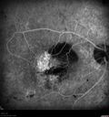
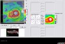
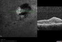
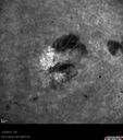
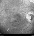
January 2, 2012. This pleasant 56-year-old man has had a complex retinal detachment repair in the right eye. I most recently removed his silicone oil October 12th. His vision now is the best it has ever been. The last he was here though, he did have some macular hemorrhaging and I brought him back. He does recall angiograms being done in the past and a discussion of serous retinal detachment before he developed a rhegmatogenous retinal detachment. He did even think he had a laser done maybe. VISUAL ACUITY: OD 4/200. IOP: 4. There is 1+ cell and flare of the anterior chamber. There is no lens. EXTENDED OPHTHALMOSCOPY: OD: Vertical C/D ratio is 0.2. There is a well-circumscribed hemorrhagic pigment epithelial detachment in the center of the fovea about 2 disc-diameter across. The retina is otherwise attached. SPECTRALIS-SD-OCT SCAN: The OCT scan shows subretinal fluid. Photos confirm clinical findings. FLUORESCEIN ANGIOGRAPHY: Fluorescein angiography shows an occult subfoveal choroidal neovascular membrane about 4 disc areas in size. INDOCYANINE GREEN ANGIOGRAPHY: Shows no leakage and no polypoidal choroidal vasculopathy. IMPRESSION: 1. WET AGE-RELATED MACULAR DEGENERATION – RIGHT EYE 2. PIGMENT EPITHELIAL DETACHMENT – RIGHT EYE 3. SUBFOVEAL CHOROIDAL NEOVASCULAR MEMBRANE - RIGHT EYE 4. HISTORY OF COMPLEX RETINAL DETACHMENT REPAIR – RIGHT EYE 5. CHRONIC VISION LOSS DISCUSSION: I explained to the patient that he does look like he has wet macular degeneration. Treating that in a vitrectomized eye is problematic, because the pharmacokinetics of the drive are unfavorable. I suggested, at least for now we leave the eye alone. He feels this is the best vision he has had in five years and I think photodynamic laser might help dry up the macula, but it might not be necessary. It is possible the disease won’t progress any further. I asked him to return for a check in six weeks or sooner should he notice a problem and see you back periodically. |
|||||||
|
|
|
|
||||
|
|
|
|
||||
|
|
|
|||||
|
|||||||
January 2, 2012. This pleasant 56-year-old man has had a complex retinal detachment repair in the right eye. I most recently removed his silicone oil October 12th. His vision now is the best it has ever been. The last he was here though, he did have some macular hemorrhaging and I brought him back. He does recall angiograms being done in the past and a discussion of serous retinal detachment before he developed a rhegmatogenous retinal detachment. He did even think he had a laser done maybe. VISUAL ACUITY: OD 4/200. IOP: 4. There is 1+ cell and flare of the anterior chamber. There is no lens. EXTENDED OPHTHALMOSCOPY: OD: Vertical C/D ratio is 0.2. There is a well-circumscribed hemorrhagic pigment epithelial detachment in the center of the fovea about 2 disc-diameter across. The retina is otherwise attached. SPECTRALIS-SD-OCT SCAN: The OCT scan shows subretinal fluid. Photos confirm clinical findings. FLUORESCEIN ANGIOGRAPHY: Fluorescein angiography shows an occult subfoveal choroidal neovascular membrane about 4 disc areas in size. INDOCYANINE GREEN ANGIOGRAPHY: Shows no leakage and no polypoidal choroidal vasculopathy. IMPRESSION: 1. WET AGE-RELATED MACULAR DEGENERATION – RIGHT EYE 2. PIGMENT EPITHELIAL DETACHMENT – RIGHT EYE 3. SUBFOVEAL CHOROIDAL NEOVASCULAR MEMBRANE - RIGHT EYE 4. HISTORY OF COMPLEX RETINAL DETACHMENT REPAIR – RIGHT EYE 5. CHRONIC VISION LOSS DISCUSSION: I explained to the patient that he does look like he has wet macular degeneration. Treating that in a vitrectomized eye is problematic, because the pharmacokinetics of the drive are unfavorable. I suggested, at least for now we leave the eye alone. He feels this is the best vision he has had in five years and I think photodynamic laser might help dry up the macula, but it might not be necessary. It is possible the disease won’t progress any further. I asked him to return for a check in six weeks or sooner should he notice a problem and see you back periodically.