|
Most viewed - NVE (neovascularization elsewhere) and Vitreous Hemorrhage
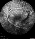
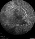
55-year-old woman has had a vitreous hemorrhage in her left eye. She had one in the right eye in the past. The vision is clearing some as the blood absorbs. VISUAL ACUITY: OD 20/32, OS 20/40. IOP: OD 13, OS 12. The posterior chamber intraocular lens is in good position in both eyes. The left eye has a 1+ posterior capsular opacity. EXTENDED OPHTHALMOSCOPY: OD: Vertical C/D ratio is 0.3. There are patchy retinal hemorrhages. OS: Vertical C/D ratio is 0.3. There is neovascularization superotemporal to the macula, which is elevated into the vitreous and clearly under tension. There is a 2+ vitreous hemorrhage. FUNDUS PHOTOGRAPHY: The photos show the retinal hemorrhages in the right eye and the neovascularization in the left eye. FLUORESCEIN ANGIOGRAPHY: Fluorescein angiography shows moderate non perfusion in the periphery of the left eye with heavy leakage from the neovascularization. In the right eye there is some leakage from the microaneurysms in the macula and there is moderate non perfusion in the periphery. SPECTRALIS-SD-OCT SCAN: The OCT scan shows no significant retinal edema in either eye. IMPRESSION: 1. PROLIFERATIVE DIABETIC RETINOPATHY – LEFT EYE 2. VITREOUS HEMORRHAGE – LEFT EYE 3. BACKGROUND DIABETIC RETINOPATHY – RIGHT EYE 4. ABSORBING VITREOUS HEMORRHAGE – LEFT EYE DISCUSSION: I explained to the patient with pan retinal laser we can substantially reduce the risk of severe vision loss in the left eye. We talked about the risks, benefits, indications, and alternatives of panretinal photocoagulation and the patient agreed to this procedure after the questions were answered. Panretinal photocoagulation was performed today without difficulty. |
|||||||
|
|
|
|
||||
|
|
|
|
||||
|
|
|
|
||||
|
|
|
|
||||
|
|
|
|
||||
|
|
|
|
||||
|
|||||||
|
|||||||
55-year-old woman has had a vitreous hemorrhage in her left eye. She had one in the right eye in the past. The vision is clearing some as the blood absorbs. VISUAL ACUITY: OD 20/32, OS 20/40. IOP: OD 13, OS 12. The posterior chamber intraocular lens is in good position in both eyes. The left eye has a 1+ posterior capsular opacity. EXTENDED OPHTHALMOSCOPY: OD: Vertical C/D ratio is 0.3. There are patchy retinal hemorrhages. OS: Vertical C/D ratio is 0.3. There is neovascularization superotemporal to the macula, which is elevated into the vitreous and clearly under tension. There is a 2+ vitreous hemorrhage. FUNDUS PHOTOGRAPHY: The photos show the retinal hemorrhages in the right eye and the neovascularization in the left eye. FLUORESCEIN ANGIOGRAPHY: Fluorescein angiography shows moderate non perfusion in the periphery of the left eye with heavy leakage from the neovascularization. In the right eye there is some leakage from the microaneurysms in the macula and there is moderate non perfusion in the periphery. SPECTRALIS-SD-OCT SCAN: The OCT scan shows no significant retinal edema in either eye. IMPRESSION: 1. PROLIFERATIVE DIABETIC RETINOPATHY – LEFT EYE 2. VITREOUS HEMORRHAGE – LEFT EYE 3. BACKGROUND DIABETIC RETINOPATHY – RIGHT EYE 4. ABSORBING VITREOUS HEMORRHAGE – LEFT EYE DISCUSSION: I explained to the patient with pan retinal laser we can substantially reduce the risk of severe vision loss in the left eye. We talked about the risks, benefits, indications, and alternatives of panretinal photocoagulation and the patient agreed to this procedure after the questions were answered. Panretinal photocoagulation was performed today without difficulty.