Most viewed - Optic Nerve Disease
|

Optic disc swelling6536 views56 year old woman
|
|

Anterior Ischemic Optic Neuropathy - Non-Arteritic - AION2676 views72-year-old woman sudden vision loss in the right eye about four days ago. She also has noticed that things are dim out of that eye. She has no headaches. She does feel a little tired. She has no pain in her jaw when she chews and she has no fevers.
OD is 20/80, PH 20/25; OS is 20/25
|
|

Anterior Ischemic Optic Neuropathy - Non-Arteritic - AION2383 views72-year-old woman sudden vision loss in the right eye about four days ago. She also has noticed that things are dim out of that eye. She has no headaches. She does feel a little tired. She has no pain in her jaw when she chews and she has no fevers.
OD is 20/80, PH 20/25; OS is 20/25
|
|
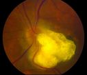
Astrocytic Hamartoma2254 viewsCalcified astrocytic hamartoma of the optic nerve
|
|

Anterior Ischemic Optic Neuropathy - Non-Arteritic - AION2028 views72-year-old woman sudden vision loss in the right eye about four days ago. She also has noticed that things are dim out of that eye. She has no headaches. She does feel a little tired. She has no pain in her jaw when she chews and she has no fevers.
OD is 20/80, PH 20/25; OS is 20/25
|
|

Coloboma of the optic Nerve1958 viewsColoboma
James L. Perron
|
|

Papilledema - Spectral Domain OCT used for Diagnosis - 14 year old Child - Line Scan over Nerve Shows Retinal Edema1894 views14-year-old was with optic nerve swelling asymptomatic - picked up during routine eye exam. OD 20/20, OS 20/32
SD-OCT is used to differentiate optic disc edema from optic nerve head drusen (which are not yet calcified in children).
|
|

Anterior Ischemic Optic Neuropathy - Non-Arteritic - AION1867 views72-year-old woman sudden vision loss in the right eye about four days ago. She also has noticed that things are dim out of that eye. She has no headaches. She does feel a little tired. She has no pain in her jaw when she chews and she has no fevers.
OD is 20/80, PH 20/25; OS is 20/25
|
|

OU Stereo Pair Optic Disc Drusen1847 viewsText book example of OU optic discs with drusen in stereo.
|
|

Papilledema - Spectral Domain OCT used for Diagnosis - 14 year old Child - Nerve Fiber Layer Scan shows swelling1818 views14-year-old was with optic nerve swelling asymptomatic - picked up during routine eye exam. OD 20/20, OS 20/32
SD-OCT is used to differentiate optic disc edema from optic nerve head drusen (which are not yet calcified in children).
|
|

Papilledema - Spectral Domain OCT used for Diagnosis - 14 year old Child - Line Scan over Nerve Shows Retinal Edema1793 views14-year-old was with optic nerve swelling asymptomatic - picked up during routine eye exam. OD 20/20, OS 20/32
SD-OCT is used to differentiate optic disc edema from optic nerve head drusen (which are not yet calcified in children).
|
|

Anterior Ischemic Optic Neuropathy - Non-Arteritic - AION1753 views72-year-old woman sudden vision loss in the right eye about four days ago. She also has noticed that things are dim out of that eye. She has no headaches. She does feel a little tired. She has no pain in her jaw when she chews and she has no fevers.
OD is 20/80, PH 20/25; OS is 20/25
|
|

Optic Nerve Drusen - - Fundus Autofluorescence - FAF1720 views72-year-old woman has had optic nerve drusen for sometime and has visual field abnormalities. OD 20/32, OS 20/32.
|
|

Optic Nerve Head Drusen Right Eye1700 views57-year-old woman has optic nerve drusen in both eyes.
VISUAL ACUITY: OU 20/20.
|
|
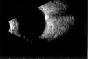
Optic Nerve Head Drusen B-Scan 1695 views57-year-old woman has optic nerve drusen in both eyes.
VISUAL ACUITY: OU 20/20.
|
|

Notching of Optic Nerve Rim Inferiorly1680 viewsProgression of glaucoma in the left eye. Past 3-years, developed a notch inferior to the optic disc in the left eye.
|
|
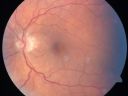
Anterior Ischemic Optic Neuropathy - Non-Arteritic - AION1623 views72-year-old woman sudden vision loss in the right eye about four days ago. She also has noticed that things are dim out of that eye. She has no headaches. She does feel a little tired. She has no pain in her jaw when she chews and she has no fevers.
OD is 20/80, PH 20/25; OS is 20/25
|
|

Swollen Nerve with Hemorraghe1617 views
|
|

Papilledema 10 year old child1601 views10-year-old child has uveitis in both eyes. She probably has tubulointerstitial nephritis-uveitis syndrome, and she apparently also independently has idiopathic elevated intracranial pressure. VISUAL ACUITY: Vision OU is 20/20.
|
|

Optic Nerve Drusen in 7 Year Old Child - Miminal Calcification1595 views7-year-old child difficulty seeing the blackboard in school. She was checked for glasses and it was noted at that time that she had unusual looking optic nerves. Vision OU is 20/50.
|
|

Optic Nerve (Disc) Drusen - Fundus Autofluorescence1591 views34 Year Old woman no headaches normal vision
|
|

Giant Cell Arteritis - 85 year old woman sudden vision loss left eye - AION - Pale Swollen Optic Nerve1570 views 85-year-old woman has felt sick and weak for about the last two months. She is having headaches, jaw claudication, and her eyes ache. Then, three days ago she had sudden vision loss in the left eye. You saw her with optic nerve swelling and suggested she come in here for evaluation. She had coronary bypass surgery in 2004.
VISUAL ACUITY: Vision OD is 20/80, OS is light perception
|
|

Giant Cell Arteritis - 85 year old woman sudden vision loss left eye - AION- Pale Swollen Optic Nerve1549 views 85-year-old woman has felt sick and weak for about the last two months. She is having headaches, jaw claudication, and her eyes ache. Then, three days ago she had sudden vision loss in the left eye. You saw her with optic nerve swelling and suggested she come in here for evaluation. She had coronary bypass surgery in 2004.
VISUAL ACUITY: Vision OD is 20/80, OS is light perception
|
|

Morning Glory Syndrome, Optic Nerve1498 viewsPatient comes in for cataract evaluation. VA 20/60, right eye and 20/150, left eye. Fundus photo shows "morning glory syndrome" of the optic nerve in the left eye.
|
|

Papilledema - Spectral Domain OCT used for Diagnosis - 14 year old Child - FAF1481 views14-year-old was with optic nerve swelling asymptomatic - picked up during routine eye exam. OD 20/20, OS 20/32
SD-OCT is used to differentiate optic disc edema from optic nerve head drusen (which are not yet calcified in children).
|
|

Bilateral Papilledema 1475 viewsYoung male presents with decreased vision in both eyes. Fundus photos reveal swollen optic nerves in both eyes.
|
|

Low Tension Glaucoma - Arrows show nerve fiber layer defect1449 views59-year-old woman has a history of glaucoma dating back to 1990. She had trabeculectomy in the left eye in 1998 and then persisted to lose vision despite normal intraocular pressures from low-tension glaucoma in the left eye. She is now on Cosopt and Travatan in both eyes.
Vision OD is 20/20, OS is 20/16. IOP: OD 9, OS 6.
|
|

Optic Nerve Head Drusen B-Scan 1445 views57-year-old woman has optic nerve drusen in both eyes.
VISUAL ACUITY: OU 20/20.
|
|

Papilledema - Spectral Domain OCT used for Diagnosis - 14 year old Child - Nerve Fiber Layer Scan shows swelling1444 views14-year-old was with optic nerve swelling asymptomatic - picked up during routine eye exam. OD 20/20, OS 20/32
SD-OCT is used to differentiate optic disc edema from optic nerve head drusen (which are not yet calcified in children).
|
|

Optic Nerve Drusen - Line Scan over Optic Nerve1437 views72-year-old woman has had optic nerve drusen for sometime and has visual field abnormalities. OD 20/32, OS 20/32.
|
|

Anterior Ischemic Optic Neuropathy - Non-Arteritic - AION1397 views72-year-old woman sudden vision loss in the right eye about four days ago. She also has noticed that things are dim out of that eye. She has no headaches. She does feel a little tired. She has no pain in her jaw when she chews and she has no fevers.
OD is 20/80, PH 20/25; OS is 20/25
|
|

Papilledema - Spectral Domain OCT used for Diagnosis - 14 year old Child - Nerve Fiber Layer Scan shows swelling1396 views14-year-old was with optic nerve swelling asymptomatic - picked up during routine eye exam. OD 20/20, OS 20/32
SD-OCT is used to differentiate optic disc edema from optic nerve head drusen (which are not yet calcified in children).
|
|

Low Tension Glaucoma1375 views59-year-old woman has a history of glaucoma dating back to 1990. She had trabeculectomy in the left eye in 1998 and then persisted to lose vision despite normal intraocular pressures from low-tension glaucoma in the left eye. She is now on Cosopt and Travatan in both eyes.
Vision OD is 20/20, OS is 20/16. IOP: OD 9, OS 6.
|
|
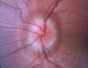
Optic Nerve Drusen in 7 Year Old Child - Miminal Calcification1374 views7-year-old child difficulty seeing the blackboard in school. She was checked for glasses and it was noted at that time that she had unusual looking optic nerves. Vision OU is 20/50.
|
|
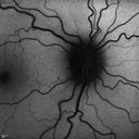
Optic Nerve (Disc) Drusen - Fundus Autofluorescence1361 views34 Year Old woman no headaches normal vision
|
|

Papilledema - Spectral Domain OCT used for Diagnosis - 14 year old Child - FAF1342 views14-year-old was with optic nerve swelling asymptomatic - picked up during routine eye exam. OD 20/20, OS 20/32
SD-OCT is used to differentiate optic disc edema from optic nerve head drusen (which are not yet calcified in children).
|
|

Papilledema - Spectral Domain OCT used for Diagnosis - 14 year old Child - Nerve Fiber Layer Scan shows swelling1338 views14-year-old was with optic nerve swelling asymptomatic - picked up during routine eye exam. OD 20/20, OS 20/32
SD-OCT is used to differentiate optic disc edema from optic nerve head drusen (which are not yet calcified in children).
|
|

Myopic - Tilted - Optic Nerves with Torsion1333 views 82-year-old man He had an injury when he was a child. The right eye unfortunately has had poor vision since he is so nearsighted, even without a lens, he sees some out of the right eye. Vision OD is 20/100, OS is 20/20.
|
|
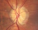
Optic Nerve Head Drusen Left Eye1325 views57-year-old woman has optic nerve drusen in both eyes.
VISUAL ACUITY: OU 20/20.
|
|

Myopic - Tilted - Optic Nerves with Torsion1321 views 82-year-old man He had an injury when he was a child. The right eye unfortunately has had poor vision since he is so nearsighted, even without a lens, he sees some out of the right eye. Vision OD is 20/100, OS is 20/20.
|
|
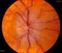
Bilateral Papilledema1287 viewsYoung female that presents with severe frequent headaches. VA is 20/15 in both eyes. Fundus exam reveals swelling of both optic nerves.
|
|

Giant Cell Arteritis - 85 year old woman sudden vision loss left eye - AION- Pale Swollen Optic Nerve1285 views 85-year-old woman has felt sick and weak for about the last two months. She is having headaches, jaw claudication, and her eyes ache. Then, three days ago she had sudden vision loss in the left eye. You saw her with optic nerve swelling and suggested she come in here for evaluation. She had coronary bypass surgery in 2004.
VISUAL ACUITY: Vision OD is 20/80, OS is light perception
|
|

Anterior Ischemic Optic Neuropathy - Non-Arteritic - AION1257 views72-year-old woman sudden vision loss in the right eye about four days ago. She also has noticed that things are dim out of that eye. She has no headaches. She does feel a little tired. She has no pain in her jaw when she chews and she has no fevers.
OD is 20/80, PH 20/25; OS is 20/25
|
|

Optic Nerve Drusen - - Nerve Fiber Layer Scan1219 views72-year-old woman has had optic nerve drusen for sometime and has visual field abnormalities. OD 20/32, OS 20/32.
|
|

Glaucoma (also Diabetes and Previous Retinal Detachment both Eyes)1213 views84-year-old man has had retinal detachment repairs in both eyes. He also has advanced optic atrophy. He also has had diabetes for thirty five years. OD 20/30, OS 20/40
|
|

Optic Nerve Drusen in 7 Year Old Child - Miminal Calcification1205 views7-year-old child difficulty seeing the blackboard in school. She was checked for glasses and it was noted at that time that she had unusual looking optic nerves. Vision OU is 20/50.
|
|

Optic Nerve Drusen - Line Scan over Optic Nerve1201 views72-year-old woman has had optic nerve drusen for sometime and has visual field abnormalities. OD 20/32, OS 20/32.
|
|
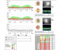
Optic Nerve Head Drusen Nerve Fiber Layer Scan1200 views57-year-old woman has optic nerve drusen in both eyes.
VISUAL ACUITY: OU 20/20.
|
|

Optic Pit OCT1195 viewsOCT Scan of an Optic Pit in OS of a 40 yr old male
|
|

Optic Nerve Drusen in 7 Year Old Child - Miminal Calcification1184 views7-year-old child difficulty seeing the blackboard in school. She was checked for glasses and it was noted at that time that she had unusual looking optic nerves. Vision OU is 20/50.
|
|

Optic Nerve Hypoplasia1170 views28-year-old man with diabetes and anomalous vessels near nerve. OD is 20/25, OS is 20/20. IOP: OD 20, OS 22.
OD: Vertical C/D ratio is 0.0. The nerve is tilted and small and there is an anomalous branching pattern of the major retinal vessels. There is a tuft of irregular telangiectatic vessels on the nasal edge of the optic nerve. There is also a buried optic nerve druse at the optic margin at 5:00 o’clock. There are rare retinal hemorrhages in the macula.
OS: Vertical C/D ratio is 0.0. Again, the nerve looks small. There is an anomalous branching pattern of the retinal vessels and rare retinal hemorrhages.
|
|

Glaucomatous optic nerve with severe cupping1167 views
|
|

Papilledema - Spectral Domain OCT used for Diagnosis - 14 year old Child1162 views14-year-old was with optic nerve swelling asymptomatic - picked up during routine eye exam. OD 20/20, OS 20/32
SD-OCT is used to differentiate optic disc edema from optic nerve head drusen (which are not yet calcified in children).
|
|

Glaucoma (also Diabetes and Previous Retinal Detachment both Eyes)1150 views84-year-old man has had retinal detachment repairs in both eyes. He also has advanced optic atrophy. He also has had diabetes for thirty five years. OD 20/30, OS 20/40
|
|

Myopic - Tilted - Optic Nerves with Torsion1144 views 82-year-old man He had an injury when he was a child. The right eye unfortunately has had poor vision since he is so nearsighted, even without a lens, he sees some out of the right eye. Vision OD is 20/100, OS is 20/20.
|
|
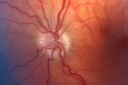
Optic Nerve Hypoplasia1142 views28-year-old man with diabetes and anomalous vessels near nerve. OD is 20/25, OS is 20/20. IOP: OD 20, OS 22.
OD: Vertical C/D ratio is 0.0. The nerve is tilted and small and there is an anomalous branching pattern of the major retinal vessels. There is a tuft of irregular telangiectatic vessels on the nasal edge of the optic nerve. There is also a buried optic nerve druse at the optic margin at 5:00 o’clock. There are rare retinal hemorrhages in the macula.
OS: Vertical C/D ratio is 0.0. Again, the nerve looks small. There is an anomalous branching pattern of the retinal vessels and rare retinal hemorrhages.
|
|

Optic Nerve (Disc) Drusen - Spectral Domain Optical Coherence Tomography Line Scan1136 views34 Year Old woman no headaches normal vision
|
|

Low Tension Glaucoma1133 views59-year-old woman has a history of glaucoma dating back to 1990. She had trabeculectomy in the left eye in 1998 and then persisted to lose vision despite normal intraocular pressures from low-tension glaucoma in the left eye. She is now on Cosopt and Travatan in both eyes.
Vision OD is 20/20, OS is 20/16. IOP: OD 9, OS 6.
|
|

Papilledema - Spectral Domain OCT used for Diagnosis - 14 year old Child1133 views14-year-old was with optic nerve swelling asymptomatic - picked up during routine eye exam. OD 20/20, OS 20/32
SD-OCT is used to differentiate optic disc edema from optic nerve head drusen (which are not yet calcified in children).
|
|

Myopic - Tilted - Optic Nerves with Torsion1113 views 82-year-old man He had an injury when he was a child. The right eye unfortunately has had poor vision since he is so nearsighted, even without a lens, he sees some out of the right eye. Vision OD is 20/100, OS is 20/20.
|
|

Papilledema - Spectral Domain OCT used for Diagnosis - 14 year old Child - IR Photo1111 views14-year-old was with optic nerve swelling asymptomatic - picked up during routine eye exam. OD 20/20, OS 20/32
SD-OCT is used to differentiate optic disc edema from optic nerve head drusen (which are not yet calcified in children).
|
|

Glaucoma (also Diabetes and Previous Retinal Detachment both Eyes)1110 views84-year-old man has had retinal detachment repairs in both eyes. He also has advanced optic atrophy. He also has had diabetes for thirty five years. OD 20/30, OS 20/40
|
|

Optic Neuritis with Suspected Harada's Disease 1109 viewsYoung female patient comes in with red and irritated eyes which were sensitive to the touch. Patient also had vision loss in both eyes. VA was 20/80, right eye, 20/60, left eye. Fundiscopic exam shows sub retinal fluid in the right eye surrounding the optic nerve. Patient will start steroid therapy to reduce any other inflammation that is active.
|
|

Optic Nerve Drusen - - Fundus Autofluorescence - FAF1105 views72-year-old woman has had optic nerve drusen for sometime and has visual field abnormalities. OD 20/32, OS 20/32.
|
|

Optic Nerve Drusen in 7 Year Old Child - Miminal Calcification1095 views7-year-old child difficulty seeing the blackboard in school. She was checked for glasses and it was noted at that time that she had unusual looking optic nerves. Vision OU is 20/50.
|
|
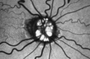
Optic Nerve Drusen - Fundus Autofluorescence - FAF1093 views72-year-old woman has had optic nerve drusen for sometime and has visual field abnormalities. OD 20/32, OS 20/32.
|
|

Papilledema 10 year old child1092 views10-year-old child has uveitis in both eyes. She probably has tubulointerstitial nephritis-uveitis syndrome, and she apparently also independently has idiopathic elevated intracranial pressure. VISUAL ACUITY: Vision OU is 20/20.
|
|
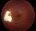
Myelinated Nerve Fiber Layer1089 views
|
|

Papilledema unspecified 1088 views
|
|

Optic Nerve Drusen in 7 Year Old Child - Miminal Calcification1083 views7-year-old child difficulty seeing the blackboard in school. She was checked for glasses and it was noted at that time that she had unusual looking optic nerves. Vision OU is 20/50.
|
|

Optic Nerve Pit Retinal Detachment - Macular Detachment - 8 Year Old - 5/200 Vision OS1083 views8-year-old child on two school exams that his vision was poor in the left eye and his last normal exam was in preschool.
VISUAL ACUITY: OD 20/30, OS 5/200. OS: Vertical C/D ratio is 0.2. There is a whitening on the temporal edge of the optic nerve consistent with an optic disc pit and there is a serous macular detachment.
|
|

Optic Nerve (Disc) Drusen - Infrared Reflectance1083 views34 Year Old woman no headaches normal vision
|
|
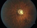
Acute Non-arteritic Anterior Ischemic Optic Neuropathy Right Eye - Previous NA-AION left eye 1079 views69-year-old man previously lost vision in the left eye from nonarteritic anterior ischemic optic neuropathy in 2006. About a week ago and noticed blurred vision in the inferonasal part of the vision in the right eye.
VISUAL ACUITY: Vision OD is 20/16, OS is 20/40
|
|

Optic Disc Drusen1074 viewsOptic Nerve Head Drusen in teenage patient.
James L. Perron, C.R.A.
|
|

Optic Nerve Head Drusen B-Scan 1072 views57-year-old woman has optic nerve drusen in both eyes.
VISUAL ACUITY: OU 20/20.
|
|

Acute Non-arteritic Anterior Ischemic Optic Neuropathy Right Eye - Previous NA-AION left eye 1072 views69-year-old man previously lost vision in the left eye from nonarteritic anterior ischemic optic neuropathy in 2006. About a week ago and noticed blurred vision in the inferonasal part of the vision in the right eye.
VISUAL ACUITY: Vision OD is 20/16, OS is 20/40
|
|

Glaucoma (also Diabetes and Previous Retinal Detachment both Eyes)1062 views84-year-old man has had retinal detachment repairs in both eyes. He also has advanced optic atrophy. He also has had diabetes for thirty five years. OD 20/30, OS 20/40
|
|

Giant Cell Arteritis - 85 year old woman sudden vision loss left eye - AION- Pale Swollen Optic Nerve1057 views 85-year-old woman has felt sick and weak for about the last two months. She is having headaches, jaw claudication, and her eyes ache. Then, three days ago she had sudden vision loss in the left eye. You saw her with optic nerve swelling and suggested she come in here for evaluation. She had coronary bypass surgery in 2004.
VISUAL ACUITY: Vision OD is 20/80, OS is light perception
|
|

Optic Nerve Head Drusen B-Scan 1055 views57-year-old woman has optic nerve drusen in both eyes.
VISUAL ACUITY: OU 20/20.
|
|

Optic Nerve Drusen - Nerve Fiber Layer Scan1051 views72-year-old woman has had optic nerve drusen for sometime and has visual field abnormalities. OD 20/32, OS 20/32.
|
|
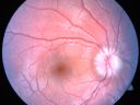
Optic Nerve Drusen in 7 Year Old Child - Miminal Calcification1050 views7-year-old child difficulty seeing the blackboard in school. She was checked for glasses and it was noted at that time that she had unusual looking optic nerves. Vision OU is 20/50.
|
|

Superior Peripapillary Hemorrhage1032 viewsPatient was seen for acute vision loss in the right eye. Patient has glaucoma. VA was 20/70 in the right eye. Had Vitrectomy back in May 2012 for ERM stripping. Also had Trabectome with cataract surgery back in December of 2012. Fundus photos presents a Superior Peripapillary Hemorrhage of the optic nerve. Patient will be followed up in one month.
|
|

Papilledema - Spectral Domain OCT used for Diagnosis - 14 year old Child1018 views14-year-old was with optic nerve swelling asymptomatic - picked up during routine eye exam. OD 20/20, OS 20/32
SD-OCT is used to differentiate optic disc edema from optic nerve head drusen (which are not yet calcified in children).
|
|
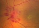
Atypical Neuroretinitis - ?Ischemic - ?Paraneoplastic - Optic Nerve Swelling and Foveal Edema and Exudate1016 views74-year-old woman has stage 4 non-small cell lung carcinoma decreasing vision in the left eye for at least the last two weeks, VA 10/200. Trace APD OS. No pain
|
|

Low Tension Glaucoma1015 views59-year-old woman has a history of glaucoma dating back to 1990. She had trabeculectomy in the left eye in 1998 and then persisted to lose vision despite normal intraocular pressures from low-tension glaucoma in the left eye. She is now on Cosopt and Travatan in both eyes.
Vision OD is 20/20, OS is 20/16. IOP: OD 9, OS 6.
|
|

Acute Non-arteritic Anterior Ischemic Optic Neuropathy Right Eye - Previous NA-AION left eye 1015 views69-year-old man previously lost vision in the left eye from nonarteritic anterior ischemic optic neuropathy in 2006. About a week ago and noticed blurred vision in the inferonasal part of the vision in the right eye.
VISUAL ACUITY: Vision OD is 20/16, OS is 20/40
|
|

Optic Nerve (Disc) Drusen - Infrared Reflectance1015 views34 Year Old woman no headaches normal vision
|
|

Optic Nerve (Disc) Drusen - Spectral Domain Optical Coherence Tomography Line Scan1012 views34 Year Old woman no headaches normal vision
|
|

Acute Non-arteritic Anterior Ischemic Optic Neuropathy Right Eye - Previous NA-AION left eye 1006 views69-year-old man previously lost vision in the left eye from nonarteritic anterior ischemic optic neuropathy in 2006. About a week ago and noticed blurred vision in the inferonasal part of the vision in the right eye.
VISUAL ACUITY: Vision OD is 20/16, OS is 20/40
|
|
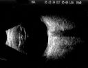
Optic Nerve Drusen in 7 Year Old Child - Miminal Calcification994 views7-year-old child difficulty seeing the blackboard in school. She was checked for glasses and it was noted at that time that she had unusual looking optic nerves. Vision OU is 20/50.
|
|

Papilledema - Spectral Domain OCT used for Diagnosis - 14 year old Child988 views14-year-old was with optic nerve swelling asymptomatic - picked up during routine eye exam. OD 20/20, OS 20/32
SD-OCT is used to differentiate optic disc edema from optic nerve head drusen (which are not yet calcified in children).
|
|

Optic Nerve (Disc) Drusen - Spectral Domain Optical Coherence Tomography Line Scan987 views34 Year Old woman no headaches normal vision
|
|

Papilledema from pseudotumor cerebri Pre Spinal Tap - VA 20/100983 views24-year-old woman was seen in the office on 2/23/2010. She has had noticed positional blurring of her vision when she bends over since around November. When she bends over and stands up straight, she notices her vision is blurred. Vision OD is 20/100, PH 20/40; OS is 20/80, PH 20/30. IOP: 19 OU.
vision was 20/25 after tap
|
|

Acute Non-arteritic Anterior Ischemic Optic Neuropathy Right Eye - Previous NA-AION left eye 978 views69-year-old man previously lost vision in the left eye from nonarteritic anterior ischemic optic neuropathy in 2006. About a week ago and noticed blurred vision in the inferonasal part of the vision in the right eye.
VISUAL ACUITY: Vision OD is 20/16, OS is 20/40
|
|

Papilledema Probable to Pseudo-tumor Cerebri976 viewsA 24-year old female who presents severe headaches and vision loss over the past 6-months. VA was 20/40 with pinhole 20/25, right eye and 20/25, left eye. Fundiscopic exam reveals 3-4 plus papilledema of both optic nerves. Patient has VF defects inferior, nasal-right eye and enlarged blind spot with an inferior arcuate defect present. Patient will get MRI and work-up with treatment in the hospital.
|
|

Optic Nerve (Disc) Drusen vs. Disc Edema Vs. AION - Spectral Domain Optical Coherence Tomography Line Scan967 views
|
|

Optic Nerve Drusen - - Nerve Fiber Layer Scan946 views72-year-old woman has had optic nerve drusen for sometime and has visual field abnormalities. OD 20/32, OS 20/32.
|
|

Acute Non-arteritic Anterior Ischemic Optic Neuropathy Right Eye - Previous NA-AION left eye 938 views69-year-old man previously lost vision in the left eye from nonarteritic anterior ischemic optic neuropathy in 2006. About a week ago and noticed blurred vision in the inferonasal part of the vision in the right eye.
VISUAL ACUITY: Vision OD is 20/16, OS is 20/40
|
|
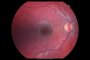
Optic Nerve Pit Retinal Detachment - Macular Detachment - 8 Year Old - 5/200 Vision OS935 views8-year-old child on two school exams that his vision was poor in the left eye and his last normal exam was in preschool.
VISUAL ACUITY: OD 20/30, OS 5/200. OS: Vertical C/D ratio is 0.2. There is a whitening on the temporal edge of the optic nerve consistent with an optic disc pit and there is a serous macular detachment.
|
|

Optic Nerve (Disc) Drusen - Spectral Domain Optical Coherence Tomography Line Scan934 views34 Year Old woman no headaches normal vision
|
|
|
|


