Most viewed - Optic Nerve Disease
|

Non-arteritic Anterior Ischemic Optic Neuropathy following Intravitreal Injection 551 views81-year-old woman has had injections in both eyes for wet age-related macular degeneration. 2 weeks ago the left eye was treated. Less than a week ago she noticed a change in vision in the left eye. She had a dull left frontal headache and noticed her vision was hazy as if there was a veil over the vision. OD is 20/25, OS is 20/25. IOP: OD 18, OS 26. The left eye has a 3+ APD
|
|

Neuroretinitis - Cat Scratch - Bartonella - Sudden Vision Loss Right Eye - 29 Year Old Female551 views
|
|

Acute AION OS - Mild disc pallor OD550 viewsSudden vision loss OS x 1 week with optic nerve swelling. Evidence of optic atrophy in right eye.
|
|
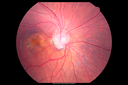
Optic Nerve Pit Retinal Detachment - SD OCT of Nerve EDI shows Pit550 views9 Year old with morning glory disc who developed nerve pit RD. Raster scans of the optic nerve on EDI setting show the communication between the subretinal space and the fluid around the cerebrospinal fluid surrounding the optic nerve sheath.
|
|

Diabetic Papillitis549 viewsPatient is about to undergo surgery for CNS Aneurysm. Patient woke up with little spots which never cleared up. Patients VA was 20/30-OD and 20/40-OS. Both eyes appeared to have disc edema with hemorrhages in the right eye. Ordered a CT of the brain to make sure the aneurysm didn't ruptured.
|
|

Acute non arteritic anterior ischemic optic neuropathy left eye - SD Optical Coherence Tomography Line Scan549 views66 y.o. woman spot in central vision in left eye started about 1 week ago since then it is not changing. VA OD: sc20/25 OS: sc20/200
|
|

548 views
|
|
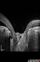
Optic Nerve Pit Retinal Detachment - SD OCT of Nerve EDI shows Pit548 views9 Year old with morning glory disc who developed nerve pit RD. Raster scans of the optic nerve on EDI setting show the communication between the subretinal space and the fluid around the cerebrospinal fluid surrounding the optic nerve sheath.
|
|

Papilledema - Sagittal Cerebral Vein Thrombosis - Lung Transplant Cystic Fibrosis 36 Year Old Man547 viewsColor Photo
|
|

546 views
|
|

Neuroretinitis - Cat Scratch - Bartonella - Sudden Vision Loss Right Eye - 29 Year Old Female546 views
|
|

Acute AION OS - Mild disc pallor OD544 viewsSudden vision loss OS x 1 week with optic nerve swelling. Evidence of optic atrophy in right eye.
|
|
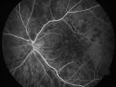
Non-arteritic Anterior Ischemic Optic Neuropathy following Intravitreal Injection 543 views81-year-old woman has had injections in both eyes for wet age-related macular degeneration. 2 weeks ago the left eye was treated. Less than a week ago she noticed a change in vision in the left eye. She had a dull left frontal headache and noticed her vision was hazy as if there was a veil over the vision. OD is 20/25, OS is 20/25. IOP: OD 18, OS 26. The left eye has a 3+ APD
|
|

Optic Nerve Pit Retinal Detachment - SD OCT of Nerve EDI shows Pit543 views9 Year old with morning glory disc who developed nerve pit RD. Raster scans of the optic nerve on EDI setting show the communication between the subretinal space and the fluid around the cerebrospinal fluid surrounding the optic nerve sheath.
|
|

Papilledema - Sagittal Cerebral Vein Thrombosis - Lung Transplant Cystic Fibrosis 36 Year Old Man542 viewsLine scan through optic nerve
|
|

Optic Nerve Hypoplasia - 43 Year Old Man - declining vision540 views43-year-old man was diagnosed with optic nerve hypoplasia at age fifteen at the National Institute of Health. His vision seems to be declining lately.
Vision OD is 20/80, OS is 20/120
|
|

Optic Disk Drusen - Pucker - Pattern Dystrophy539 viewsAutofluorescence from Drusen and Macular Dystrophy
|
|

Optic Nerve Pit Retinal Detachment - SD OCT of Nerve EDI shows Pit539 views9 Year old with morning glory disc who developed nerve pit RD. Raster scans of the optic nerve on EDI setting show the communication between the subretinal space and the fluid around the cerebrospinal fluid surrounding the optic nerve sheath.
|
|

Acute AION OS - Mild disc pallor OD536 viewsSudden vision loss OS x 1 week with optic nerve swelling. Evidence of optic atrophy in right eye.
|
|

Neuroretinitis - Cat Scratch - Bartonella - Sudden Vision Loss Right Eye - 29 Year Old Female535 views
|
|

Syphilitic Optic Neuritis - possible Gumma535 views82 year old woman with vision loss in the left eye 20/32 and positive FTA-ABS (negative RPR). - this may be a gumma
|
|

Optic Nerve Pit Retinal Detachment - SD OCT of Nerve EDI shows Pit534 views9 Year old with morning glory disc who developed nerve pit RD. Raster scans of the optic nerve on EDI setting show the communication between the subretinal space and the fluid around the cerebrospinal fluid surrounding the optic nerve sheath.
|
|

Syphilitic Optic Neuritis - possible Gumma534 views82 year old woman with vision loss in the left eye 20/32 and positive FTA-ABS (negative RPR). - this may be a gumma
|
|

Optic Atrophy - Probably Dominant532 views76-year-old woman decreased vision for at least 40 years. She said she was told she had optic atrophy at Bascom Palmer in the 1970s. She does not know if she has optic atrophy in her family. VA 20/400 in each eye.
|
|

Non-arteritic Anterior Ischemic Optic Neuropathy following Intravitreal Injection 529 views81-year-old woman has had injections in both eyes for wet age-related macular degeneration. 2 weeks ago the left eye was treated. Less than a week ago she noticed a change in vision in the left eye. She had a dull left frontal headache and noticed her vision was hazy as if there was a veil over the vision. OD is 20/25, OS is 20/25. IOP: OD 18, OS 26. The left eye has a 3+ APD
|
|
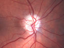
Acute AION OS - Mild disc pallor OD529 viewsSudden vision loss OS x 1 week with optic nerve swelling. Evidence of optic atrophy in right eye.
|
|

Optic Atrophy - Probably Dominant528 views76-year-old woman decreased vision for at least 40 years. She said she was told she had optic atrophy at Bascom Palmer in the 1970s. She does not know if she has optic atrophy in her family. VA 20/400 in each eye.
|
|
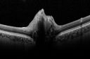
Optic Disk Drusen Advanced Nerve Fiber Loss527 views
|
|

Optic Disk Drusen Advanced Nerve Fiber Loss526 views
|
|

524 views
|
|

Neuroretinitis - Cat Scratch - Bartonella - Sudden Vision Loss Right Eye - 29 Year Old Female524 views
|
|

Non-arteritic Anterior Ischemic Optic Neuropathy following Intravitreal Injection 523 views81-year-old woman has had injections in both eyes for wet age-related macular degeneration. 2 weeks ago the left eye was treated. Less than a week ago she noticed a change in vision in the left eye. She had a dull left frontal headache and noticed her vision was hazy as if there was a veil over the vision. OD is 20/25, OS is 20/25. IOP: OD 18, OS 26. The left eye has a 3+ APD
|
|

Nerve Fiber Defect from Optic Disk Drusen523 views31 year old male - asymptomatic
|
|

Autosomal Dominant Optic Atrophy - Father523 views52 year old man Vision has not been correctable for many years.
EXTENDED HPI: The patient has been seen for a while and his vision is not normal. Â
VA OD: sc20/100 PH20/50-1 Nsc20/400
VA OS: sc20/63 PH20/50-2 NscJ16
IOP: TP: OD:10 OS:9 Time:02:29 PM
Two siblings and his son have the same problem
|
|

Optic Nerve Pit Retinal Detachment - SD OCT of Nerve EDI shows Pit522 views9 Year old with morning glory disc who developed nerve pit RD. Raster scans of the optic nerve on EDI setting show the communication between the subretinal space and the fluid around the cerebrospinal fluid surrounding the optic nerve sheath.
|
|

Optic Disk Drusen - Pucker - Pattern Dystrophy521 views
|
|

Neuroretinitis - Cat Scratch - Bartonella - Sudden Vision Loss Right Eye - 29 Year Old Female521 views
|
|
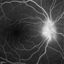
520 views
|
|

Acute AION OS - Mild disc pallor OD519 viewsSudden vision loss OS x 1 week with optic nerve swelling. Evidence of optic atrophy in right eye.
|
|
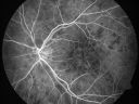
Non-arteritic Anterior Ischemic Optic Neuropathy following Intravitreal Injection 518 views81-year-old woman has had injections in both eyes for wet age-related macular degeneration. 2 weeks ago the left eye was treated. Less than a week ago she noticed a change in vision in the left eye. She had a dull left frontal headache and noticed her vision was hazy as if there was a veil over the vision. OD is 20/25, OS is 20/25. IOP: OD 18, OS 26. The left eye has a 3+ APD
|
|

Optic Disk Drusen Advanced Nerve Fiber Loss518 views
|
|
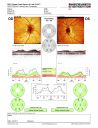
517 views
|
|

Papilledema Probable to Pseudo-tumor Cerebri517 viewsA 24-year old female who presents severe headaches and vision loss over the past 6-months. VA was 20/40 with pinhole 20/25, right eye and 20/25, left eye. Fundiscopic exam reveals 3-4 plus papilledema of both optic nerves. Patient has VF defects inferior, nasal-right eye and enlarged blind spot with an inferior arcuate defect present. Patient will get MRI and work-up with treatment in the hospital.
|
|

Nerve Fiber Defect from Optic Disk Drusen517 views31 year old male - asymptomatic
|
|

Nerve Fiber Defect from Optic Disk Drusen517 views31 year old male - asymptomatic
|
|

Autosomal Dominant Optic Atrophy - Father517 views52 year old man Vision has not been correctable for many years.
EXTENDED HPI: The patient has been seen for a while and his vision is not normal. Â
VA OD: sc20/100 PH20/50-1 Nsc20/400
VA OS: sc20/63 PH20/50-2 NscJ16
IOP: TP: OD:10 OS:9 Time:02:29 PM
Two siblings and his son have the same problem
|
|

Acute AION OS - Mild disc pallor OD516 viewsSudden vision loss OS x 1 week with optic nerve swelling. Evidence of optic atrophy in right eye.
|
|

515 views
|
|

515 views
|
|

Acute AION OS - Mild disc pallor OD515 viewsSudden vision loss OS x 1 week with optic nerve swelling. Evidence of optic atrophy in right eye.
|
|

Optic Nerve Hypoplasia - 43 Year Old Man - declining vision514 views43-year-old man was diagnosed with optic nerve hypoplasia at age fifteen at the National Institute of Health. His vision seems to be declining lately.
Vision OD is 20/80, OS is 20/120
|
|
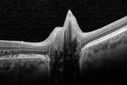
Optic Disk Drusen Advanced Nerve Fiber Loss514 views
|
|
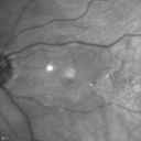
Optic Disk Drusen - Pucker - Pattern Dystrophy513 views
|
|

Optic Nerve Pit Retinal Detachment - SD OCT of Nerve EDI shows Pit513 views9 Year old with morning glory disc who developed nerve pit RD. Raster scans of the optic nerve on EDI setting show the communication between the subretinal space and the fluid around the cerebrospinal fluid surrounding the optic nerve sheath.
|
|
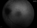
Diabetic Papillitis512 viewsPatient is about to undergo surgery for CNS Aneurysm. Patient woke up with little spots which never cleared up. Patients VA was 20/30-OD and 20/40-OS. Both eyes appeared to have disc edema with hemorrhages in the right eye. Ordered a CT of the brain to make sure the aneurysm didn't ruptured.
|
|

Optic Nerve Pit Maculopathy512 views10 year old child who had inner retinal fenestration (see youtube video on scohen125 channel) and did very well.
|
|

Neuroretinitis - Cat Scratch - Bartonella - Sudden Vision Loss Right Eye - 29 Year Old Female511 views
|
|

Acute AION OS - Mild disc pallor OD509 viewsSudden vision loss OS x 1 week with optic nerve swelling. Evidence of optic atrophy in right eye.
|
|

Toxic Optic Neuropathy - Ethambutol506 views15 months of Ethambutol with rapid vision loss OU. VA 20/100 OD (post cataract sugery ) 20/200 OS with dense cataract. There is temporal RNFL loss in both eyes
|
|

Diabetic Papillitis505 viewsPatient is about to undergo surgery for CNS Aneurysm. Patient woke up with little spots which never cleared up. Patients VA was 20/30-OD and 20/40-OS. Both eyes appeared to have disc edema with hemorrhages in the right eye. Ordered a CT of the brain to make sure the aneurysm didn't ruptured.
|
|

Toxic Optic Neuropathy - Ethambutol505 views15 months of Ethambutol with rapid vision loss OU. VA 20/100 OD (post cataract sugery ) 20/200 OS with dense cataract. There is temporal RNFL loss in both eyes
|
|

Autosomal Dominant Optic Atrophy - Son505 views12 year old man. His vision was not correctable to 20/20. There was concern about possible macula problem. His was diagnosed with Streff syndrome in NY.
VA OD: Dcc20/63 PH20/50-2 NccJ2
VA OS: Dcc20/63 PH20/50-2 NccJ3
IOP: TP: OD:10 OS:15 Time:02:24 PM
The diagnostic criteria for Streff syndrome are not well established, and the validity of this condition has not been recognized by The American Academy of Ophthalmology, The American Academy of Pediatric Ophthalmology, The American Academy of Optometry or The American academy of Pediatrics.
|
|

Acute AION OS - Mild disc pallor OD504 viewsSudden vision loss OS x 1 week with optic nerve swelling. Evidence of optic atrophy in right eye.
|
|
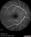
Syphilitic Optic Neuritis - possible Gumma504 views82 year old woman with vision loss in the left eye 20/32 and positive FTA-ABS (negative RPR). - this may be a gumma
|
|

Optic Nerve Pit Maculopathy504 views10 year old child who had inner retinal fenestration (see youtube video on scohen125 channel) and did very well.
|
|

Optic Nerve Pit Retinal Detachment - SD OCT of Nerve EDI shows Pit503 views9 Year old with morning glory disc who developed nerve pit RD. Raster scans of the optic nerve on EDI setting show the communication between the subretinal space and the fluid around the cerebrospinal fluid surrounding the optic nerve sheath.
|
|

Optic Nerve Pit Maculopathy503 views10 year old child who had inner retinal fenestration (see youtube video on scohen125 channel) and did very well.
|
|

Optic Nerve Pit Maculopathy502 views10 year old child who had inner retinal fenestration (see youtube video on scohen125 channel) and did very well.
|
|

Autosomal Dominant Optic Atrophy - Father502 views52 year old man Vision has not been correctable for many years.
EXTENDED HPI: The patient has been seen for a while and his vision is not normal. Â
VA OD: sc20/100 PH20/50-1 Nsc20/400
VA OS: sc20/63 PH20/50-2 NscJ16
IOP: TP: OD:10 OS:9 Time:02:29 PM
Two siblings and his son have the same problem
|
|
501 views
|
|

Optic Disk Drusen Advanced Nerve Fiber Loss500 views
|
|

Syphilitic Optic Neuritis - possible Gumma500 views82 year old woman with vision loss in the left eye 20/32 and positive FTA-ABS (negative RPR). - this may be a gumma
|
|

Non-arteritic Anterior Ischemic Optic Neuropathy following Intravitreal Injection 499 views81-year-old woman has had injections in both eyes for wet age-related macular degeneration. 2 weeks ago the left eye was treated. Less than a week ago she noticed a change in vision in the left eye. She had a dull left frontal headache and noticed her vision was hazy as if there was a veil over the vision. OD is 20/25, OS is 20/25. IOP: OD 18, OS 26. The left eye has a 3+ APD
|
|

496 views
|
|

Optic Nerve Pit Retinal Detachment - SD OCT of Nerve EDI shows Pit496 views9 Year old with morning glory disc who developed nerve pit RD. Raster scans of the optic nerve on EDI setting show the communication between the subretinal space and the fluid around the cerebrospinal fluid surrounding the optic nerve sheath.
|
|

Optic Disk Drusen Advanced Nerve Fiber Loss495 views
|
|

Autosomal Dominant Optic Atrophy - Father495 views52 year old man Vision has not been correctable for many years.
EXTENDED HPI: The patient has been seen for a while and his vision is not normal. Â
VA OD: sc20/100 PH20/50-1 Nsc20/400
VA OS: sc20/63 PH20/50-2 NscJ16
IOP: TP: OD:10 OS:9 Time:02:29 PM
Two siblings and his son have the same problem
|
|

Papilledema - Sagittal Cerebral Vein Thrombosis - Lung Transplant Cystic Fibrosis 36 Year Old Man494 viewsLine scan through optic nerve
|
|

Optic Disk Drusen - Pucker - Pattern Dystrophy493 views
|
|

492 views
|
|
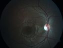
Autosomal Dominant Optic Atrophy - Son491 views12 year old man. His vision was not correctable to 20/20. There was concern about possible macula problem. His was diagnosed with Streff syndrome in NY.
VA OD: Dcc20/63 PH20/50-2 NccJ2
VA OS: Dcc20/63 PH20/50-2 NccJ3
IOP: TP: OD:10 OS:15 Time:02:24 PM
The diagnostic criteria for Streff syndrome are not well established, and the validity of this condition has not been recognized by The American Academy of Ophthalmology, The American Academy of Pediatric Ophthalmology, The American Academy of Optometry or The American academy of Pediatrics.
|
|

Papilledema Probable to Pseudo-tumor Cerebri490 viewsA 24-year old female who presents severe headaches and vision loss over the past 6-months. VA was 20/40 with pinhole 20/25, right eye and 20/25, left eye. Fundiscopic exam reveals 3-4 plus papilledema of both optic nerves. Patient has VF defects inferior, nasal-right eye and enlarged blind spot with an inferior arcuate defect present. Patient will get MRI and work-up with treatment in the hospital.
|
|

490 views
|
|

489 views
|
|

488 views
|
|

Bilateral Optic Nerve Drusen486 views
|
|

Neuroretinitis - Cat Scratch - Bartonella - Sudden Vision Loss Right Eye - 29 Year Old Female486 views
|
|

Optic Nerve Pit Maculopathy485 views10 year old child who had inner retinal fenestration (see youtube video on scohen125 channel) and did very well.
|
|
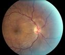
Optic Neuritis with Suspected Harada's Disease 484 viewsYoung female patient comes in with red and irritated eyes which were sensitive to the touch. Patient also had vision loss in both eyes. VA was 20/80, right eye, 20/60, left eye. Fundiscopic exam shows sub retinal fluid in the right eye surrounding the optic nerve. Patient will start steroid therapy to reduce any other inflammation that is active.
|
|
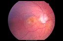
Optic Nerve Pit Maculopathy484 views10 year old child who had inner retinal fenestration (see youtube video on scohen125 channel) and did very well.
|
|

483 views
|
|

Autosomal Dominant Optic Atrophy - Father483 views52 year old man Vision has not been correctable for many years.
EXTENDED HPI: The patient has been seen for a while and his vision is not normal. Â
VA OD: sc20/100 PH20/50-1 Nsc20/400
VA OS: sc20/63 PH20/50-2 NscJ16
IOP: TP: OD:10 OS:9 Time:02:29 PM
Two siblings and his son have the same problem
|
|

Acute AION OS - Mild disc pallor OD482 viewsSudden vision loss OS x 1 week with optic nerve swelling. Evidence of optic atrophy in right eye.
|
|

Syphilitic Optic Neuritis - possible Gumma482 views82 year old woman with vision loss in the left eye 20/32 and positive FTA-ABS (negative RPR). - this may be a gumma
|
|
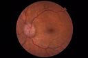
Papilledema - Benign Intracranial hypertension - Idiopathic intracranial hypertension482 views35 year old obese female. Normal vision and headache. Opening pressure was 30 cm water (normal 10-20). Nerve swelling responded to diamox.
|
|

Papilledema - Benign Intracranial hypertension - Idiopathic intracranial hypertension482 views35 year old obese female. Normal vision and headache. Opening pressure was 30 cm water (normal 10-20). Nerve swelling responded to diamox.
|
|

Superior Peripapillary Hemorrhage481 viewsPatient was seen for acute vision loss in the right eye. Patient has glaucoma. VA was 20/70 in the right eye. Had Vitrectomy back in May 2012 for ERM stripping. Also had Trabectome with cataract surgery back in December of 2012. Fundus photos presents a Superior Peripapillary Hemorrhage of the optic nerve. Patient will be followed up in one month.
|
|

Papilledema Probable to Pseudo-tumor Cerebri481 viewsA 24-year old female who presents severe headaches and vision loss over the past 6-months. VA was 20/40 with pinhole 20/25, right eye and 20/25, left eye. Fundiscopic exam reveals 3-4 plus papilledema of both optic nerves. Patient has VF defects inferior, nasal-right eye and enlarged blind spot with an inferior arcuate defect present. Patient will get MRI and work-up with treatment in the hospital.
|
|
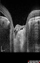
Optic Nerve Pit Maculopathy480 views10 year old child who had inner retinal fenestration (see youtube video on scohen125 channel) and did very well.
|
|

479 views
|
|
|
|


