Most viewed - Macular Disease
|

Plaquenil Toxicity both Eyes - Partial Bull's Eye - Discontinued 6 Years ago SD-OCT (Spectral domain optical coherence tomography)4350 views82-year-old woman was on Plaquenil from 1976 from 2005, 200 mg a day. It was discontinued because of abnormal visual fields 6 years ago. OD 20/32, OS 20/40
|
|

Plaquenil Toxicity both Eyes - Partial Bull's Eye - Discontinued 6 Years ago SD-OCT (Spectral domain optical coherence tomography)3834 views82-year-old woman was on Plaquenil from 1976 from 2005, 200 mg a day. It was discontinued because of abnormal visual fields 6 years ago. OD 20/32, OS 20/40
|
|

Plaquenil Toxicity both Eyes - Partial Bull's Eye - Discontinued 6 Years ago SD-OCT (Spectral domain optical coherence tomography)3676 views82-year-old woman was on Plaquenil from 1976 from 2005, 200 mg a day. It was discontinued because of abnormal visual fields 6 years ago. OD 20/32, OS 20/40
|
|

Reticular Macular Disease - Reticular Pseudodrusen and Soft Macular Drusen - 57 Year Old Woman3128 views57-year-old woman was seen in the office on 2/22/2011. She has macular drusen in both eyes. I saw her back in 2006. Since then she does notice slight increased distortion. OD is 20/25, OS is 20/25
|
|

Plaquenil Toxicity both Eyes - Partial Bull's Eye - Discontinued 6 Years ago SD-OCT (Spectral domain optical coherence tomography)2857 views82-year-old woman was on Plaquenil from 1976 from 2005, 200 mg a day. It was discontinued because of abnormal visual fields 6 years ago. OD 20/32, OS 20/40
|
|

Plaquenil Toxicity both Eyes - Partial Bull's Eye - Discontinued 6 Years ago Fundus Autofluorescence2693 views82-year-old woman was on Plaquenil from 1976 from 2005, 200 mg a day. It was discontinued because of abnormal visual fields 6 years ago. OD 20/32, OS 20/40
|
|
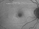
Plaquenil Toxicity both Eyes - Partial Bull's Eye - Discontinued 6 Years ago Fundus Autofluorescence2482 views82-year-old woman was on Plaquenil from 1976 from 2005, 200 mg a day. It was discontinued because of abnormal visual fields 6 years ago. OD 20/32, OS 20/40
|
|

Reticular Macular Disease - Reticular Pseudodrusen and Soft Macular Drusen - 57 Year Old Woman2298 views57-year-old woman was seen in the office on 2/22/2011. She has macular drusen in both eyes. I saw her back in 2006. Since then she does notice slight increased distortion. OD is 20/25, OS is 20/25
|
|
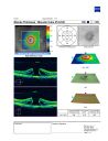
Large mac hole OCT2269 views76 yof with large mac hole
VA: CF at 3ft
chance of spontaneous closure low; surgery option available
|
|
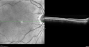
Plaquenil Toxicity both Eyes - Partial Bull's Eye - Discontinued 6 Years ago SD-OCT (Spectral domain optical coherence tomography)2263 views82-year-old woman was on Plaquenil from 1976 from 2005, 200 mg a day. It was discontinued because of abnormal visual fields 6 years ago. OD 20/32, OS 20/40
|
|

80 year old woman with Reticular Drusen (pseudodrusen) 20/30 Vision2171 views80 year old woman with asteroid hyalosis and macular degeneration.
OPHTHALMIC EXAMINATION: Visual acuity with correction 20/30-3 OD and 20/30-2
|
|

ANGIOID STREAKS2123 viewsA 29 year old woman was referred to the ophthalmologist for an eye exam since the dermatologist had noted “chicken-skin†papules in the neck skin. PAD showed for pseudoxathoma elasticum typical pathology. The eye exam disclosed angioid streaks, asymptomatic so far.
Magnus Gjötterberg MD
International membrer 1440
S:t Erik Eye Hospital
SE-11282
Stockholm
Sweden
magnusgjotterberg@hem.utfors.se
magnus.gjotterberg@sankterik.se.
Photographer/technician
Malin Langhals
|
|

Reticular Macular Disease (Pseudo-drusen) Both Eyes - Wet AMD OS - Dry AMD OD SD-OCT2089 views84-year-old woman has wet age-related macular degeneration in the left eye and dry macular degeneration in the right eye. She takes the eye vitamins and her vision is stable since she was treated three months ago with Avastin. OD 20/50, OS 20/32
|
|

Reticular Macular Disease - Reticular Pseudodrusen and Soft Macular Drusen - 57 Year Old Woman2079 views57-year-old woman was seen in the office on 2/22/2011. She has macular drusen in both eyes. I saw her back in 2006. Since then she does notice slight increased distortion. OD is 20/25, OS is 20/25
|
|

Plaquenil Toxicity both Eyes - Partial Bull's Eye - Discontinued 6 Years ago SD-OCT (Spectral domain optical coherence tomography)2037 views82-year-old woman was on Plaquenil from 1976 from 2005, 200 mg a day. It was discontinued because of abnormal visual fields 6 years ago. OD 20/32, OS 20/40
|
|

Plaquenil Toxicity both Eyes - Partial Bull's Eye - Discontinued 6 Years ago Fundus Autofluorescence2034 views82-year-old woman was on Plaquenil from 1976 from 2005, 200 mg a day. It was discontinued because of abnormal visual fields 6 years ago. OD 20/32, OS 20/40
|
|

Pigment migration in dry age-related macular degeneration2016 views80 year old female. Dry AMD with GA in the left eye and pigment migration visible on OCT scan.
VA 20/40 OD, 20/160 OS
|
|

Plaquenil Toxicity both Eyes - Partial Bull's Eye - Discontinued 6 Years ago Fundus Autofluorescence1980 views82-year-old woman was on Plaquenil from 1976 from 2005, 200 mg a day. It was discontinued because of abnormal visual fields 6 years ago. OD 20/32, OS 20/40
|
|

Reticular Macular Disease (Pseudo-drusen) Both Eyes - Wet AMD OS - Dry AMD OD SD-OCT1932 views84-year-old woman has wet age-related macular degeneration in the left eye and dry macular degeneration in the right eye. She takes the eye vitamins and her vision is stable since she was treated three months ago with Avastin. OD 20/50, OS 20/32
|
|

Plaquenil Toxicity - Bulls Eye Maculopathy1868 views70-year-old woman with systemic Lupus erythematosus and clotting problems. She was on the Plaquenil for about eight years and then off the Plaquenil for the last eight years because she developed macular toxicity. Although her vision was hazy, it was stable. Recent deceased vision left eye: OD 20/60, OS 20/100. IOP: OD 18, OS 19.
|
|

Plaquenil Toxicity both Eyes - Partial Bull's Eye - Discontinued 6 Years ago SD-OCT (Spectral domain optical coherence tomography)1763 views82-year-old woman was on Plaquenil from 1976 from 2005, 200 mg a day. It was discontinued because of abnormal visual fields 6 years ago. OD 20/32, OS 20/40
|
|

Acute Central Serous Retinopathy - Classic Smokestack on Fluorescein Angiogram 1694 views34-year-old man noticed about five days ago decreased vision in the right eye. He was having difficulty playing video games and seeing with that eye. He thought maybe his refractive error was shifting.
VISUAL ACUITY: Vision OD is 20/40, OS is 20/16
|
|

Basal Laminar Drusen - Cuticular Drusen - Equatorial Drusen - 70 Year Old Woman - Macular Photo Right Eye1681 views70 year old woman has had macular drusen since her thirties and she said her sister has a similar problem. Recently the vision in the left eye has substantially declined. OD 20/16, OS 10/200.
|
|

Reticular Macular Disease - Reticular Pseudodrusen and Soft Macular Drusen - 57 Year Old Woman1666 views57-year-old woman was seen in the office on 2/22/2011. She has macular drusen in both eyes. I saw her back in 2006. Since then she does notice slight increased distortion. OD is 20/25, OS is 20/25
|
|

Central Serous Retinopathy Acute - Indocyanine Green Angiogram - Leakage Choriocapillaris 1637 views42-year-old man was seen in the office on October 5, 2011. He had noticed starting in August after a course of antibiotic and steroids, that he developed new spots in his vision in the right eye. He may have had an episode like this sometime in the past. He did take steroids a few years ago and his vision did change at that time, but then returned.
VISUAL ACUITY: OD 20/32, OS 20/32
|
|

late phase mushroom cloud, acute CSR1634 viewsFocus is on the peri-foveal vessels illustrates the extremely bullous RD from this mushroom cloud of leakage. The RD was substantially improved after 2 weeks of oral rifampin. Visual acuity improved from 20/200 to 20/70 OD.
|
|

Drusen Maculopathy - Confluent Subfoveal Soft Hydrophobic Drusen - Yellow Drusen in Center of the Macula1633 views
|
|

Acute Central Serous Retinopathy - Classic Smokestack on Fluorescein Angiogram - Antimated .GIF1623 views34-year-old man noticed about five days ago decreased vision in the right eye. He was having difficulty playing video games and seeing with that eye. He thought maybe his refractive error was shifting.
VISUAL ACUITY: Vision OD is 20/40, OS is 20/16
|
|

Plaquenil Toxicity both Eyes - Partial Bull's Eye - Discontinued 6 Years ago 1600 views82-year-old woman was on Plaquenil from 1976 from 2005, 200 mg a day. It was discontinued because of abnormal visual fields 6 years ago. OD 20/32, OS 20/40
|
|

Plaquenil Toxicity both Eyes - Partial Bull's Eye - Discontinued 6 Years ago InfraRed Image1573 views82-year-old woman was on Plaquenil from 1976 from 2005, 200 mg a day. It was discontinued because of abnormal visual fields 6 years ago. OD 20/32, OS 20/40
|
|

Plaquenil Toxicity both Eyes - Partial Bull's Eye - Discontinued 6 Years ago InfraRed Image1566 views82-year-old woman was on Plaquenil from 1976 from 2005, 200 mg a day. It was discontinued because of abnormal visual fields 6 years ago. OD 20/32, OS 20/40
|
|

Equatorial Drusen Fundus Photo Left Eye1558 views
|
|

Reticular Macular Disease (Pseudo-drusen) Both Eyes - Wet AMD OS - Dry AMD OD SD-OCT1546 views84-year-old woman has wet age-related macular degeneration in the left eye and dry macular degeneration in the right eye. She takes the eye vitamins and her vision is stable since she was treated three months ago with Avastin. OD 20/50, OS 20/32
|
|

Juxtafoveal Retinal Telangiectasia (Telangiectasis) - MacTel - Striking Gray Ring in Macula1533 views59-year-old woman decreasing vision in the right eye for a few years. She has had diabetes for five years. OD 20/100, Pinhole 20/40. OS 20/20
|
|

Plaquenil Toxicity both Eyes - Partial Bull's Eye - Discontinued 6 Years ago 1517 views82-year-old woman was on Plaquenil from 1976 from 2005, 200 mg a day. It was discontinued because of abnormal visual fields 6 years ago. OD 20/32, OS 20/40
|
|

Basal Laminar Drusen - Cuticular Drusen - Equatorial Drusen - 70 Year Old Woman 1515 views70 year old woman has had macular drusen since her thirties and she said her sister has a similar problem. Recently the vision in the left eye has substantially declined. OD 20/16, OS 10/200.
|
|
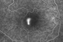
Acute Central Serous Retinopathy - Classic Smokestack on Fluorescein Angiogram 1512 views34-year-old man noticed about five days ago decreased vision in the right eye. He was having difficulty playing video games and seeing with that eye. He thought maybe his refractive error was shifting.
VISUAL ACUITY: Vision OD is 20/40, OS is 20/16
|
|

Acute Central Serous Retinopathy - Classic Smokestack on Fluorescein Angiogram 1498 views34-year-old man noticed about five days ago decreased vision in the right eye. He was having difficulty playing video games and seeing with that eye. He thought maybe his refractive error was shifting.
VISUAL ACUITY: Vision OD is 20/40, OS is 20/16
|
|

Central Serous Retinopathy Acute - Fundus Autofluorescence1495 views42-year-old man was seen in the office on October 5, 2011. He had noticed starting in August after a course of antibiotic and steroids, that he developed new spots in his vision in the right eye. He may have had an episode like this sometime in the past. He did take steroids a few years ago and his vision did change at that time, but then returned.
VISUAL ACUITY: OD 20/32, OS 20/32
|
|

Juxtafoveal Telangiectasis - MacTel - Retinal Telangiectasis - Both Eyes - Crystals1494 views61-year-old man has idiopathic juxtafoveal retinal telangiectasis type II in both eyes OD is 20/40, OS is 20/25
|
|

Posterior Staphyloma and Geographic Atrophy Myopic Degeneration1462 views54-year-old man . He is a high myope. He has posterior staphylomas. He has had gradually changing vision for the last month or two. OD is 20/40, OS is 20/50.
|
|

Retinal Pigment Epithelial Detachment with no Sub-Retinal Fluid...1459 viewsA 38-year old male who comes in with blurred vision in the left eye. VA is 20/30. Notices a defect inferior of his central vision. Did an Fluorescence Angiogram to determine an RPE with no sub retinal fluid. Also OCT confirms. Patient was injected with Avastin.
|
|

Acute Central Serous Retinopathy - Classic Smokestack on Fluorescein Angiogram 1447 views34-year-old man noticed about five days ago decreased vision in the right eye. He was having difficulty playing video games and seeing with that eye. He thought maybe his refractive error was shifting.
VISUAL ACUITY: Vision OD is 20/40, OS is 20/16
|
|

Solar Retinopathy1440 viewsFoveal defect due to sun staring
|
|

Acute Central Serous Retinopathy - Classic Smokestack on Fluorescein Angiogram 1420 views34-year-old man noticed about five days ago decreased vision in the right eye. He was having difficulty playing video games and seeing with that eye. He thought maybe his refractive error was shifting.
VISUAL ACUITY: Vision OD is 20/40, OS is 20/16
|
|

Acute Central Serous Retinopathy - Classic Smokestack on Fluorescein Angiogram - OCT1414 views34-year-old man noticed about five days ago decreased vision in the right eye. He was having difficulty playing video games and seeing with that eye. He thought maybe his refractive error was shifting.
VISUAL ACUITY: Vision OD is 20/40, OS is 20/16
|
|
Acute Central Serous Retinopathy - Classic Smokestack on Fluorescein Angiogram - Animated .GIF1408 views34-year-old man noticed about five days ago decreased vision in the right eye. He was having difficulty playing video games and seeing with that eye. He thought maybe his refractive error was shifting.
VISUAL ACUITY: Vision OD is 20/40, OS is 20/16
|
|

Central Serous Retinopathy - Acute1402 views49-year-old man sees a spot in the central vision with a red halo for 2 weeks OD is 20/50, OS is 20/20
|
|

Central Serous Retinopathy Acute - Fundus Autofluorescence1399 views42-year-old man was seen in the office on October 5, 2011. He had noticed starting in August after a course of antibiotic and steroids, that he developed new spots in his vision in the right eye. He may have had an episode like this sometime in the past. He did take steroids a few years ago and his vision did change at that time, but then returned.
VISUAL ACUITY: OD 20/32, OS 20/32
|
|

Acute Central Serous Retinopathy - Classic Smokestack on Fluorescein Angiogram 1395 views34-year-old man noticed about five days ago decreased vision in the right eye. He was having difficulty playing video games and seeing with that eye. He thought maybe his refractive error was shifting.
VISUAL ACUITY: Vision OD is 20/40, OS is 20/16
|
|

Reticular Macular Disease (Pseudo-drusen) Both Eyes - Occult CNVM and Macular Edema Right Eye 1392 viewsSpectral Domain Optical Coherence Tomography
|
|

Reticular Macular Disease (Pseudo-drusen) Both Eyes - Wet AMD OS - Dry AMD OD SD-OCT1390 views84-year-old woman has wet age-related macular degeneration in the left eye and dry macular degeneration in the right eye. She takes the eye vitamins and her vision is stable since she was treated three months ago with Avastin. OD 20/50, OS 20/32
|
|

Coats' Disease1388 views11 year old male seen for trauma to OS from a volleyball. After examination, it was determined the pt had Coat's Disease. VA 20/20 OD 20/60 OS. FA confirmed the diagnosis.
|
|

Acute Central Serous Retinopathy - Classic Smokestack on Fluorescein Angiogram 1386 views34-year-old man noticed about five days ago decreased vision in the right eye. He was having difficulty playing video games and seeing with that eye. He thought maybe his refractive error was shifting.
VISUAL ACUITY: Vision OD is 20/40, OS is 20/16
|
|

80 year old woman with Reticular Drusen (pseudodrusen) 20/30 Vision1383 views80 year old woman with asteroid hyalosis and macular degeneration.
OPHTHALMIC EXAMINATION: Visual acuity with correction 20/30-3 OD and 20/30-2
|
|

OCT Image of Tractional Maculopathy1381 views
|
|

Basal Laminar Drusen - Cuticular Drusen - 19 Year Old with Lupus Nephritis1380 views19-year-old. She has a history of developing severe systemic lupus erythematosus in 2001. She was in renal failure and had biopsy proven Class 2 lupus nephritis. She is currently on Plaquenil 400 mg for six years, as well as Prednisolone orally, every other day. OD 20/20, OS 20/25.
|
|

Macular Telangiectasis (Group 2a Juxtafoveal Telangiectasis) Decreased Fundus Autofluorescence1372 views63-year-old woman has juxtafoveal retinal telangiectasis in both eyes. She notices her vision a little worse with more distortion and change over the last six months.
VISUAL ACUITY: OD 20/40, OS 20/40.
|
|

Plaquenil Toxicity - Bulls Eye Maculopathy1369 views70-year-old woman with systemic Lupus erythematosus and clotting problems. She was on the Plaquenil for about eight years and then off the Plaquenil for the last eight years because she developed macular toxicity. Although her vision was hazy, it was stable. Recent deceased vision left eye: OD 20/60, OS 20/100. IOP: OD 18, OS 19.
|
|

Hyperpigmentation of the Retinal Pigment Epithelium in Dry AMD1365 views87-year-old woman has wet age-related macular degeneration in the left eye. Vision OD is 20/25, OS is 20/400
|
|

Epiretinal Membrane1360 viewsEpiretinal Membrane
|
|

Acute Central Serous Retinopathy - Classic Smokestack on Fluorescein Angiogram 1358 views34-year-old man noticed about five days ago decreased vision in the right eye. He was having difficulty playing video games and seeing with that eye. He thought maybe his refractive error was shifting.
VISUAL ACUITY: Vision OD is 20/40, OS is 20/16
|
|

Hyperpigmentation of the Retinal Pigment Epithelium Right Eye (Treated wet AMD OS)1355 views85-year-old man has wet age-related macular degeneration in the left eye treated for one year most recently one year ago. OD is 20/30, OS is 20/60
|
|

Acute Central Serous Retinopathy - Classic Smokestack on Fluorescein Angiogram 1349 views34-year-old man noticed about five days ago decreased vision in the right eye. He was having difficulty playing video games and seeing with that eye. He thought maybe his refractive error was shifting.
VISUAL ACUITY: Vision OD is 20/40, OS is 20/16
|
|

Acute Central Serous Retinopathy - Classic Smokestack on Fluorescein Angiogram 1348 views34-year-old man noticed about five days ago decreased vision in the right eye. He was having difficulty playing video games and seeing with that eye. He thought maybe his refractive error was shifting.
VISUAL ACUITY: Vision OD is 20/40, OS is 20/16
|
|

New Subfoveal Classic CNVM OS - Wet AMD - Macular Scar Congenital Toxoplasmosis Right Eye 1344 views77-year-old woman as had poor vision in the right eye since birth from congenital toxoplasmosis of her macula in the right eye. Two to three weeks ago when she noticed vision loss in the left eye. VISUAL ACUITY: OD 2/200, OS 20/200.
|
|

Central Serous Retinopathy Acute - Fluorescein Angiogram1341 views42-year-old man was seen in the office on October 5, 2011. He had noticed starting in August after a course of antibiotic and steroids, that he developed new spots in his vision in the right eye. He may have had an episode like this sometime in the past. He did take steroids a few years ago and his vision did change at that time, but then returned.
VISUAL ACUITY: OD 20/32, OS 20/32
|
|

Acute Central Serous Retinopathy - Classic Smokestack on Fluorescein Angiogram 1337 views34-year-old man noticed about five days ago decreased vision in the right eye. He was having difficulty playing video games and seeing with that eye. He thought maybe his refractive error was shifting.
VISUAL ACUITY: Vision OD is 20/40, OS is 20/16
|
|

Acute Central Serous Retinopathy - Classic Smokestack on Fluorescein Angiogram 1336 views34-year-old man noticed about five days ago decreased vision in the right eye. He was having difficulty playing video games and seeing with that eye. He thought maybe his refractive error was shifting.
VISUAL ACUITY: Vision OD is 20/40, OS is 20/16
|
|
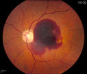
Retinal Hemorrhage 1336 viewsFundus photography shows central retinal hemorrhage. Patient was treated with Avastin Injection and will be followed up in 4-weeks.
|
|

Posterior Staphyloma and Geographic Atrophy Myopic Degeneration1330 views54-year-old man . He is a high myope. He has posterior staphylomas. He has had gradually changing vision for the last month or two. OD is 20/40, OS is 20/50.
|
|

Adult Vitelliform Macular Dystrophy - Pattern Dystrophy1328 views69-year-old woman sees a dark spot centrally. When she looks at things she loses letters and numbers and is having more and more difficulty balancing her checkbook. OD is 20/80, OS is 20/40
|
|

Central Serous Retinopathy Acute - Fundus Autofluorescence1326 views42-year-old man was seen in the office on October 5, 2011. He had noticed starting in August after a course of antibiotic and steroids, that he developed new spots in his vision in the right eye. He may have had an episode like this sometime in the past. He did take steroids a few years ago and his vision did change at that time, but then returned.
VISUAL ACUITY: OD 20/32, OS 20/32
|
|

Basal Laminar Drusen - Cuticular Drusen - Equatorial Drusen - 70 Year Old Woman - Photo Montage Right Eye1323 views70 year old woman has had macular drusen since her thirties and she said her sister has a similar problem. Recently the vision in the left eye has substantially declined. OD 20/16, OS 10/200.
|
|

Acute Central Serous Retinopathy - Classic Smokestack on Fluorescein Angiogram 1321 views34-year-old man noticed about five days ago decreased vision in the right eye. He was having difficulty playing video games and seeing with that eye. He thought maybe his refractive error was shifting.
VISUAL ACUITY: Vision OD is 20/40, OS is 20/16
|
|

Acute Central Serous Retinopathy - Classic Smokestack on Fluorescein Angiogram 1320 views34-year-old man noticed about five days ago decreased vision in the right eye. He was having difficulty playing video games and seeing with that eye. He thought maybe his refractive error was shifting.
VISUAL ACUITY: Vision OD is 20/40, OS is 20/16
|
|

Wet AMD Fresh Submacular Hemorrhage - Displaced with Vitrectomy and Subretinal TPA1318 views85-year-old woman has wet age-related macular degeneration in her better eye and she was reading with it until two days ago when she had sudden severe vision loss in the eye. Her last injection with Avastin 2 weeks ago 20/160 (Pre-Surgical Displacement)
|
|

AMD - Wet - Fibrovascular Hemorrhagic PED - Occult CNVM1315 views87-year-old woman for one month OS: 20/60 Fibrovascular PED and Hemorrhage
|
|

Acute Central Serous Retinopathy - Classic Smokestack on Fluorescein Angiogram 1310 views34-year-old man noticed about five days ago decreased vision in the right eye. He was having difficulty playing video games and seeing with that eye. He thought maybe his refractive error was shifting.
VISUAL ACUITY: Vision OD is 20/40, OS is 20/16
|
|

Macular Pucker1310 viewsPatient comes in for followup on macular pucker, right eye. VA is 20/50 in the right eye...Fundus photo shows prominent pucker centrally pulling temporally in the right eye.
|
|

Macular Degeneration with Hemorrhage1298 viewsPatient comes in for follow up exam for wet AMD. Fundus photography reveals retinal hemorrhage temporally to the macula. Patient was not treated. Wait to see if the blood will absorb on its own.
|
|

Angioid Streaks - Peau D'Orange - Pseudoxanthoma Elasticum - Asymptomatic Extrafoveal Choroidal Neovascular Membrane1296 views60-year-old woman has pseudoxanthoma elasticum since 2008. Her sister has the same problem and lost vision in both eyes. She had a problem with blood vessel growth in the left eye for which she had injections of Avastin in August, September, and October of 2011 (5 months ago). Subsequent to that, the macula has been dry and you have just been watching her carefully. She is in Florida for a short time but will ultimately be spending nine months out of the year here. Both of her eyes have good vision at
|
|
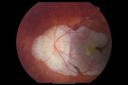
Posterior Staphyloma and Geographic Atrophy Myopic Degeneration1286 views54-year-old man . He is a high myope. He has posterior staphylomas. He has had gradually changing vision for the last month or two. OD is 20/40, OS is 20/50.
|
|

Reticular Macular Disease (Pseudo-drusen) Both Eyes - Wet AMD OS - Dry AMD OD SD-OCT1282 views84-year-old woman has wet age-related macular degeneration in the left eye and dry macular degeneration in the right eye. She takes the eye vitamins and her vision is stable since she was treated three months ago with Avastin. OD 20/50, OS 20/32
|
|

Juxtafoveal Telangiectasis - MacTel - Retinal Telangiectasis - Both Eyes - Crystals1278 views61-year-old man has idiopathic juxtafoveal retinal telangiectasis type II in both eyes OD is 20/40, OS is 20/25
|
|

Plaquenil Toxicity - Bull's Eye Maculopathy - 25 Years Therapy - Fundus Autofluorescence1277 views73-year-old woman has been on Plaquenil for twenty five years. Plaquenil has now been stopped for 6 months. VA continues to decline as does visual function - 20/80 OD , 20/40 OS
|
|

Plaquenil Toxicity - Bulls Eye Maculopathy1275 views70-year-old woman with systemic Lupus erythematosus and clotting problems. She was on the Plaquenil for about eight years and then off the Plaquenil for the last eight years because she developed macular toxicity. Although her vision was hazy, it was stable. Recent deceased vision left eye: OD 20/60, OS 20/100. IOP: OD 18, OS 19.
|
|

Acute Central Serous Retinopathy - Classic Smokestack on Fluorescein Angiogram 1274 views34-year-old man noticed about five days ago decreased vision in the right eye. He was having difficulty playing video games and seeing with that eye. He thought maybe his refractive error was shifting.
VISUAL ACUITY: Vision OD is 20/40, OS is 20/16
|
|

Acute Central Serous Retinopathy - Classic Smokestack on Fluorescein Angiogram 1268 views34-year-old man noticed about five days ago decreased vision in the right eye. He was having difficulty playing video games and seeing with that eye. He thought maybe his refractive error was shifting.
VISUAL ACUITY: Vision OD is 20/40, OS is 20/16
|
|

Acute Central Serous Retinopathy - Classic Smokestack on Fluorescein Angiogram 1264 views34-year-old man noticed about five days ago decreased vision in the right eye. He was having difficulty playing video games and seeing with that eye. He thought maybe his refractive error was shifting.
VISUAL ACUITY: Vision OD is 20/40, OS is 20/16
|
|

Patterned Pigment Dystrophy of the Fovea 2 year later VA still good1261 views
|
|

Angioid Streaks and Treated CNVM1260 viewsMaculopathy associated to angioid streaks in a 45 years old patient, man,
treated for related CNV
|
|

Cuticular Drusen (Basal Laminar Drusen) and Bilateral Chronic Subfoveal Fluid (Serous Retinal Detachment) unresponsive to monthly Avastin OCT OS EDI1257 views66-year-old man with bilateral subfoveal fluid unresponsive to one year of monthly avastin therapy.
OD 20/160, Pinhole 20/80. OS 20/63, Pinhole 20/50. OCT scan shows subretinal fluid with normal choroidal thickness. Fluorescein angiogram shows cuticular/basal laminar drusen. ICG shows no foci of choriocapillaris leakage.
|
|

Patterned Pigment Dystrophy of the Fovea 79 YO Woman VA 20/20 Right 20/30 Left1256 views
|
|

Macular Telangiectasis - visible telangiectasia on color photos1251 viewsMulticolor photos too show pathology. This patient had 20/50 vision with pinhole to 20/25
|
|

Hyperpigmentation of the Retinal Pigment Epithelium Right Eye (Treated wet AMD OS)1244 views85-year-old man has wet age-related macular degeneration in the left eye treated for one year most recently one year ago. OD is 20/30, OS is 20/60
|
|

Equatorial Drusen Fundus Photo Left Eye1243 views
|
|

Central Serous Retinopathy Acute - Fundus Autofluorescence1243 views42-year-old man was seen in the office on October 5, 2011. He had noticed starting in August after a course of antibiotic and steroids, that he developed new spots in his vision in the right eye. He may have had an episode like this sometime in the past. He did take steroids a few years ago and his vision did change at that time, but then returned.
VISUAL ACUITY: OD 20/32, OS 20/32
|
|

Hyperpigmentation of Retinal Pigment Epithlium in Dry AMD (hyperplasia)1240 views87-year-old woman has wet age-related macular degeneration in the left eye. She had treatment up north but there has been a gap in therapy and the vision in the left eye is poor. Vision OD is 20/25, OS is 20/400
|
|

PXE with Angioid Streaks1240 viewsPXE with angioid streaks OS (12/09/2011)
|
|
| 5015 files on 51 page(s) |
1 |
 |
|
|
|
|
|
|
|
|
|


