am meisten angesehen - Macular Disease
|

Reticular Macular Disease (Pseudo-drusen) Both Eyes - Occult CNVM and Macular Edema Right Eye 642 x angesehenSpectral Domain Optical Coherence Tomography
|
|

Polypoidal Choroidal Vasculopathy with Pulsating ICG polyp left eye642 x angesehen
|
|

Outer Retinal Holes - Probably Solar Retinopathy (history is negative)642 x angesehen
|
|
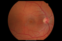
Macular Hole Closed with Vitrectomy - Preop641 x angesehen
|
|

Plaquenil Toxicity with New Symptomatic Scotoma Left Eye641 x angesehen64-year-old female decreased vision OS over the past 2 months. She describes a blurred central scotoma. She has rheumatoid arthritis and is on Plaquenil 200 mg BID for 15 years. She does have yearly visual field tests done with last visual field test performed 3 years ago which was within normal limits OD 20/40-2 OS 20/40-1
|
|

Hyperpigmentation of Retinal Pigment Epithlium in Dry AMD (hyperplasia)641 x angesehen87-year-old woman has wet age-related macular degeneration in the left eye. She had treatment up north but there has been a gap in therapy and the vision in the left eye is poor. Vision OD is 20/25, OS is 20/400
|
|

Myopic Macular Degeneration - Staphyloma - Recent Vision Loss Right Eye - Possible CNVM641 x angesehen83-year-old woman is a high myope with myopic macular degeneration. She had a retinal detachment repair in the right eye in 1968. I saw her in June of 2003. She had posterior colobomas and areas of pigment epithelial atrophy. She has noticed over the last six weeks substantial declining vision in the right eye. Prior to that, the vision in the two eyes was about the same. She uses Xalatan in the left eye and Alphagan in both eyes.
VISUAL ACUITY: Vision OD is 20/160, OS is 20/40
|
|

Cuticular Drusen (Basal Laminar Drusen) and Bilateral Chronic Subfoveal Fluid (Serous Retinal Detachment) unresponsive to monthly Avastin OCT OD VIDEO TAKES A LONG LONG TIME TO LOAD641 x angesehen66-year-old man with bilateral subfoveal fluid unresponsive to one year of monthly avastin therapy.
OD 20/160, Pinhole 20/80. OS 20/63, Pinhole 20/50. OCT scan shows subretinal fluid with normal choroidal thickness. Fluorescein angiogram shows cuticular/basal laminar drusen. ICG shows no foci of choriocapillaris leakage.
|
|

Reticular Macular Disease (Pseudo-drusen) Both Eyes - Occult CNVM and Macular Edema Right Eye 641 x angesehenFluorescein Angiogram
|
|
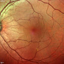
Macular Pucker 20/40 vision - minimal symptoms for 1 year Right eye641 x angesehenMulticolor
|
|

IPCV - idiopathic polypoidal choroidal vasculopathy - juxtapapillary CNVM with hemorrhage both eyes (also choroidal nevus right eye) - Subretinal hemorrhage both eyes641 x angesehen86 year old woman with shadow in vision OD and new hemmorhage OU.FA shows juxtapapillary CNVM
|
|

Travatan Induced CME Left eye - 1 month post stopping travatan CME is gone641 x angesehen63 year old woman 4 years post ECCE and 2 years post PPV for floaters. She developed CME in the left eye after 1 year of travatan. The travatan was stopped and the CME and serous retinal detachment resolved.
|
|

Wet AMD - Subfoveal CNVM - Excellent Response to Avastin640 x angesehen76-year-old woman two to three months of vision loss OD
Vision started at 20/200 and with Avastin improved to 20/25. She has required monthly injections and as of October 2010 has been treated about 16 times. Her left eye remains good at 20/20.
The OCT from post-treatment as well as pretreatment images are in this album.
|
|
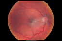
Macular Pucker in eye following giant retinal tear RD repair pre-op pucker photo640 x angesehennow that the bubble is gone, but he is starting to notice distortion of central vision. He had a giant retinal tear retinal detachment repair emergently done on November 8, 2007, and subsequent laser on November 15th.
VISUAL ACUITY: Vision OD 20/400.
6 weeks post-op VA 20/40
|
|

Juxtapapillary CNVM and Serous Macular Detachment Wet AMD Rx Laser640 x angesehen85-year-old man OD is 20/25, OS is 20/100. IOP: OD 21, OS 22.
OS: Vertical C/D ratio is 0.1. There is a posterior vitreous separation. There is a hemorrhagic pigment epithelial detachment on the superior pole of the optic nerve, extending a disc-and-a-half diameter off the nerve, with adjacent exudates. There is a serous macular detachment involving the center of the fovea.
VA improved from 20/100 to 20/50 in four months from laser.
|
|
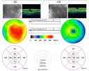
Macular Pucker Pre-op 20/200 vision (history of retinal tears)640 x angesehen66-year-old man has had laser to retinal tears in both eyes in 1999 and in 2001. His right eye has had rapidly declining vision over the last month. VISUAL ACUITY: OD: 20/200
|
|
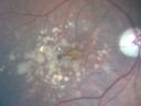
Drusen Maculopathy 85 year old Male640 x angesehen85-year-old man has age-related macular degeneration in both eyes with drusen maculopathy. OD is 20/40, OS is 20/400
|
|

Pigment Epithelial Detachment with Subretinal Fluid and Exudate 20/400 Vision640 x angesehen87-year-old woman has wet age-related macular degeneration in the left eye. She had treatment up north but there has been a gap in therapy and the vision in the left eye is poor. Vision OD is 20/25, OS is 20/400
|
|

Pattern Dystrophy - Foveal Atrophy640 x angesehen73 year old female with vision loss - looks like macular hole
|
|

Plaquenil Toxicity - Bulls Eye Maculopathy639 x angesehen70-year-old woman with systemic Lupus erythematosus and clotting problems. She was on the Plaquenil for about eight years and then off the Plaquenil for the last eight years because she developed macular toxicity. Although her vision was hazy, it was stable. Recent deceased vision left eye: OD 20/60, OS 20/100. IOP: OD 18, OS 19.
|
|

Pigment Epithelial Detachment with Subretinal Fluid and Exudate 20/400 Vision639 x angesehen87-year-old woman has wet age-related macular degeneration in the left eye. She had treatment up north but there has been a gap in therapy and the vision in the left eye is poor. Vision OD is 20/25, OS is 20/400
|
|

Cuticular Drusen (Basal Laminar Drusen) and Bilateral Chronic Subfoveal Fluid (Serous Retinal Detachment) unresponsive to monthly Avastin Late FA OD639 x angesehen66-year-old man with bilateral subfoveal fluid unresponsive to one year of monthly avastin therapy.
OD 20/160, Pinhole 20/80. OS 20/63, Pinhole 20/50. OCT scan shows subretinal fluid with normal choroidal thickness. Fluorescein angiogram shows cuticular/basal laminar drusen. ICG shows no foci of choriocapillaris leakage.
|
|

Asymptomatic Juxtapapillary Choroidal Neovascular Membrane - Reticular Macular Disease (Wet AMD) - FAF639 x angesehen
|
|
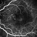
Polypoidal Choroidal Vasculopathy with Pulsating ICG polyp left eye639 x angesehen
|
|

Macular Edema and dry AMD - excellent example of why FA and ICG are necessary in management of wet AMD639 x angesehenThis 72 year old woman has an OCT that looks like wet AMD. Only her FA and ICG show that she has inflammatory macular edema. She had no clinical signs or symptoms of uveitis. Nevertheless, the macular edema responded to subtenon's kenalog.
|
|
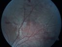
Macular Hole - Traumatic - 11 year old boy (also retinal whitening Berlins Edema)638 x angesehen11-year-old boy with a history of blunt trauma to the left eye yesterday. He reports he lost vision immediately but it has gotten somewhat better. He reports he sees “red streamers†and he has very blurry visual acuity., 20/200 OS
|
|
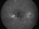
Hyperpigmentation of the Retinal Pigment Epithelium in Dry AMD638 x angesehen87-year-old woman has wet age-related macular degeneration in the left eye. Vision OD is 20/25, OS is 20/400
|
|

Macular Hole Left Eye - progression from VMT - Stage II macular hole - Spontaneously Closed Macular Hole638 x angesehen
|
|

Macular Pucker Pre-OP 20/200 637 x angesehen
|
|

Drusen Maculopathy - Macular Degeneration (Dry) - 50 year old African-American Woman637 x angesehen50-year-old woman has drusen and maculopathy. VISUAL ACUITY: OD 20/30, OS 20/25.
|
|

New Subfoveal Classic CNVM OS - Wet AMD - Macular Scar Congenital Toxoplasmosis Right Eye 637 x angesehen77-year-old woman as had poor vision in the right eye since birth from congenital toxoplasmosis of her macula in the right eye. Two to three weeks ago when she noticed vision loss in the left eye. VISUAL ACUITY: OD 2/200, OS 20/200.
|
|

Macular Hole - Traumatic - 11 year old boy (also retinal whitening Berlins Edema)636 x angesehen11-year-old boy with a history of blunt trauma to the left eye yesterday. He reports he lost vision immediately but it has gotten somewhat better. He reports he sees “red streamers†and he has very blurry visual acuity., 20/200 OS
|
|

Plaquenil (Hydroxycholorquine) Toxicity - Bull's Eye Maculopathy - 73 year old womand 25 Years therapy - 64 inches tall - 110 lbs636 x angesehen73-year-old woman has been on Plaquenil for twenty five years. She noticed loss of paracentral vision a few months ago. VISUAL ACUITY: OD 20/70, OS 20/30. Her color vision was 0 out 10 correct in both eyes and she also missed the controls.
|
|

Wet AMD - PED with exudate 20/400 87 Year old636 x angesehen87-year-old woman has wet age-related macular degeneration in the left eye. Vision OD is 20/25, OS is 20/400
|
|

Myopic Traction Maculopathy - Vitreoretinal Traction - Possible Occult Neovascular Membrane636 x angesehen58-year-old woman was seen in the office on June 30, 2011. In 2002, her vision was 20/30. Subsequent to that she had her left eye made her distance eye, which was dominate. She had LASIK surgery on the right eye to make that her near eye and she was getting along well until about a year ago when she noticed substantial vision loss in the left eye only. VISUAL ACUITY: The near eye is 20/200, but she reads J2. OS 20/100- and J16.
|
|
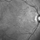
Psuedomacular Hole - Epiretinal Membrane - Vision unchanged 20/40 for 6 months636 x angesehen
|
|

Wet AMD 62 year old male smoker 635 x angesehen62-year-old man has wet age-related macular degeneration in the right eye. He has been undergoing treatment with Lucentis for that. Then his left eye developed hazy vision a few weeks ago while he was overseas and now his vision is hazy in both eyes. OD 20/40, OS 20/30. FA shows a classic subfoveal choroidal neovascular membrane in the left eye, which is leaking and in the right eye, which is dry. He is going to stop smoking today. FU: VA 20/30 OU with 2 years of every 6 weeks bilateral avastin!
|
|
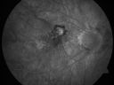
Myopic Choroidal Neovascular Membrane 54 year old woman 20/20635 x angesehen54-year-old woman has had problems in her vision in the right eye for about the last three months. Inferotemporally she sees a gray area, which is distorted. OD is 20/20, OS is 20/20
|
|

Myopic Choroidal Neovascular Membrane 54 year old woman 20/20635 x angesehen54-year-old woman has had problems in her vision in the right eye for about the last three months. Inferotemporally she sees a gray area, which is distorted. OD is 20/20, OS is 20/20
|
|

Patterned Pigment Dystrophy of the Fovea 79 YO Woman VA 20/20 Right 20/30 Left635 x angesehen
|
|

Choriodal Neovascular Membrane - Subfoveal - Pseudo-Retinitis Pigmentosa - 50 years post Retinal Detachment Repair635 x angesehen86-year-old with a history of decreased vision in the right eye. The patient reports she has lost vision in the right eye over the past two months or so. She reports the vision was always compromised in that eye since retinal detachment surgery 50 years ago with a scleral buckler. Vision dropped from 20/50 to 20/200 right eye in 2 months.
|
|

Macular Pucker - 1 year after Vitrectomy - 20/25 Vision - Postoperative Images635 x angesehen60-year-old man had vitrectomy for a macular pucker in the right eye a year ago. His vision is good.
VISUAL ACUITY: Vision OD is 20/25 Eye is now pseudophakic
|
|

Reticular Macular Disease (Pseudo-drusen) Both Eyes - Occult CNVM and Macular Edema Right Eye 635 x angesehenIndocyanine Green Angiogram
|
|
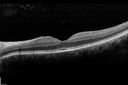
Macular Pucker 20/40 vision - minimal symptoms for 1 year Right eye635 x angesehen SD OCT line Scan Fellow Eye 20/16
|
|
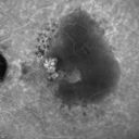
Polypoidal Choroidal Vasculopathy with Pulsating ICG polyp left eye635 x angesehen
|
|

Chronic CSR with mild vision loss - Macular Atrophy Right Eye635 x angesehen55 year old man has not been to an eye doctor in years. VA 20/40 OD and 20/25 OS. OCT shows outer retinal atrophy and choroidal thickening. FAF shows guttering. FA shows no active lesions. ICG shows patchy mild leakage visible at about 5 minutes.
|
|
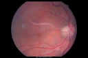
Retinal fold following removal of severe macular pucker635 x angesehen75 year old man had macula on inferior retinal detachment repair. He then developed a 5/200 dense macular pucker. After the pucker was removed the vision returned to 20/40 but there was a persistent superior retinal fold that looks a little like a snake.
|
|

Macular Hole Closed with Vitrectomy - Preop634 x angesehen
|
|

Group 2a Juxtafoveal Retinal Telangiectasis (type 2)634 x angesehen58-year-old man decreased vision right eye and not better since cataract surgery one year ago. OD 20/40, OS 20/40.
|
|

Plaquenil Toxicity - Bull's Eye Maculopathy - 25 Years Therapy OCT Posterior Pole MAP634 x angesehen73-year-old woman has been on Plaquenil for twenty five years. Plaquenil has now been stopped for 6 months. VA continues to decline as does visual function - 20/80 OD , 20/40 OS
|
|

634 x angesehen
|
|

Wet Age-related Macular Degeneration (AMD) Radiating Chorioretinal Folds from Contracting Macular Scar634 x angesehen93-year-old man has wet age-related macular degeneration in his left eye with a subfoveal pigment epithelial detachment.
Vision OS is 20/40. There are striking radiating chorioretinal folds emanating off of the scar, which suggests it is contracting.
|
|

Wet AMD - New Small Hemorrhage Both Eyes - Mature CNVM visible on Early Indocyanine Green Angiogram634 x angesehen
|
|
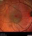
Chronic Central Serous Retinopathy - False Color634 x angesehen
|
|
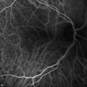
Cartwheel CNVM - Classic - wet AMD - evidence of possible old CSR - ICG is normal - shows nothing634 x angesehen75 y.o Decreased Vision right eye for one year. VA OD: sc20/125+2 OS 20/25. Autofluorescence and SD OCT show evidence of old serous retinal detachment superior to the fovea suggesting a history of CSR. The macular lesion on FA shows a classic cartwheel appearance
|
|

Polypoidal Choroidal Vasculopathy with Pulsating ICG polyp left eye634 x angesehen
|
|

Wet AMD - Subfoveal CNVM - Excellent Response to Avastin633 x angesehen76-year-old woman two to three months of vision loss OD
Vision started at 20/200 and with Avastin improved to 20/25. She has required monthly injections and as of October 2010 has been treated about 16 times. Her left eye remains good at 20/20.
The OCT from post-treatment as well as pretreatment images are in this album.
|
|
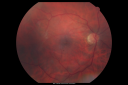
Macular Pucker in eye following giant retinal tear RD repair post-op Fundus Photo633 x angesehennow that the bubble is gone, but he is starting to notice distortion of central vision. He had a giant retinal tear retinal detachment repair emergently done on November 8, 2007, and subsequent laser on November 15th.
VISUAL ACUITY: Vision OD 20/400.
6 weeks post-op VA 20/40
|
|

Macular Pucker 20/40 Vision - Pigment Bottom of Photo from Old Retinal Detachment 633 x angesehen59-year-old woman blurry vision in the right eye for about the last one or two years. She had cataract surgery in 1981, a retinal detachment repair with scleral buckle in 1988 in the right eye, and then she had lens implants in both eyes, anterior chamber lenses, in 1994. She did start taking Latisse for eyelashes about a year and a half ago.
VISUAL ACUITY: Vision OD is 20/40, OS is 20/20
(4 months later with a short course of antiinflammatory drops and new glasses her vision improved to 20/25)
|
|

633 x angesehen
|
|
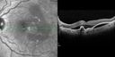
Polypoidal Choroidal Vasculopathy with Pulsating ICG polyp left eye633 x angesehen
|
|

Subfoveal CNVM wet AMD early fa632 x angesehenShe has noticed decreased vision in the left eye. It is hard to say for how long, maybe a few months. She had her last eye exam about a year ago. She thoughts perhaps it was cataracts, but you saw a problem with the macula and suggested she come here for evaluation.
VISUAL ACUITY: Vision OD is 20/25, OS is 2/200
|
|

632 x angesehen
|
|

Macular Degeneration - Choroidal Laser Left eye - New Subfoveal CNVM left eye - Did well with Avastin632 x angesehen69-year-old woman new vision loss right eye and old macular scar left eye. OD 20/40, OS 20/200
|
|
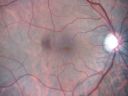
Macular Pucker - 1 year after Vitrectomy - 20/25 Vision - Postoperative Images632 x angesehen60-year-old man had vitrectomy for a macular pucker in the right eye a year ago. His vision is good.
VISUAL ACUITY: Vision OD is 20/25 Eye is now pseudophakic
|
|

Asymptomatic Occult Subfoveal CNVM in Good Eye Visible on ICG632 x angesehen
|
|

Adult Vitelliform Macular Dystrophy - Pattern Dystrophy632 x angesehenSD OCT shows material to be subretinal not sub-RPE as you would see in typical AMD
|
|

Wet AMD - Subfoveal CNVM - Excellent Response to Avastin631 x angesehen76-year-old woman two to three months of vision loss OD
Vision started at 20/200 and with Avastin improved to 20/25. She has required monthly injections and as of October 2010 has been treated about 16 times. Her left eye remains good at 20/20.
The OCT from post-treatment as well as pretreatment images are in this album.
|
|
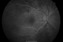
Cystoid Macular Edema - Xalatan - ECCE was done 20 years previously631 x angesehen71 Year old woman who noticed about a month ago the central vision in the right eye fogged up. OD 20/200 POSSIBLE XALATAN INDUCED CYSTOID MACULAR EDEMA
|
|
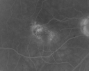
Juxtafoveal Telangiectasis - (MacTel) - Stage 4 both Eyes631 x angesehen57-year-old woman with a history of decreased vision. The patient reports that her vision has been gradually decreasing in the left more so than the right eye. 20/50 OD and 20/60
|
|

Acute Central Serous Chorioretinopathy with Symptoms in Hazy Vision Both Eyes631 x angesehen48-year-old man with hazy vision in the right eye for about three days. He has a little more of a headache than usual. He is under a fair amount of stress. He does not take any steroids that he knows of. He does use a nasal spray, which he is not sure if that is a steroid spray or not. He is going to call us with that medicine. I checked his blood pressure today is was 115/95.
VISUAL ACUITY: OD 20/15, OS 20/25
|
|
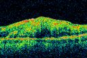
Post Retinal Detachment Repair Macular Pucker - VA 20/400 Pre-op 20/25 Post-op631 x angesehen72 year old woman who is 6 months post macula-on retinal detachment repair developed a 20/400 macular pucker. 8 months after surgery vision had improved to 20/25
|
|

Angioid Streaks and Macular Degeneration - CNVM OS - Band Keratopathy 631 x angesehen79-year-old man has wet age-related macular degeneration in the left eye, for which he is having Lucentis treatment. OD 20/40, Pinhole 20/32. OS 20/160, Pinhole 20/50. He has band keratopathy sparing the visual axis in both eyes. ICG shows angioid streaks both eye.
|
|

Serous Macular Detachment Both Eyes - Wet AMD vs. CSR - 62 Year Old Man - SD - OCT631 x angesehen
|
|

Macular Edema and dry AMD - excellent example of why FA and ICG are necessary in management of wet AMD631 x angesehenThis 72 year old woman has an OCT that looks like wet AMD. Only her FA and ICG show that she has inflammatory macular edema. She had no clinical signs or symptoms of uveitis. Nevertheless, the macular edema responded to subtenon's kenalog.
|
|

Cystoid Macular Edema 10 years after Cataract Surgery631 x angesehenNo overt uveitis but the patient had aching in the eyes and responded to topical anti-inflammatory drops with recurrence of the CME on discontinuation of topical therapy
|
|

Confluent Soft Drusen with Subetinal Fluid - Right Eye631 x angesehenAsymptomatic 85 year old man. OCT shows pocket of subfoveal fluid in the center of the macula. This could be mechanical from the drusen preventing the retina from settling down. Testing did not reveal a CNVM and subsequent OCT 6 weeks later showed no change in structure.
|
|

Macular Pucker in eye following giant retinal tear RD repair post-op macular photo630 x angesehennow that the bubble is gone, but he is starting to notice distortion of central vision. He had a giant retinal tear retinal detachment repair emergently done on November 8, 2007, and subsequent laser on November 15th.
VISUAL ACUITY: Vision OD 20/400.
6 weeks post-op VA 20/40
|
|
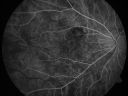
Myopic Choroidal Neovascular Membrane 54 year old woman 20/20630 x angesehen54-year-old woman has had problems in her vision in the right eye for about the last three months. Inferotemporally she sees a gray area, which is distorted. OD is 20/20, OS is 20/20
|
|

Wet Macular Degeneration Left Eye with Gutter (atypical) and Geographic Atrophy Right Eye630 x angesehen80-year-old man has wet age-related macular degeneration in his left eye. He has an atypical angiogram and that there is a gutter under the choroidal neovascular membrane suggesting still leakage of subretinal fluid. OS 20/400.
|
|

630 x angesehen
|
|

Wet AMD - PED with exudate 20/400 87 Year old630 x angesehen87-year-old woman has wet age-related macular degeneration in the left eye. Vision OD is 20/25, OS is 20/400
|
|

Non-Foveal Geographic Atrophy Both Eyes630 x angesehen81-year-old man has age-related macular degeneration in both eyes. Vision OD is 20/40, OS is 20/80.
|
|

Juxtafoveal Telangiectasis (MacTel) and Asymptomatic Extrafoveal Juxtapapillary Choroidal Neovascular Membrane630 x angesehen84-year-old woman has juxtafoveal retinal telangiectasis in both eyes. The right eye has an asymptomatic extra foveal juxtapapillary choroidal neovascular membrane. OD 20/400, OS 20/63.
|
|

Reticular Macular Disease (Pseudo-drusen) Both Eyes - Occult CNVM and Macular Edema Right Eye 630 x angesehenFluorescein Angiogram
|
|

Wet AMD - Subfoveal CNVM - Excellent Response to Avastin629 x angesehen76-year-old woman two to three months of vision loss OD
Vision started at 20/200 and with Avastin improved to 20/25. She has required monthly injections and as of October 2010 has been treated about 16 times. Her left eye remains good at 20/20.
The OCT from post-treatment as well as pretreatment images are in this album.
|
|
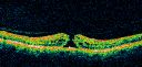
Macular Hole Closed with Vitrectomy - Preop629 x angesehen
|
|

Idiopathic Central Serous Retinopathy - Bilateral - Acute 4 months later VA 20/40 OU629 x angesehenNo treatment and fluid under fovea in left eye is competely absorbed at 4 months.
|
|

Vision Loss, Blood, and Cartwheel CNVM OD with no fluid on OCT Scan629 x angesehen86-year-old man has had changing vision. He says his vision has been gradually declining since I saw him in June. You saw a hemorrhage in the eye and suggested he comes in here for an evaluation. He is on Combigan and Travatan in the right eye for his glaucoma.
VISUAL ACUITY: Vision OD is 6/200 OS is 8/200 The macula in the right eye is wet even though interestingly the OCT does not show subretinal fluid.
|
|
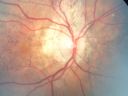
Angioid Streaks - Pseudoxanthoma Elasticum - Supressed CNVM629 x angesehen54-year-old woman had angioid streaks and macular degeneration in both eyes. She has had intravitreal Avastin in the right eye about every three months and her vision in that eye is good.
VISUAL ACUITY: Vision OD is 20/30, OS is 2/200
|
|

Acute Central Serous Retinopathy - CSR - Steroid Induced (probably) - Good Vision - ICG Fellow Eye629 x angesehen42-year-old man was seen in the office on October 5, 2011. He had noticed starting in August after a course of antibiotic and steroids, that he developed new spots in his vision in the right eye. He may have had an episode like this sometime in the past. He did take steroids a few years ago and his vision did change at that time, but then returned.
VISUAL ACUITY: OD 20/32, OS 20/32. The OCT scan of the right eye confirms subretinal fluid.
|
|

Macular Pucker in eye following giant retinal tear RD repair post-op OCT map628 x angesehennow that the bubble is gone, but he is starting to notice distortion of central vision. He had a giant retinal tear retinal detachment repair emergently done on November 8, 2007, and subsequent laser on November 15th.
VISUAL ACUITY: Vision OD 20/400.
6 weeks post-op VA 20/40
|
|

Macular Pucker in eye following giant retinal tear RD repair photo of tear628 x angesehennow that the bubble is gone, but he is starting to notice distortion of central vision. He had a giant retinal tear retinal detachment repair emergently done on November 8, 2007, and subsequent laser on November 15th.
VISUAL ACUITY: Vision OD 20/400.
6 weeks post-op VA 20/40
|
|

Central Serous Retinopathy - Acute628 x angesehen49-year-old man sees a spot in the central vision with a red halo for 2 weeks OD is 20/50, OS is 20/20
|
|

628 x angesehen
|
|

Macular Degeneration - Choroidal Laser Left eye - New Subfoveal CNVM left eye - Did well with Avastin628 x angesehen69-year-old woman new vision loss right eye and old macular scar left eye. OD 20/40, OS 20/200
|
|

Chronic Pseudophakic Cystoid Macular Edema - Haptic Migrated into Anterior Chamber Through Peripheral Iridotomy628 x angesehen57-year-old man with congenital nystagmus. He had cataract surgery done 30 years ago and has decreased vision left eye for 6 months. OD is 20/50, OS is 20/100
|
|

Chronic Pseudophakic Cystoid Macular Edema - Haptic Migrated into Anterior Chamber Through Peripheral Iridotomy628 x angesehen57-year-old man with congenital nystagmus. He had cataract surgery done 30 years ago and has decreased vision left eye for 6 months. OD is 20/50, OS is 20/100
|
|

Reticular Macular Disease (Pseudo-drusen) Both Eyes - Occult CNVM and Macular Edema Right Eye 628 x angesehenInfra-Red
|
|

Choroidal Effusions and Serous retinal detachment with Systemic Testosterone and Cialis in 75 year old Man628 x angesehenIR of good eye looks normal
|
|
| 5015 Dateien auf 51 Seite(n) |
 |
|
|
|
|
10 | |
|
|
|
|


