Most viewed - Macular Disease
|

Central Serous Retinopathy 25 year old professional baseball player - Atypical - Choroidal Hypo-perfusion?559 views
|
|

Asymptomatic Juxtapapillary Choroidal Neovascular Membrane - Reticular Macular Disease (Wet AMD) 559 views
|
|

Cystoid macular edema 4 months following cataract surgery with serous retinal detachment559 views68 year old with 2 months of vision loss OS 20/100. ECCE was 4 months ago.
|
|

Macular Telangiectasia and dry AMD - Simulating wet AMD559 viewsThis patient was told different things by different retina specialists. Her case is complex because she has OCT findings of Macular Telangiectasis and dry AMD. This makes it look like she has wet AMD. Her vision is stable.
|
|

Confluent Soft Drusen with Subetinal Fluid - Right Eye559 viewsAsymptomatic 85 year old man. OCT shows pocket of subfoveal fluid in the center of the macula. This could be mechanical from the drusen preventing the retina from settling down. Testing did not reveal a CNVM and subsequent OCT 6 weeks later showed no change in structure.
|
|

Macular Pucker Post-op 20/30 Vision558 viewsVa 20/30 after surgery
|
|

Angioid Streaks - New Extrafoveal Choroidal Neovascular Membrane Right Eye558 views61-year-old woman twelve days ago, saw a spot to the right of her central vision. OD is 20/25, OS is 20/25
Treated with one year of avastin with good result and no vision loss.
|
|

Plaquenil (Hydroxycholorquine) Toxicity - Bull's Eye Maculopathy - 73 year old womand 25 Years therapy - 64 inches tall - 110 lbs558 views73-year-old woman has been on Plaquenil for twenty five years. She noticed loss of paracentral vision a few months ago. VISUAL ACUITY: OD 20/70, OS 20/30. Her color vision was 0 out 10 correct in both eyes and she also missed the controls.
|
|

Acute Central Serous Retinopathy (Chorioretinopathy) Right Eye558 views45-year-old man decreased vision in the right eye starting about two days ago. VISUAL ACUITY: Vision OD is 20/60, OS is 20/20.
|
|

Equatorial Drusen558 views76-year-old man with equatorial drusen
VISUAL ACUITY: Vision OD is 20/40, OS is 20/25.
|
|

Central Serous Retinopathy 25 year old professional baseball player - Atypical - Choroidal Hypo-perfusion?558 views
|
|

Asymptomatic Juxtapapillary Choroidal Neovascular Membrane - Reticular Macular Disease (Wet AMD) 558 views
|
|

Vitreomacular traction - recent vision loss Right Eye 558 views
|
|
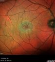
Macular Telangiectasia - Stage 3 right eye 20/40 vision558 viewsMulti-Color Image
|
|

Polypoidal Choroidal Vasculopathy with Pulsating ICG polyp left eye558 views
|
|

Dry AMD - Confluent Drusen Sparing Center of Macula which has Atrophy558 views65 year old woman, VA 20/40 OD; 20/80 OS. The center of the macula has few or no drusen with predominantly non-geographic atrophy
|
|

Juxtapapillary CNVM and Serous Macular Detachment Wet AMD Rx Laser557 views85-year-old man OD is 20/25, OS is 20/100. IOP: OD 21, OS 22.
OS: Vertical C/D ratio is 0.1. There is a posterior vitreous separation. There is a hemorrhagic pigment epithelial detachment on the superior pole of the optic nerve, extending a disc-and-a-half diameter off the nerve, with adjacent exudates. There is a serous macular detachment involving the center of the fovea.
VA improved from 20/100 to 20/50 in four months from laser.
|
|

Macular Degeneration - Choroidal Laser Left eye - New Subfoveal CNVM left eye - Did well with Avastin557 views69-year-old woman new vision loss right eye and old macular scar left eye. OD 20/40, OS 20/200
|
|

Patterned Pigment Dystrophy of the Fovea 79 YO Woman VA 20/20 Right 20/30 Left557 views
|
|

557 views
|
|

Acute Central Serous Retinopathy (Chorioretinopathy) Right Eye557 views45-year-old man decreased vision in the right eye starting about two days ago. VISUAL ACUITY: Vision OD is 20/60, OS is 20/20.
|
|

Plaquenil Toxicity - Bull's Eye Maculopathy - 25 Years Therapy - InfraRed Image557 views73-year-old woman has been on Plaquenil for twenty five years. Plaquenil has now been stopped for 6 months. VA continues to decline as does visual function - 20/80 OD , 20/40 OS
|
|
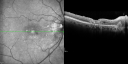
Juxtafoveal Telangiectasis (MacTel) and Asymptomatic Extrafoveal Juxtapapillary Choroidal Neovascular Membrane557 views84-year-old woman has juxtafoveal retinal telangiectasis in both eyes. The right eye has an asymptomatic extra foveal juxtapapillary choroidal neovascular membrane. OD 20/400, OS 20/63.
|
|

Macular Telangiectasis (MacTel) - Blue Reflectance557 viewsGray ring is more visible before the retina is exposed to bright light. This could be from bleaching of intraretinal chromophores if they are the source of the gray ring.
|
|

Chronic CSC Both Eyes - Recent vision loss Right Eye Treated with PDT557 views50 year old man The right eye has had vision loss for a few weeks. He had previous episodes seen by me in 2012 OS and 2008 OD. This was 2018. VA OS has been poor for several years.
VA OD: Dcc20/100
VA OS: Dcc20/200-3
The patient had PDT to leaky area superotemporal to the fovea 1/2 dose and within 3 months was back up to 20/25. The last two OCT scans show fluid absorbing
|
|
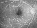
Cystoid Macular Edema - Xalatan - ECCE was done 20 years previously556 views71 Year old woman who noticed about a month ago the central vision in the right eye fogged up. OD 20/200 POSSIBLE XALATAN INDUCED CYSTOID MACULAR EDEMA
|
|

Patterned Pigment Dystrophy of the Fovea 79 YO Woman VA 20/20 Right 20/30 Left556 views
|
|

Macular Pucker - 3 Weeks Vision Loss - 20/50 Vision - Preoperative Images556 viewsPREOP:
59-year-old man new onset floaters about a year ago. Subsequent to that his vision was not quite right in the right eye and then he noticed, starting about three weeks ago, substantial change in the vision. VISUAL ACUITY: Vision OD is 20/50, OS is 20/16
|
|

New Subfoveal Classic CNVM OS - Wet AMD - Macular Scar Congenital Toxoplasmosis Right Eye 556 views77-year-old woman as had poor vision in the right eye since birth from congenital toxoplasmosis of her macula in the right eye. Two to three weeks ago when she noticed vision loss in the left eye. VISUAL ACUITY: OD 2/200, OS 20/200.
|
|

Polypoidal Choroidal Vasculopathy Right Eye - Disciform Scar Left Eye556 views84-year-old woman has polypoidal choroidal vasculopathy in both eyes with macular degeneration. She has macular scars in both eyes and she remains with remarkably good vision in the right eye despite the scarring.
VISUAL ACUITY: OD 20/30, OS 1/200
Avastin Rx OD
|
|

Hyperpigmentation of the Retinal Pigment Epithelium Right Eye (Treated wet AMD OS)556 views85-year-old man has wet age-related macular degeneration in the left eye treated for one year most recently one year ago. OD is 20/30, OS is 20/60
|
|

Reticular Macular Disease (Pseudo-drusen) Both Eyes - Occult CNVM and Macular Edema Right Eye 556 viewsFluorescein Angiogram
|
|
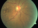
Polypoidal Choroidal Vasculopathy with Pulsating ICG polyp left eye556 views
|
|

Pseudophakic CME with serous retinal detachment - FA shows leakage in both eyes556 views68 year old man with 2 months of vision loss. FA shows CME, notice that the nerve also leaks. This is typical for pseudophakic inflammatory cystoid macular edema
|
|

Vitreomacular traction - spontaneous release556 views80 year old man with transient vision loss from 20/20 - 20/30 from vitreomacular traction. No surgery or procedure was done.
|
|

Patterned Pigment Dystrophy of the Fovea 79 YO Woman VA 20/20 Right 20/30 Left555 views
|
|

Macular Hemorrahge and Probable CNVM Right eye for 2 weeks VA 20/400 - 20 Year old555 views20-year-old man noticed vision loss in the right eye about two weeks ago and if anything it seems to be getting worse. He is a high myope. VISUAL ACUITY: OD 20/400
|
|

Myopic Traction Maculopathy - Vitreoretinal Traction - Possible Occult Neovascular Membrane555 views58-year-old woman was seen in the office on June 30, 2011. In 2002, her vision was 20/30. Subsequent to that she had her left eye made her distance eye, which was dominate. She had LASIK surgery on the right eye to make that her near eye and she was getting along well until about a year ago when she noticed substantial vision loss in the left eye only. VISUAL ACUITY: The near eye is 20/200, but she reads J2. OS 20/100- and J16.
|
|

Choroidal Effusions and Serous retinal detachment with Systemic Testosterone and Cialis in 75 year old Man555 viewsIR shows choroidal effusion as black (confocal system)
|
|

Multifocal Pattern Dystrophy555 views51 year old with normal vision - images look a lot like fundus flavimaculatus
|
|

Macular Telangiectasia and dry AMD - Simulating wet AMD555 viewsThis patient was told different things by different retina specialists. Her case is complex because she has OCT findings of Macular Telangiectasis and dry AMD. This makes it look like she has wet AMD. Her vision is stable.
|
|

Outer Retinal Tubulation555 views78 year old man chronic wet AMD - 20/200
|
|

Macular Telangiectasis - Very Mild555 views52 year old female with normal vision and suspicious OCT referred. Multimodal imaging shows very mild mactel (loss of macular pigment, ring on multicolor image and very mild OCT findings)
|
|

Retinal fold following removal of severe macular pucker555 views75 year old man had macula on inferior retinal detachment repair. He then developed a 5/200 dense macular pucker. After the pucker was removed the vision returned to 20/40 but there was a persistent superior retinal fold that looks a little like a snake.
|
|

Cystoid Macular Edema - Xalatan - ECCE was done 20 years previously554 views71 Year old woman who noticed about a month ago the central vision in the right eye fogged up. OD 20/200 POSSIBLE XALATAN INDUCED CYSTOID MACULAR EDEMA
|
|

554 views
|
|

Patterned Pigment Dystrophy of the Fovea 79 YO Woman VA 20/20 Right 20/30 Left554 views
|
|

Macular Pucker Post-OP 20/30554 views
|
|

Cuticular Drusen (Basal Laminar Drusen) and Bilateral Chronic Subfoveal Fluid (Serous Retinal Detachment) unresponsive to monthly Avastin Infrared OD554 views66-year-old man with bilateral subfoveal fluid unresponsive to one year of monthly avastin therapy.
OD 20/160, Pinhole 20/80. OS 20/63, Pinhole 20/50. OCT scan shows subretinal fluid with normal choroidal thickness. Fluorescein angiogram shows cuticular/basal laminar drusen. ICG shows no foci of choriocapillaris leakage.
|
|
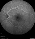
Macular Pucker 20/40 vision - minimal symptoms for 1 year Right eye554 viewsFA shows leakage from traction from pucker in the right eye - cystoid macular edema
|
|

Hydroxychlorquine (Plaquenil) Toxicity - Taiwanese 56 year old female (Asian)554 views56 year old female on 400 mg plaquenil for 22 years presents with vision loss. VA 20/50 both eyes. Plaquenil was stopped. The vision in the left eye continued to deteriorate to 3/200. 5'2" 127 lb (6.6 mg/kg body weight)
|
|

Cystoid Macular Edema 10 years after Cataract Surgery554 viewsNo overt uveitis but the patient had aching in the eyes and responded to topical anti-inflammatory drops with recurrence of the CME on discontinuation of topical therapy
|
|

Macular Pucker553 views. This pleasant 59-year-old man had vitrectomy for a macular pucker in the right eye in July. His vision is gradually improving.
VISUAL ACUITY: Vision OD is 20/30. IOP: OD 22. There is 2+ nuclear sclerosis.
EXTENDED OPHTHALMOSCOPY:
OD: Vertical C/D ratio is 0.5. The macula is smooth and the retina is attached.
OCT SCAN: The OCT scan shows an average central foveal thickness of 349 microns, which compares favorably to the pre-op thickness of 482 microns.
|
|

Patterned Pigment Dystrophy of the Fovea 79 YO Woman VA 20/20 Right 20/30 Left553 views
|
|

Plaquenil (Hydroxycholorquine) Toxicity - Bull's Eye Maculopathy - 73 year old womand 25 Years therapy - 64 inches tall - 110 lbs553 views73-year-old woman has been on Plaquenil for twenty five years. She noticed loss of paracentral vision a few months ago. VISUAL ACUITY: OD 20/70, OS 20/30. Her color vision was 0 out 10 correct in both eyes and she also missed the controls.
|
|

Juxtafoveal Telangiectasis - After Hemorrhage Absorbed Right Eye with Avastin Injections553 views73-year-old woman has juxtafoveal retinal telangiectasis in both eyes. The right eye has stage 5 disease. Her last Avastin treatment in the right eye was in 6 months ago. The right eye has been the worst of her two eyes for years.
VISUAL ACUITY: Vision OD is 20/200, OS is 20/50.
|
|

Myopic Traction Maculopathy - Vitreoretinal Traction - Possible Occult Neovascular Membrane553 views58-year-old woman was seen in the office on June 30, 2011. In 2002, her vision was 20/30. Subsequent to that she had her left eye made her distance eye, which was dominate. She had LASIK surgery on the right eye to make that her near eye and she was getting along well until about a year ago when she noticed substantial vision loss in the left eye only. VISUAL ACUITY: The near eye is 20/200, but she reads J2. OS 20/100- and J16.
|
|

Myopic Traction Maculopathy - Vitreoretinal Traction - Possible Occult Neovascular Membrane553 views58-year-old woman was seen in the office on June 30, 2011. In 2002, her vision was 20/30. Subsequent to that she had her left eye made her distance eye, which was dominate. She had LASIK surgery on the right eye to make that her near eye and she was getting along well until about a year ago when she noticed substantial vision loss in the left eye only. VISUAL ACUITY: The near eye is 20/200, but she reads J2. OS 20/100- and J16.
|
|
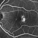
Group 2a Juxtafoveal Telangiectasis - MacTel - Stage 5 left eye - Macular Hole Right eye553 views76-year-old woman has juxtafoveal retinal telangiectasis. She had Stage five neovascularization in the left eye I treated with injections between September of 2007 and October of 2008 with Avastin (3 years ago). OCT scan today shows macular hole right eye. VISUAL ACUITY: OD 20/400, OS 20/40.
|
|
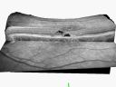
Group 2a Juxtafoveal Telangiectasis - MacTel - Stage 5 left eye - Macular Hole Right eye553 views76-year-old woman has juxtafoveal retinal telangiectasis. She had Stage five neovascularization in the left eye I treated with injections between September of 2007 and October of 2008 with Avastin (3 years ago). OCT scan today shows macular hole right eye. VISUAL ACUITY: OD 20/400, OS 20/40.
|
|

Lumigan (Bimatoprost) Cystoid Macular Edema - Eye Better 1 month after Stopping Lumigan553 views 82-year-old man had cataract surgery in 1993. He has had glaucoma for seven or eight years for a long time. He is taking Lumigan only in the left eye. About a year ago he started in the right eye and then starting just about a few months ago he has noticed intermittent blurring in the vision in the right eye. Vision 20/25 OD, 20/20 OS
|
|
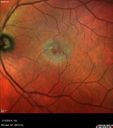
Macular Telangiectasia - Stage 5 left eye - 20/40 vision after 2 years of Avastin 553 viewsMulticolor Image
|
|
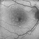
Polypoidal Choroidal Vasculopathy with Pulsating ICG polyp left eye553 views
|
|

Adult Vitelliform Macular Dystrophy - Pattern Dystrophy553 views
|
|

Juxtapapillary CNVM - Hemorrhage553 views83 year old female VA 20/200 improved to 20/32 within 2 months with Lucentis
|
|

Angioid Streaks and Macular Degeneration - CNVM OS - Band Keratopathy 552 views79-year-old man has wet age-related macular degeneration in the left eye, for which he is having Lucentis treatment. OD 20/40, Pinhole 20/32. OS 20/160, Pinhole 20/50. He has band keratopathy sparing the visual axis in both eyes. ICG shows angioid streaks both eye.
|
|

Chronic Central Serous Retinopathy - Fluorescein Angiogram552 views
|
|
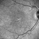
Polypoidal Choroidal Vasculopathy with Pulsating ICG polyp left eye552 views
|
|

Outer Retinal Holes - Probably Solar Retinopathy (history is negative)552 views
|
|

Confluent Soft Drusen with Subetinal Fluid - Right Eye552 viewsAsymptomatic 85 year old man. OCT shows pocket of subfoveal fluid in the center of the macula. This could be mechanical from the drusen preventing the retina from settling down. Testing did not reveal a CNVM and subsequent OCT 6 weeks later showed no change in structure.
|
|

Cystoid Macular Edema following Cataract surgery and vitrectomy for floaters552 views73 year old woman who had decreased vision 4 months following combined vitrectomy and cataract surgery. VA 20/63 improved in 1 month to 20/25 with ketorolac and prednisolone drops.
|
|

CNV secondary to Angioid Streaks, PXE551 views
|
|

Central Serous Retinopathy 25 year old professional baseball player - Atypical - Choroidal Hypo-perfusion?551 views
|
|

Macular Hole Left Eye - progression from VMT - Stage II macular hole - Spontaneously Closed Macular Hole551 viewsMacular hole is closing and vision has improved to 20/40
|
|

Wet AMD OD and Lamellar Macular hole OS551 viewsWet AMD in right and better eye failed extend to 2 months but was able to stop therapy once he started on systemic Avastin for cancer. The last image shows the macula dry 9 months without local therapy. The left eye has a lamellar macular hole.
|
|

Presumed Ocular Histoplasmosis with new CNVM in right and better eye - VA 20/80 OD; 20/800 OS551 viewsFluorescein Angiogram
|
|

Cystoid macular edema 4 months following cataract surgery with serous retinal detachment551 views68 year old with 2 months of vision loss OS 20/100. ECCE was 4 months ago.
|
|

Macular Telangiectasia and dry AMD - Simulating wet AMD551 viewsThis patient was told different things by different retina specialists. Her case is complex because she has OCT findings of Macular Telangiectasis and dry AMD. This makes it look like she has wet AMD. Her vision is stable.
|
|

Cystoid Macular Edema following Cataract surgery and vitrectomy for floaters551 views73 year old woman who had decreased vision 4 months following combined vitrectomy and cataract surgery. VA 20/63 improved in 1 month to 20/25 with ketorolac and prednisolone drops.
|
|

Basal Laminar (cuticular) Drusen both eyes 60 Year Old Woman550 views60-year-old woman with minimal vision loss. Vision OD is 20/30 OS 20/40
|
|

Vitreomacular Traction 1 month - 20/60 vision - Fellow Eye Normal - 80 Year Old Man 550 views80-year-old man has a macular pucker in the right eye with vitreomacular traction and vision loss for 1 month. His vision seems about stable to him. OD 20/60.
(these images are from initial presentation)
|
|

Acute Central Serous Retinopathy - CSR - Steroid Induced (probably) - Good Vision 550 views42-year-old man was seen in the office on October 5, 2011. He had noticed starting in August after a course of antibiotic and steroids, that he developed new spots in his vision in the right eye. He may have had an episode like this sometime in the past. He did take steroids a few years ago and his vision did change at that time, but then returned.
VISUAL ACUITY: OD 20/32, OS 20/32. The OCT scan of the right eye confirms subretinal fluid.
|
|

Central Serous Retinopathy 25 year old professional baseball player - Atypical - Choroidal Hypo-perfusion?550 views
|
|

Reticular Macular Disease (Pseudo-drusen) Both Eyes - Occult CNVM and Macular Edema Right Eye 550 viewsFundus AutofluorescenceFundus Autofluorescence
|
|

Cystoid Macular Edema 10 years after Cataract Surgery550 viewsNo overt uveitis but the patient had aching in the eyes and responded to topical anti-inflammatory drops with recurrence of the CME on discontinuation of topical therapy
|
|

Asymptomatic wet AMD - Subfoveal CNM both eyes550 views82 year old woman - 20/25 OU. Images show occult subfoveal CNVM both eyes best seen on late ICG. The left eye also has probable polypoidal choroidal vasculopathy. No treatment was recommended on the first visit - follow-up at 1 month.
|
|

Central Serous Retinopathy - Subretinal Fibrin550 views66 year old female with 20 year history of single episode of CSR. Now with vision loss for a month but improving - VA 20/63 OD; 20/32 OS
|
|

Retinal fold following removal of severe macular pucker550 views75 year old man had macula on inferior retinal detachment repair. He then developed a 5/200 dense macular pucker. After the pucker was removed the vision returned to 20/40 but there was a persistent superior retinal fold that looks a little like a snake.
|
|

Small Retinal Pigment Epithelial Rip and Poor Vision (20/400)549 views80-year-old woman vision in the right eye has substantially declined.
OD: 20/400; OS: 20/50
|
|

Angioid Streaks - New Extrafoveal Choroidal Neovascular Membrane Right Eye549 views61-year-old woman twelve days ago, saw a spot to the right of her central vision. OD is 20/25, OS is 20/25
Treated with one year of avastin with good result and no vision loss.
|
|

Acute Central Serous Chorioretinopathy with Symptoms in Hazy Vision Both Eyes549 views48-year-old man with hazy vision in the right eye for about three days. He has a little more of a headache than usual. He is under a fair amount of stress. He does not take any steroids that he knows of. He does use a nasal spray, which he is not sure if that is a steroid spray or not. He is going to call us with that medicine. I checked his blood pressure today is was 115/95.
VISUAL ACUITY: OD 20/15, OS 20/25
|
|

549 views
|
|

Chronic Central Serous Retinopathy - Fluorescein Angiogram549 views
|
|

Chronic Central Serous Retinopathy - Fluorescein Angiogram549 views
|
|

Polypoidal Choroidal Vasculopathy with Pulsating ICG polyp left eye549 views
|
|

Vitrectomy with membrane peel549 viewsIntraoperative images showing the peeling of an epiretinal membrane - macular pucker - with a 25 gauge forcepts.
|
|

Cystoid Macular Edema following Cataract surgery and vitrectomy for floaters549 views73 year old woman who had decreased vision 4 months following combined vitrectomy and cataract surgery. VA 20/63 improved in 1 month to 20/25 with ketorolac and prednisolone drops.
|
|

Acute Bilateral Central Serous Chorioretinopathy - Female - Smokestack549 views39 year old female She had no prior epsisodes of vision loss and then a week ago she noticed decreased vision in the right eye. She has not had any health problems and she is not pregnant. Phendimetrazine started 3 months ago. She is not on steroids. VA OD: sc20/63-1 NscJ16 VA OS: sc20/125-1 Dcc20/80 NscJ16 IOP: TP: OD:14 OS:14 Time:11:23 AM
|
|

Small Retinal Pigment Epithelial Rip and Poor Vision (20/400)548 views80-year-old woman vision in the right eye has substantially declined.
OD: 20/400; OS: 20/5080-year-old woman vision in the right eye has substantially declined.
OD: 20/400; OS: 20/50
|
|

Macular Pucker 20/40 vision and recent cataract surgery548 views 73-year-old man distortion and vision loss since ECCE 6 months ago OS. Patient taking anti-inflammatory drops. VISUAL ACUITY: OD 20/30, OS 20/40.
|
|
| 5013 files on 51 page(s) |
 |
|
|
|
|
15 | |
|
|
|
|


