Enimvaadatud - Macular Disease
|

Chronic Central Serous Chorioretinopathy Both Eyesvaatamisi: 51260 year old man with 20 year history of chronic CSR. He has had laser and PDT in the past. The left eye has been 20/100 for decades. The right eye is now declining. Was 20/32 and is down to 20/50. There is chronic low grade subretinal fluid away from the center in the right eye. (focal laser in the right eye in 2005, left eye 2001 at BPEI). Refampin tried in 2012, Aldactone in 2013. PDT in 204 and 2017 Images are from 2017
|
|

Juxtafoveal Telangiectasis and Newly Decreasing Visionvaatamisi: 51170-year-old woman followed for 10 years with IJFT with newly decreasing vision OU OD: 20/160; OD: 20/80
|
|
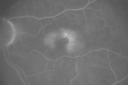
Juxtfoveal Telangiectasis Group 2a - Type II - MacTel - also Juxtapapillary Asymptomatic CNVM Right Eyevaatamisi: 51182-year-old woman who has juxtafoveal retinal telangiectasis in both eyes. She recently had cataract surgery in the right eye and the vision in the right eye is still limited. OD 20/100, OS 20/40.
|
|

Macular Pucker Pre-OP 20/200 vaatamisi: 511
|
|

Juxtafoveal Telangiectasis - After Hemorrhage Absorbed Right Eye with Avastin Injectionsvaatamisi: 51173-year-old woman has juxtafoveal retinal telangiectasis in both eyes. The right eye has stage 5 disease. Her last Avastin treatment in the right eye was in 6 months ago. The right eye has been the worst of her two eyes for years.
VISUAL ACUITY: Vision OD is 20/200, OS is 20/50.
|
|

Wet AMD controlled with Avastin 20/60 Visionvaatamisi: 51179-year-old woman has wet age-related macular degeneration in both eyes. The left eye had intravitreal injections of Avastin starting in May of 2009. Her vision remains good in the left eye and poor in the right eye.
VISUAL ACUITY: OD 8/200, OS 20/60.
|
|

Chronic Pseudophakic Cystoid Macular Edema - Haptic Migrated into Anterior Chamber Through Peripheral Iridotomyvaatamisi: 51157-year-old man with congenital nystagmus. He had cataract surgery done 30 years ago and has decreased vision left eye for 6 months. OD is 20/50, OS is 20/100
|
|
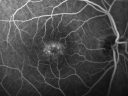
Juxtafoveal Telangiectasis - Stage 5 Left Eye - New Blood and Neovascularization MacTel vaatamisi: 51160-year-old man has juxtafoveal retinal telangiectasis recent vision loss OD 20/50, OS 20/70 : Hemorrhage OS and new SRNVM
|
|
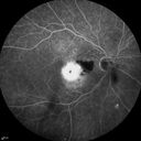
Cartwheel CNVM - Classic - wet AMD - evidence of possible old CSR - FA shows cartwheel shaped CNVMvaatamisi: 51175 y.o Decreased Vision right eye for one year. VA OD: sc20/125+2 OS 20/25. Autofluorescence and SD OCT show evidence of old serous retinal detachment superior to the fovea suggesting a history of CSR. The macular lesion on FA shows a classic cartwheel appearance
|
|
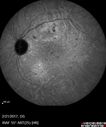
Multifocal Bilateral Central Serous Retinopathy in African American Malevaatamisi: 51167 year old african american male with : Blurred Vision OD. Duration of Problem: 4-6 weeks. VA 20/32 OD 20/20 OS
|
|

Basal Laminar (cuticular) Drusen both eyes 60 Year Old Womanvaatamisi: 51060-year-old woman with minimal vision loss. Vision OD is 20/30 OS 20/40
|
|

Myopic Macular Degeneration - Staphyloma - Recent Vision Loss Right Eye - Possible CNVMvaatamisi: 51083-year-old woman is a high myope with myopic macular degeneration. She had a retinal detachment repair in the right eye in 1968. I saw her in June of 2003. She had posterior colobomas and areas of pigment epithelial atrophy. She has noticed over the last six weeks substantial declining vision in the right eye. Prior to that, the vision in the two eyes was about the same. She uses Xalatan in the left eye and Alphagan in both eyes.
VISUAL ACUITY: Vision OD is 20/160, OS is 20/40
|
|

Central Serous Retinopathy - Steroid Induced - Serous Retinal Detachmentvaatamisi: 51038-year-old man has noticed a spot in the right eye. The vision was a little bit off for a few weeks and then just recently he noticed that it was the right eye. He did take high dose oral steroids for about two weeks in early March and he was using an intermittent steroid inhaler, but he has not used anything recently. He is under a lot of stress.
VISUAL ACUITY: OD 20/100, OS 20/50.
|
|

Basal Laminar Drusen - Cuticular Drusen - Equatorial Drusen - 70 Year Old Woman vaatamisi: 51070 year old woman has had macular drusen since her thirties and she said her sister has a similar problem. Recently the vision in the left eye has substantially declined. OD 20/16, OS 10/200.
|
|
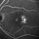
Group 2a Juxtafoveal Telangiectasis - MacTel - Stage 5 left eye - Macular Hole Right eyevaatamisi: 51076-year-old woman has juxtafoveal retinal telangiectasis. She had Stage five neovascularization in the left eye I treated with injections between September of 2007 and October of 2008 with Avastin (3 years ago). OCT scan today shows macular hole right eye. VISUAL ACUITY: OD 20/400, OS 20/40.
|
|

Lumigan (Bimatoprost) Cystoid Macular Edema - Eye Better 1 month after Stopping Lumiganvaatamisi: 510 82-year-old man had cataract surgery in 1993. He has had glaucoma for seven or eight years for a long time. He is taking Lumigan only in the left eye. About a year ago he started in the right eye and then starting just about a few months ago he has noticed intermittent blurring in the vision in the right eye. Vision 20/25 OD, 20/20 OS
|
|

Reticular Macular Disease (Pseudo-drusen) Both Eyes - Occult CNVM and Macular Edema Right Eye vaatamisi: 510Fluorescein Angiogram
|
|

Cuticular Drusen - 26 year old female - Glomerulonephritisvaatamisi: 510VA 20/25 OU. Images are over 4 years
|
|

hydroxychloroquine (Plaquenil) Toxicity - Taiwanese 56 year old female (Asian)vaatamisi: 51056 year old female on 400 mg plaquenil for 22 years presents with vision loss. VA 20/50 both eyes. Plaquenil was stopped. The vision in the left eye continued to deteriorate to 3/200. 5'2" 127 lb (6.6 mg/kg body weight)
|
|

Cystoid Macular Edema 10 years after Cataract Surgeryvaatamisi: 510No overt uveitis but the patient had aching in the eyes and responded to topical anti-inflammatory drops with recurrence of the CME on discontinuation of topical therapy
|
|

Cystoid Macular Edema 10 years after Cataract Surgeryvaatamisi: 510No overt uveitis but the patient had aching in the eyes and responded to topical anti-inflammatory drops with recurrence of the CME on discontinuation of topical therapy
|
|
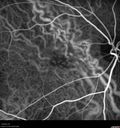
Confluent Soft Drusen with Subetinal Fluid - Right Eyevaatamisi: 510Asymptomatic 85 year old man. OCT shows pocket of subfoveal fluid in the center of the macula. This could be mechanical from the drusen preventing the retina from settling down. Testing did not reveal a CNVM and subsequent OCT 6 weeks later showed no change in structure.
|
|

Prefoveal Opercula and early Lamellar Macular Holevaatamisi: 51073 year old man with 20/32 vision and pre-foveal opercula. The OCT shows very early lamellar hole with lamellar hole epiretinal proliferation
|
|
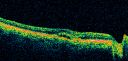
Macular Scar Left Eye from Age-Related Macular Degenerationvaatamisi: 50981-year-old woman has a macular scar in the left eye and then unfortunately developed a central retinal artery occlusion in the right eye OD 20/100, OS 6/200.
|
|

Chronic Pseudophakic Cystoid Macular Edema in a Patient with Diabetesvaatamisi: 50974-year-old woman has chronic cystoid macular edema in the left eye. She has had vision loss intermittently in that eye for 2 years, the right eye, unfortunately, lost vision from a central retinal vein occlusion in 6 years ago.
|
|

80 Year Old Woman Macular Degeneration Disciform Scar - Fibrosisvaatamisi: 509VA 20/100
|
|

Juxtafoveal Telangiectasis - After Hemorrhage Absorbed Right Eye with Avastin Injectionsvaatamisi: 50973-year-old woman has juxtafoveal retinal telangiectasis in both eyes. The right eye has stage 5 disease. Her last Avastin treatment in the right eye was in 6 months ago. The right eye has been the worst of her two eyes for years.
VISUAL ACUITY: Vision OD is 20/200, OS is 20/50.
|
|
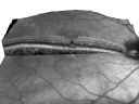
Juxtafoveal Telangiectasis - Stage 5 Left Eye - New Blood and Neovascularization MacTel VIDEO TAKES TIME TO LOADvaatamisi: 50960-year-old man has juxtafoveal retinal telangiectasis recent vision loss OD 20/50, OS 20/70 : Hemorrhage OS and new SRNVM
|
|
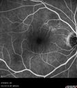
Vitreomacular traction - recent vision loss left eye - FAvaatamisi: 509
|
|

New wet AMD OS with subretinal hemorrhage and recent vision loss, Scar ODvaatamisi: 509
|
|

IPCV - idiopathic polypoidal choroidal vasculopathy - juxtapapillary CNVM with hemorrhage both eyes (also choroidal nevus right eye) - Subretinal hemorrhage both eyesvaatamisi: 50986 year old woman with shadow in vision OD and new hemmorhage OU.
|
|

Myopic Traction Maculopathy - Long Term Follow-upvaatamisi: 50988-92 year old woman with about 20/40 vision and myopic tractional maculopathy. Her vision fluctuates a little. The serial OCT's show no progression.
|
|

Cuticular Drusen - 26 year old female - Glomerulonephritisvaatamisi: 509VA 20/25 OU. Images are over 4 years
|
|

Macular Telangiectasis - visible telangiectasia on color photosvaatamisi: 509Multicolor photos too show pathology. This patient had 20/50 vision with pinhole to 20/25
|
|

Central Serous Chorioretinopathy - Moderatevaatamisi: 50949 year old man with episodes in 2011, 2017, and 2018 - now with some paracentral vision loss in the right eye and subretinal fluid.
|
|

Macular Telangiectasia (MacTel) and Pattern Dystrophyvaatamisi: 50971 year old man with diabetes for 5 years and blurred vision in the left eye. VA 20/25 OD, 20/50 OS. OCT OS shows MacTel
|
|

Myopic Macular Degeneration - Staphyloma - Recent Vision Loss Right Eye - Possible CNVMvaatamisi: 50883-year-old woman is a high myope with myopic macular degeneration. She had a retinal detachment repair in the right eye in 1968. I saw her in June of 2003. She had posterior colobomas and areas of pigment epithelial atrophy. She has noticed over the last six weeks substantial declining vision in the right eye. Prior to that, the vision in the two eyes was about the same. She uses Xalatan in the left eye and Alphagan in both eyes.
VISUAL ACUITY: Vision OD is 20/160, OS is 20/40
|
|
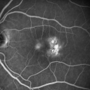
Group 2a Juxtafoveal Telangiectasis - MacTel - Stage 5 left eye - Macular Hole Right eyevaatamisi: 50876-year-old woman has juxtafoveal retinal telangiectasis. She had Stage five neovascularization in the left eye I treated with injections between September of 2007 and October of 2008 with Avastin (3 years ago). OCT scan today shows macular hole right eye. VISUAL ACUITY: OD 20/400, OS 20/40.
|
|

Cuticular Drusen (Basal Laminar Drusen) and Bilateral Chronic Subfoveal Fluid (Serous Retinal Detachment) unresponsive to monthly Avastin Very Early ICG ODvaatamisi: 50866-year-old man with bilateral subfoveal fluid unresponsive to one year of monthly avastin therapy.
OD 20/160, Pinhole 20/80. OS 20/63, Pinhole 20/50. OCT scan shows subretinal fluid with normal choroidal thickness. Fluorescein angiogram shows cuticular/basal laminar drusen. ICG shows no foci of choriocapillaris leakage.
|
|

Acute Central Serous Retinopathy - CSR - Steroid Induced (probably) - Good Vision - ICG Affected Eye - Choroidal Leakagevaatamisi: 50842-year-old man was seen in the office on October 5, 2011. He had noticed starting in August after a course of antibiotic and steroids, that he developed new spots in his vision in the right eye. He may have had an episode like this sometime in the past. He did take steroids a few years ago and his vision did change at that time, but then returned.
VISUAL ACUITY: OD 20/32, OS 20/32. The OCT scan of the right eye confirms subretinal fluid.
|
|

Cystoid macular edema 4 months following cataract surgery with serous retinal detachmentvaatamisi: 508CME which is chronic will often cause a serous reitnal detachment because of loss of the integrity of the external limiting membrane
|
|

Cuticular Drusen - 26 year old female - Glomerulonephritisvaatamisi: 508VA 20/25 OU. Images are over 4 years - FA shows starry sky early images - This is because of loss of RPE over the drusen creating very small window defects.
|
|

hydroxychloroquine (Plaquenil) Toxicity - Taiwanese 56 year old female (Asian)vaatamisi: 50856 year old female on 400 mg plaquenil for 22 years presents with vision loss. VA 20/50 both eyes. Plaquenil was stopped. The vision in the left eye continued to deteriorate to 3/200. 5'2" 127 lb (6.6 mg/kg body weight)
|
|

Multifocal Pattern Dystrophyvaatamisi: 50851 year old with normal vision - images look a lot like fundus flavimaculatus
|
|

Central Serous Retinopathy - Subretinal Fibrinvaatamisi: 50866 year old female with 20 year history of single episode of CSR. Now with vision loss for a month but improving - VA 20/63 OD; 20/32 OS
|
|

Dry AMD - Confluent Drusen Sparing Center of Macula which has Atrophyvaatamisi: 50865 year old woman, VA 20/40 OD; 20/80 OS. The center of the macula has few or no drusen with predominantly non-geographic atrophy
|
|

Vitreomacular Traction -> Macular Hole -> Aborted Macular holevaatamisi: 508Progression of VMT in both eyes over time
|
|

Macular Degeneration - Choroidal Laser Left eye - New Subfoveal CNVM left eye - Did well with Avastinvaatamisi: 50769-year-old woman new vision loss right eye and old macular scar left eye. OD 20/40, OS 20/200
|
|

Idiopathic Central Serous Retinopathy - Bilateral - Acute vaatamisi: 50749-year-old man decreasing vision in the left eye for one week. He is not taking any steroid or steroid-related products. His blood pressure as far as he knows is under control.
VISUAL ACUITY: OD 20/30, OS 20/400
|
|

Macular Pucker 20/40 vision and recent cataract surgeryvaatamisi: 507 73-year-old man distortion and vision loss since ECCE 6 months ago OS. Patient taking anti-inflammatory drops. VISUAL ACUITY: OD 20/30, OS 20/40.
|
|

vaatamisi: 507
|
|

Pattern Dystrophy - Adult Vitelliform - Both Eyesvaatamisi: 50760-year-old woman noticing less distortion and less blind spots.
OD is 20/20, OS is 20/40
|
|

Cuticular Drusen (Basal Laminar Drusen) and Bilateral Chronic Subfoveal Fluid (Serous Retinal Detachment) unresponsive to monthly Avastin - Fill ICG FA ODvaatamisi: 50766-year-old man with bilateral subfoveal fluid unresponsive to one year of monthly avastin therapy.
OD 20/160, Pinhole 20/80. OS 20/63, Pinhole 20/50. OCT scan shows subretinal fluid with normal choroidal thickness. Fluorescein angiogram shows cuticular/basal laminar drusen. ICG shows no foci of choriocapillaris leakage.
|
|

Serous Macular Detachment Both Eyes - Wet AMD vs. CSR - 62 Year Old Man ICGvaatamisi: 507
|
|

vaatamisi: 507
|
|
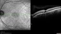
Macular Telangiectasia - Stage 5 left eye - 20/40 vision after 2 years of Avastin vaatamisi: 507Spectral Domain OCT Line Scan
|
|

Outer Retinal Holes - Probably Solar Retinopathy (history is negative)vaatamisi: 507
|
|

New wet AMD OS with subretinal hemorrhage and recent vision loss, Scar ODvaatamisi: 507
|
|

Confluent Soft Drusen with Subetinal Fluid - Right Eyevaatamisi: 507Asymptomatic 85 year old man. OCT shows pocket of subfoveal fluid in the center of the macula. This could be mechanical from the drusen preventing the retina from settling down. Testing did not reveal a CNVM and subsequent OCT 6 weeks later showed no change in structure.
|
|

Macular Hole - Asymptomaticvaatamisi: 50771 year old female did not notice 20/200 macular hole. Post surgery vision improved to 20/30
|
|

Chronic CSC Both Eyes - Recent vision loss Right Eye Treated with PDTvaatamisi: 50750 year old man The right eye has had vision loss for a few weeks. He had previous episodes seen by me in 2012 OS and 2008 OD. This was 2018. VA OS has been poor for several years.
VA OD: Dcc20/100
VA OS: Dcc20/200-3
The patient had PDT to leaky area superotemporal to the fovea 1/2 dose and within 3 months was back up to 20/25. The last two OCT scans show fluid absorbing
|
|

Concentric Geographic Atrophyvaatamisi: 50776 year old man Gradual vision loss
20/32 OD; 20/40 OS
No medicines, Non-smoker
Working and Driving
|
|
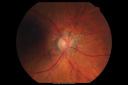
Angioid Streaks New Vision Change in Good Right Eye Treated with Lucentisvaatamisi: 50653-year-old man has angioid streaks. He has a macular scar in the left eye from choroidal neovasculari-zation that was treated with photodynamic laser 5-8 years ago. His vision in the right eye had been fine until about a week ago when he noticed temporally in the right eye an area that does not look normal and looks a little distorted.
VISUAL ACUITY: Vision OD is 20/30, OS is 20/400. Treatment was started within a month and patient responded well and is now 3 years out requiring every 3 month Lucentis
|
|
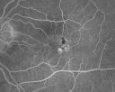
Juxtafoveal Telangiectasis - (MacTel) - Stage 4 both Eyesvaatamisi: 50657-year-old woman with a history of decreased vision. The patient reports that her vision has been gradually decreasing in the left more so than the right eye. 20/50 OD and 20/60
|
|

Macular Pucker Pre-op 20/200 vision (history of retinal tears)vaatamisi: 50666-year-old man has had laser to retinal tears in both eyes in 1999 and in 2001. His right eye has had rapidly declining vision over the last month. VISUAL ACUITY: OD: 20/200
|
|

Idiopathic Central Serous Retinopathy - Bilateral - Acute vaatamisi: 50649-year-old man decreasing vision in the left eye for one week. He is not taking any steroid or steroid-related products. His blood pressure as far as he knows is under control.
VISUAL ACUITY: OD 20/30, OS 20/400
|
|
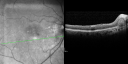
Juxtafoveal Telangiectasis (MacTel) and Asymptomatic Extrafoveal Juxtapapillary Choroidal Neovascular Membranevaatamisi: 50684-year-old woman has juxtafoveal retinal telangiectasis in both eyes. The right eye has an asymptomatic extra foveal juxtapapillary choroidal neovascular membrane. OD 20/400, OS 20/63.
|
|
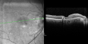
Juxtafoveal Telangiectasis (MacTel) and Asymptomatic Extrafoveal Juxtapapillary Choroidal Neovascular Membranevaatamisi: 50684-year-old woman has juxtafoveal retinal telangiectasis in both eyes. The right eye has an asymptomatic extra foveal juxtapapillary choroidal neovascular membrane. OD 20/400, OS 20/63.
|
|
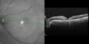
Juxtafoveal Telangiectasis (MacTel) and Asymptomatic Extrafoveal Juxtapapillary Choroidal Neovascular Membranevaatamisi: 50684-year-old woman has juxtafoveal retinal telangiectasis in both eyes. The right eye has an asymptomatic extra foveal juxtapapillary choroidal neovascular membrane. OD 20/400, OS 20/63.
|
|

vaatamisi: 50687 year old male seen for neovascular macular degeneration. Patient had huge disciform scar and lipid.
|
|

Central Serous Retinopathy 25 year old professional baseball player - Atypical - Choroidal Hypo-perfusion?vaatamisi: 506
|
|

Asymptomatic Juxtapapillary Choroidal Neovascular Membrane - Reticular Macular Disease (Wet AMD) vaatamisi: 506
|
|
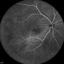
Serous Macular Detachment Both Eyes - Wet AMD vs. CSR - 62 Year Old Man - Fluorescein angiogramvaatamisi: 506
|
|

Macular Hole Left Eye - progression from VMT - Stage II macular hole - Spontaneously Closed Macular Holevaatamisi: 506
|
|

Presumed Ocular Histoplasmosis with new CNVM in right and better eye - VA 20/80 OD; 20/800 OSvaatamisi: 506Fluorescein Angiogram
|
|

Myopic Traction Maculopathy - Long Term Follow-upvaatamisi: 50688-92 year old woman with about 20/40 vision and myopic tractional maculopathy. Her vision fluctuates a little. The serial OCT's show no progression.
|
|

Confluent Soft Drusen with Subetinal Fluid - Right Eyevaatamisi: 506Asymptomatic 85 year old man. OCT shows pocket of subfoveal fluid in the center of the macula. This could be mechanical from the drusen preventing the retina from settling down. Testing did not reveal a CNVM and subsequent OCT 6 weeks later showed no change in structure.
|
|

Confluent Soft Drusen with Subetinal Fluid - Right Eyevaatamisi: 506Asymptomatic 85 year old man. OCT shows pocket of subfoveal fluid in the center of the macula. This could be mechanical from the drusen preventing the retina from settling down. Testing did not reveal a CNVM and subsequent OCT 6 weeks later showed no change in structure.
|
|

Central Serous Retinopathy - Subretinal Fibrinvaatamisi: 50666 year old female with 20 year history of single episode of CSR. Now with vision loss for a month but improving - VA 20/63 OD; 20/32 OS
|
|

Dry AMD - Confluent Drusen Sparing Center of Macula which has Atrophyvaatamisi: 50665 year old woman, VA 20/40 OD; 20/80 OS. The center of the macula has few or no drusen with predominantly non-geographic atrophy
|
|

Submacular Hemorrhage - wet AMDvaatamisi: 50684 year old man with 20/200 from submacular hemorrhage. Vision improved to 20/32 with only Avastin therapy (no surgical intervention). The initial FAF is interesting because parts of the blood hyper auto fluoresce.
|
|

Chronic CSR with mild vision loss - Macular Atrophy Right Eyevaatamisi: 50655 year old man has not been to an eye doctor in years. VA 20/40 OD and 20/25 OS. OCT shows outer retinal atrophy and choroidal thickening. FAF shows guttering. FA shows no active lesions. ICG shows patchy mild leakage visible at about 5 minutes.
|
|

Prefoveal Opercula and early Lamellar Macular Holevaatamisi: 50673 year old man with 20/32 vision and pre-foveal opercula. The OCT shows very early lamellar hole with lamellar hole epiretinal proliferation
|
|

Chronic Central Serous Retinopathy - Gutter in Both Eyes - Treated with PDT - Visudynevaatamisi: 50569-year-old man with blurred vision in the right eye for about three weeks. He had a similar episode about twenty years ago which lasted about six weeks. He is not taking any steroids. He takes Lupron injections for his prostate cancer every three months.
VISUAL ACUITY: Vision OD is 20/80
FU: Treated with PDT after 6 months of observation with success. Treatment was done to area between nerve and fovea at 1/2 fluence.
|
|

Chronic Central Serous Retinopathy - Gutter in Both Eyes - Treated with PDT - Visudynevaatamisi: 50569-year-old man with blurred vision in the right eye for about three weeks. He had a similar episode about twenty years ago which lasted about six weeks. He is not taking any steroids. He takes Lupron injections for his prostate cancer every three months.
VISUAL ACUITY: Vision OD is 20/80
FU: Treated with PDT after 6 months of observation with success. Treatment was done to area between nerve and fovea at 1/2 fluence.
|
|

Small Retinal Pigment Epithelial Rip and Poor Vision (20/400)vaatamisi: 50580-year-old woman vision in the right eye has substantially declined.
OD: 20/400; OS: 20/50
|
|

Juxtafoveal Telangiectasis - (MacTel) - Stage 4 both Eyesvaatamisi: 50557-year-old woman with a history of decreased vision. The patient reports that her vision has been gradually decreasing in the left more so than the right eye. 20/50 OD and 20/60
|
|

vaatamisi: 505
|
|

Chorioretinal Scar with Radiating Chorioretinal Foldsvaatamisi: 50579-year-old woman has wet age-related macular degeneration in both eyes. The left eye had intravitreal injections of Avastin starting in May of 2009. Her vision remains good in the left eye and poor in the right eye.
VISUAL ACUITY: OD 8/200, OS 20/60.
|
|

Polypoidal Choroidal Vasculopathy Right Eye - Disciform Scar Left Eyevaatamisi: 50584-year-old woman has polypoidal choroidal vasculopathy in both eyes with macular degeneration. She has macular scars in both eyes and she remains with remarkably good vision in the right eye despite the scarring.
VISUAL ACUITY: OD 20/30, OS 1/200
Avastin Rx OD
|
|

Asymptomatic Juxtapapillary Choroidal Neovascular Membrane - Reticular Macular Disease (Wet AMD) vaatamisi: 505
|
|
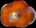
Epiretinal Membranevaatamisi: 505Patient with a history of pars planitis comes in with decreased vision in the left eye. VA is 20/30 in the left eye. Fundus photos show a rather large epiretinal membrane covering the optic nerve in the left eye.
|
|
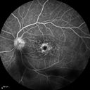
Cystoid Macular Edema 10 years after Cataract Surgeryvaatamisi: 505No overt uveitis but the patient had aching in the eyes and responded to topical anti-inflammatory drops with recurrence of the CME on discontinuation of topical therapy
|
|

Central Serous Chorioretinopathy - Moderatevaatamisi: 50549 year old man with episodes in 2011, 2017, and 2018 - now with some paracentral vision loss in the right eye and subretinal fluid.
|
|

Concentric Geographic Atrophyvaatamisi: 50576 year old man Gradual vision loss
20/32 OD; 20/40 OS
No medicines, Non-smoker
Working and Driving
|
|

Wet Macular Degeneration with Juxtapapillary Neovascular Membrane and Choroidal Laser Treatmentvaatamisi: 50487-year-old woman has wet age-related macular degeneration in the left eye with an extrafoveal choroidal neovascular membrane OD is 20/25, OS is 20/25
|
|

vaatamisi: 504
|
|

Macular Pucker - Diabetic - Severe non-foveal Edema OSvaatamisi: 50474-year-old man has a macular pucker in the left eye and diabetic retinopathy.
VISUAL ACUITY: Vision OD is 20/25, OS is 20/40.
|
|
vaatamisi: 504
|
|
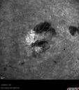
Post-Retinal Detachment Repair Occult Subfoveal CNVMvaatamisi: 504This pleasant 56-year-old man has had a complex retinal detachment repair in the right eye. I most recently removed his silicone oil 3 months ago. His vision now is the best it has ever been.
VISUAL ACUITY: OD 4/200
|
|
|
|


