Más vistos - Macular Disease
|
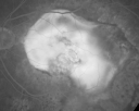
Macular Scar Left Eye from Age-Related Macular Degenerationvista 489 veces81-year-old woman has a macular scar in the left eye and then unfortunately developed a central retinal artery occlusion in the right eye OD 20/100, OS 6/200.
|
|

Myopic Macular Degeneration - Staphyloma - Recent Vision Loss Right Eye - Possible CNVMvista 489 veces83-year-old woman is a high myope with myopic macular degeneration. She had a retinal detachment repair in the right eye in 1968. I saw her in June of 2003. She had posterior colobomas and areas of pigment epithelial atrophy. She has noticed over the last six weeks substantial declining vision in the right eye. Prior to that, the vision in the two eyes was about the same. She uses Xalatan in the left eye and Alphagan in both eyes.
VISUAL ACUITY: Vision OD is 20/160, OS is 20/40
|
|

Central Serous Retinopathy - Steroid Induced - Serous Retinal Detachmentvista 489 veces38-year-old man has noticed a spot in the right eye. The vision was a little bit off for a few weeks and then just recently he noticed that it was the right eye. He did take high dose oral steroids for about two weeks in early March and he was using an intermittent steroid inhaler, but he has not used anything recently. He is under a lot of stress.
VISUAL ACUITY: OD 20/100, OS 20/50.
|
|

Vitreomacular traction with traction over retinal vessels in non-diabetic - Fluorescein leakage in areas of traction - Progressing vision loss left eyevista 489 veces80-year-old woman had vitrectomy surgery done in the right eye in 2007. Vision has been poor since despite a good anatomical result. Recently her vision in the left eye has been declining. The vision has declined in the left eye. Vision OD is 20/160, OS is 20/80
|
|
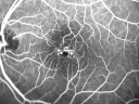
Juxtafoveal Telangiectasis - Stage 5 Left Eye - New Blood and Neovascularization MacTel vista 489 veces60-year-old man has juxtafoveal retinal telangiectasis recent vision loss OD 20/50, OS 20/70 : Hemorrhage OS and new SRNVM
|
|
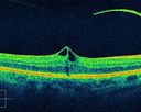
Macular Puckervista 489 vecesOCT scan of MP
|
|

Macular Degenerationvista 489 vecesPatient complains of spot in the right eye. VA 20/400 in the right eye. Fundus photo shows macular degeneration changes with small layer of hemorrhage on the fovea in the right eye.
|
|

Adult Vitelliform Macular Dystrophy - Pattern Dystrophyvista 489 vecesBright subfoveal FAF from lipofuscin
|
|

Cuticular Drusen - 26 year old female - Glomerulonephritisvista 489 vecesVA 20/25 OU. Images are over 4 years
|
|

Cystoid Macular Edema 10 years after Cataract Surgeryvista 489 vecesNo overt uveitis but the patient had aching in the eyes and responded to topical anti-inflammatory drops with recurrence of the CME on discontinuation of topical therapy
|
|

Multifocal Pattern Dystrophyvista 489 veces51 year old with normal vision - images look a lot like fundus flavimaculatus
|
|

Travatan Induced CME Left eyevista 489 veces63 year old woman 4 years post ECCE and 2 years post PPV for floaters. She developed CME in the left eye after 1 year of travatan. The travatan was stopped and the CME and serous retinal detachment resolved.
|
|

Travatan Induced CME Left eyevista 489 veces63 year old woman 4 years post ECCE and 2 years post PPV for floaters. She developed CME in the left eye after 1 year of travatan. The travatan was stopped and the CME and serous retinal detachment resolved.
|
|

Central Serous Retinopathy - Subretinal Fibrinvista 489 veces66 year old female with 20 year history of single episode of CSR. Now with vision loss for a month but improving - VA 20/63 OD; 20/32 OS
|
|

Stage 4 Macular Holevista 489 veces57 year old female with 20/160 vision and stage 4 macular hole
|
|

Outer Retinal Tubulationvista 489 veces78 year old man chronic wet AMD - 20/200
|
|

Pattern Dystrophy - Foveal Atrophyvista 489 veces73 year old female with vision loss - looks like macular hole
|
|
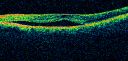
Chronic Central Serous Retinopathy - Gutter in Both Eyes - Treated with PDT - Visudynevista 488 veces69-year-old man with blurred vision in the right eye for about three weeks. He had a similar episode about twenty years ago which lasted about six weeks. He is not taking any steroids. He takes Lupron injections for his prostate cancer every three months.
VISUAL ACUITY: Vision OD is 20/80
FU: Treated with PDT after 6 months of observation with success. Treatment was done to area between nerve and fovea at 1/2 fluence.
|
|

Small Retinal Pigment Epithelial Rip and Poor Vision (20/400)vista 488 veces80-year-old woman vision in the right eye has substantially declined.
OD: 20/400; OS: 20/50
|
|

Wet Macular Degeneration Left Eye with Gutter (atypical) and Geographic Atrophy Right Eyevista 488 veces80-year-old man has wet age-related macular degeneration in his left eye. He has an atypical angiogram and that there is a gutter under the choroidal neovascular membrane suggesting still leakage of subretinal fluid. OS 20/400.
|
|

vista 488 veces
|
|

Myopic Macular Degeneration - Staphyloma - Recent Vision Loss Right Eye - Possible CNVMvista 488 veces83-year-old woman is a high myope with myopic macular degeneration. She had a retinal detachment repair in the right eye in 1968. I saw her in June of 2003. She had posterior colobomas and areas of pigment epithelial atrophy. She has noticed over the last six weeks substantial declining vision in the right eye. Prior to that, the vision in the two eyes was about the same. She uses Xalatan in the left eye and Alphagan in both eyes.
VISUAL ACUITY: Vision OD is 20/160, OS is 20/40
|
|

Post-Retinal Detachment Repair Occult Subfoveal CNVMvista 488 vecesThis pleasant 56-year-old man has had a complex retinal detachment repair in the right eye. I most recently removed his silicone oil 3 months ago. His vision now is the best it has ever been.
VISUAL ACUITY: OD 4/200
|
|
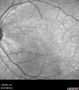
Macular Pucker 20/40 vision - minimal symptoms for 1 year Right eyevista 488 vecesIR
|
|

New wet AMD OS with subretinal hemorrhage and recent vision loss, Scar ODvista 488 veces
|
|

Cuticular Drusen - 26 year old female - Glomerulonephritisvista 488 vecesVA 20/25 OU. Images are over 4 years
|
|
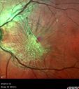
Macular Pucker 20/32 with increased distortion for a monthvista 488 veces
|
|
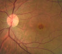
Stage 4 Macular Holevista 488 veces57 year old female with 20/160 vision and stage 4 macular hole
|
|

Choriodal Neovascular Membrane - Subfoveal - Pseudo-Retinitis Pigmentosa - 50 years post Retinal Detachment Repairvista 487 veces86-year-old with a history of decreased vision in the right eye. The patient reports she has lost vision in the right eye over the past two months or so. She reports the vision was always compromised in that eye since retinal detachment surgery 50 years ago with a scleral buckler. Vision dropped from 20/50 to 20/200 right eye in 2 months.
|
|
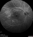
Macular Pucker 20/40 vision - minimal symptoms for 1 year Right eyevista 487 vecesFA shows leakage from traction from pucker in the right eye - cystoid macular edema
|
|
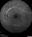
Macular Pucker 20/40 vision - minimal symptoms for 1 year Right eyevista 487 vecesFA shows leakage from traction from pucker in the right eye - cystoid macular edema
|
|

vista 487 veces
|
|

Macular Hole Left Eye - progression from VMT - Stage II macular hole - Spontaneously Closed Macular Holevista 487 vecesVision has dropped to 20/50 and there is a full thickness macular hole
|
|

Cystoid Macular Edema 10 years after Cataract Surgeryvista 487 vecesNo overt uveitis but the patient had aching in the eyes and responded to topical anti-inflammatory drops with recurrence of the CME on discontinuation of topical therapy
|
|

Travatan Induced CME Left eyevista 487 veces63 year old woman 4 years post ECCE and 2 years post PPV for floaters. She developed CME in the left eye after 1 year of travatan. The travatan was stopped and the CME and serous retinal detachment resolved.
|
|

Dry AMD - Confluent Drusen Sparing Center of Macula which has Atrophyvista 487 veces65 year old woman, VA 20/40 OD; 20/80 OS. The center of the macula has few or no drusen with predominantly non-geographic atrophy
|
|
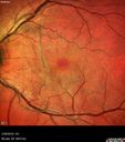
Spontaneously Peeled Macular Pucker 20/20 Visionvista 487 veces
|
|

Vitreomacular Traction 3 months - VIDEO - Takes Time to Load (be patient)vista 486 veces80-year-old man has a macular pucker in the right eye with vitreomacular traction and vision loss for 3 months. His vision seems about stable to him. OD 20/60.
|
|

Chronic Pseudophakic Cystoid Macular Edema - Haptic Migrated into Anterior Chamber Through Peripheral Iridotomyvista 486 veces57-year-old man with congenital nystagmus. He had cataract surgery done 30 years ago and has decreased vision left eye for 6 months. OD is 20/50, OS is 20/100
|
|
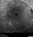
Macular Pucker 20/40 vision - minimal symptoms for 1 year Right eyevista 486 vecesFA shows leakage from traction from pucker in the right eye - cystoid macular edema
|
|

vista 486 veces
|
|

New wet AMD OS with subretinal hemorrhage and recent vision loss, Scar ODvista 486 veces
|
|

Cystoid macular edema 4 months following cataract surgery with serous retinal detachmentvista 486 veces68 year old with 2 months of vision loss OS 20/100. ECCE was 4 months ago.
|
|

Myopic Traction Maculopathy - Long Term Follow-upvista 486 veces88-92 year old woman with about 20/40 vision and myopic tractional maculopathy. Her vision fluctuates a little. The serial OCT's show no progression.
|
|

hydroxychloroquine (Plaquenil) Toxicity - Taiwanese 56 year old female (Asian)vista 486 veces56 year old female on 400 mg plaquenil for 22 years presents with vision loss. VA 20/50 both eyes. Plaquenil was stopped. The vision in the left eye continued to deteriorate to 3/200. 5'2" 127 lb (6.6 mg/kg body weight)
|
|

Central Serous Retinopathy - Subretinal Fibrinvista 486 veces66 year old female with 20 year history of single episode of CSR. Now with vision loss for a month but improving - VA 20/63 OD; 20/32 OS
|
|

Chronic CSR with mild vision loss - Macular Atrophy Right Eyevista 486 veces55 year old man has not been to an eye doctor in years. VA 20/40 OD and 20/25 OS. OCT shows outer retinal atrophy and choroidal thickening. FAF shows guttering. FA shows no active lesions. ICG shows patchy mild leakage visible at about 5 minutes.
|
|

Pattern Dystrophy - Foveal Atrophyvista 486 veces73 year old female with vision loss - looks like macular hole
|
|
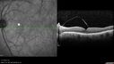
Vitreomacular Traction -> Macular Hole -> Aborted Macular holevista 486 vecesProgression of VMT in both eyes over time
|
|

Non-exudative wet AMD right eye (treatment naive quiescent)vista 486 veces82 year old man. ICG shows CNVM in the right eye. This stayed quiet for 3 years and then started leaking and responded well to Avastin.
|
|

Macular Telangiectasis and Vision loss following vitrectomy with membrane peelvista 486 veces76 year old man with MacTel who had membrane peel in the right eye 10 years ago with permanent vision loss.
VA OD: sc20/200-4 and VA OS: sc20/25-1
|
|

Macular Telangiectasia - Mildvista 486 veces56 year old female with good vision and mild macular telangiectasia. VA 20/40 OD 20/16 OS
|
|

Macular Degeneration - Choroidal Laser Left eye - New Subfoveal CNVM left eye - Did well with Avastinvista 485 veces69-year-old woman new vision loss right eye and old macular scar left eye. OD 20/40, OS 20/200
|
|

Central Serous Retinopathy - Steroid Induced - Serous Retinal Detachmentvista 485 veces38-year-old man has noticed a spot in the right eye. The vision was a little bit off for a few weeks and then just recently he noticed that it was the right eye. He did take high dose oral steroids for about two weeks in early March and he was using an intermittent steroid inhaler, but he has not used anything recently. He is under a lot of stress.
VISUAL ACUITY: OD 20/100, OS 20/50.
|
|

Vitreomacular traction with traction over retinal vessels in non-diabetic - Fluorescein leakage in areas of traction - Progressing vision loss left eyevista 485 veces80-year-old woman had vitrectomy surgery done in the right eye in 2007. Vision has been poor since despite a good anatomical result. Recently her vision in the left eye has been declining. The vision has declined in the left eye. Vision OD is 20/160, OS is 20/80
|
|

Lumigan (Bimatoprost) Cystoid Macular Edema - Eye Better 1 month after Stopping Lumiganvista 485 veces
|
|

Choroidal Effusions and Serous retinal detachment with Systemic Testosterone and Cialis in 75 year old Manvista 485 vecesB- Scan shows choroidal effusions in the left eye only
|
|

Cuticular Drusen - 26 year old female - Glomerulonephritisvista 485 vecesVA 20/25 OU. Images are over 4 years
|
|

Multifocal Bilateral Central Serous Retinopathy in African American Malevista 485 veces67 year old african american male with : Blurred Vision OD. Duration of Problem: 4-6 weeks. VA 20/32 OD 20/20 OS
|
|

Central Serous Retinopathy - Subretinal Fibrinvista 485 veces66 year old female with 20 year history of single episode of CSR. Now with vision loss for a month but improving - VA 20/63 OD; 20/32 OS
|
|

Dry AMD - Confluent Drusen Sparing Center of Macula which has Atrophyvista 485 veces65 year old woman, VA 20/40 OD; 20/80 OS. The center of the macula has few or no drusen with predominantly non-geographic atrophy
|
|

Chronic CSR with mild vision loss - Macular Atrophy Right Eyevista 485 veces55 year old man has not been to an eye doctor in years. VA 20/40 OD and 20/25 OS. OCT shows outer retinal atrophy and choroidal thickening. FAF shows guttering. FA shows no active lesions. ICG shows patchy mild leakage visible at about 5 minutes.
|
|
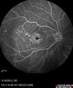
Pseudophakic Cystoid Macular Edema Both Eyesvista 485 veces81 year old man with vision loss about 4 months following ECCE. Did not respond to topical therapy but did fine with PST kenalog
|
|

Chronic CSC Both Eyes - Recent vision loss Right Eye Treated with PDTvista 485 veces50 year old man The right eye has had vision loss for a few weeks. He had previous episodes seen by me in 2012 OS and 2008 OD. This was 2018. VA OS has been poor for several years.
VA OD: Dcc20/100
VA OS: Dcc20/200-3
The patient had PDT to leaky area superotemporal to the fovea 1/2 dose and within 3 months was back up to 20/25. The last two OCT scans show fluid absorbing
|
|
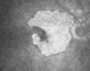
Geographic Atrophy and Age-Related Macular Degeneration - Choriocapillaris is Largely Still Intactvista 484 veces85-year-old woman has age-related macular degeneration in both eyes with geographic atrophy. She has noticed her vision getting slightly worse in the past 6 months. VISUAL ACUITY: OD 20/70, OS 20/400
|
|

Acute Central Serous Chorioretinopathy with Symptoms in Hazy Vision Both Eyesvista 484 veces48-year-old man with hazy vision in the right eye for about three days. He has a little more of a headache than usual. He is under a fair amount of stress. He does not take any steroids that he knows of. He does use a nasal spray, which he is not sure if that is a steroid spray or not. He is going to call us with that medicine. I checked his blood pressure today is was 115/95.
VISUAL ACUITY: OD 20/15, OS 20/25
|
|
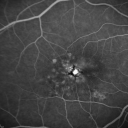
Group 2a Juxtafoveal Telangiectasis - MacTel - Stage 5 left eye - Macular Hole Right eyevista 484 veces76-year-old woman has juxtafoveal retinal telangiectasis. She had Stage five neovascularization in the left eye I treated with injections between September of 2007 and October of 2008 with Avastin (3 years ago). OCT scan today shows macular hole right eye. VISUAL ACUITY: OD 20/400, OS 20/40.
|
|

Serous Macular Detachment Both Eyes - Wet AMD vs. CSR - 62 Year Old Man ICG vista 484 veces
|
|

Cuticular Drusen - 26 year old female - Glomerulonephritisvista 484 vecesVA 20/25 OU. Images are over 4 years. FA shows starry sky early images - This is because of loss of RPE over the drusen creating very small window defects.
|
|

Asymptomatic wet AMD - Subfoveal CNM both eyesvista 484 veces82 year old woman - 20/25 OU. Images show occult subfoveal CNVM both eyes best seen on late ICG. The left eye also has probable polypoidal choroidal vasculopathy. No treatment was recommended on the first visit - follow-up at 1 month.
|
|

Pattern Dystrophy - Adult Vitellifrom (Best)vista 484 veces71 year old female - lesions nasal to the fovea in both eyes. (20/40 OU)
|
|

Juxtapapillary CNVM - Hemorrhagevista 484 veces83 year old female VA 20/200 improved to 20/32 within 2 months with Lucentis
|
|

Myopic Degeneration - Inferior staphyloma with severe atrophyvista 484 veces76 year old with gradual vision loss. 20/32 OD; 20/40 OS
|
|

Myopic CNVM in the left eye. New Classic subfoveal lesionvista 484 veces64 year old female. She had retinal detachment repair in the right eye in 1996 (25 years ago). The left eye has new distortion for a week or two.
VA 20/40 OU.
Left eye has a very small subfoveal CNVM
|
|

Concentric Geograophic Atrophy - Age-related Macular Degeneration - Charles Bonnet Syndrome - Bulls Eyevista 483 veces88-year-old woman has age-related with vision gradually declining over the last year. She sees spots in her vision, like red spots and dark spots and she even notices during the daytime, for example when she was looking at her minister, it looked like there were little flowering pots coming out of his head.
VISUAL ACUITY: OD 20/200, OS 20/400.
|
|

Central Serous Retinopathy - Steroid Induced - Serous Retinal Detachmentvista 483 veces38-year-old man has noticed a spot in the right eye. The vision was a little bit off for a few weeks and then just recently he noticed that it was the right eye. He did take high dose oral steroids for about two weeks in early March and he was using an intermittent steroid inhaler, but he has not used anything recently. He is under a lot of stress.
VISUAL ACUITY: OD 20/100, OS 20/50.
|
|

Group 2a Juxtafoveal Telangiectasis - MacTel - Stage 5 left eye - Macular Hole Right eyevista 483 veces76-year-old woman has juxtafoveal retinal telangiectasis. She had Stage five neovascularization in the left eye I treated with injections between September of 2007 and October of 2008 with Avastin (3 years ago). OCT scan today shows macular hole right eye. VISUAL ACUITY: OD 20/400, OS 20/40.
|
|

Central Serous Retinopathy 25 year old professional baseball player - Atypical - Choroidal Hypo-perfusion?vista 483 veces
|
|

Wet AMD OD and Lamellar Macular hole OSvista 483 vecesWet AMD in right and better eye failed extend to 2 months but was able to stop therapy once he started on systemic Avastin for cancer. The last image shows the macula dry 9 months without local therapy. The left eye has a lamellar macular hole.
|
|

Vitreomacular traction - 20/50 vision and symptomatic with metamorphopsiavista 483 veces
|
|

Central Serous Retinopathy - Subretinal Fibrinvista 483 veces66 year old female with 20 year history of single episode of CSR. Now with vision loss for a month but improving - VA 20/63 OD; 20/32 OS
|
|

Cystoid Macular Edema following Cataract surgery and vitrectomy for floatersvista 483 veces73 year old woman who had decreased vision 4 months following combined vitrectomy and cataract surgery. VA 20/63 improved in 1 month to 20/25 with ketorolac and prednisolone drops.
|
|

Cystoid Macular Edema following Cataract surgery and vitrectomy for floatersvista 483 veces73 year old woman who had decreased vision 4 months following combined vitrectomy and cataract surgery. VA 20/63 improved in 1 month to 20/25 with ketorolac and prednisolone drops.
|
|

Macular Telangiectasis with Symptomatic Blind Spotsvista 483 veces55 year old man with non-neovascular MacTel. VA 20/20 right eye 20/50 left eye. The color images show gray areas temporally. The FAF shows decreased macular pigment. The OCT shows outer retinal atrophy.
|
|

Vitreomacular traction OU - Aborted in 3 monthsvista 483 veces62 year old female with blurred vision in both eyes for 2 months intermittently. She was referred for macular edema.
A 20/32 OD, 20/25 OS
OCT showed VMT in one eye and edema in the other
3 months later in the last 2 OCT scans
VA 20/20 OD, 20/16 OS and VMT had released.
|
|

Central Serous Chorioretinopathy - Moderatevista 483 veces49 year old man with episodes in 2011, 2017, and 2018 - now with some paracentral vision loss in the right eye and subretinal fluid.
|
|

Myopic CNVM in the left eye. New Classic subfoveal lesionvista 483 veces64 year old female. She had retinal detachment repair in the right eye in 1996 (25 years ago). The left eye has new distortion for a week or two.
VA 20/40 OU.
Left eye has a very small subfoveal CNVM
|
|

Idiopathic Central Serous Retinopathy - Bilateral - Acute vista 482 veces49-year-old man decreasing vision in the left eye for one week. He is not taking any steroid or steroid-related products. His blood pressure as far as he knows is under control.
VISUAL ACUITY: OD 20/30, OS 20/400
|
|

Central Serous Retinopathy 25 year old professional baseball player - Atypical - Choroidal Hypo-perfusion?vista 482 veces
|
|

Central Serous Retinopathy 25 year old professional baseball player - Atypical - Choroidal Hypo-perfusion?Central Serous Retinopathy 25 year old professional baseball player - Atypical - Choroidal Hypo-perfusion?vista 482 veces
|
|

Serous Macular Detachment Both Eyes - Wet AMD vs. CSR - 62 Year Old Man - Fluorescein angiogramvista 482 veces
|
|

Macular Hole Left Eye - progression from VMT - Stage II macular hole - Spontaneously Closed Macular Holevista 482 veces
|
|

Choroidal Effusions and Serous retinal detachment with Systemic Testosterone and Cialis in 75 year old Manvista 482 vecesB- Scan shows choroidal effusions in the left eye only
|
|

Choroidal Effusions and Serous retinal detachment with Systemic Testosterone and Cialis in 75 year old Manvista 482 veces2 months following presentation, vision is better, 20/40 OU
|
|

Wet AMD OD and Lamellar Macular hole OSvista 482 vecesWet AMD in right and better eye failed extend to 2 months but was able to stop therapy once he started on systemic Avastin for cancer. The last image shows the macula dry 9 months without local therapy. The left eye has a lamellar macular hole.
|
|

Wet AMD 1 month after Eylea - macula is dry and vision is bettervista 482 veces
|
|

Pseudophakic CME with serous retinal detachment - FA shows leakage in both eyesvista 482 veces68 year old man with 2 months of vision loss. FA shows CME, notice that the nerve also leaks. This is typical for pseudophakic inflammatory cystoid macular edema
|
|

Macular Telangiectasia and dry AMD - Simulating wet AMDvista 482 vecesThis patient was told different things by different retina specialists. Her case is complex because she has OCT findings of Macular Telangiectasis and dry AMD. This makes it look like she has wet AMD. Her vision is stable.
|
|

Central Serous Retinopathy - Subretinal Fibrinvista 482 veces66 year old female with 20 year history of single episode of CSR. Now with vision loss for a month but improving - VA 20/63 OD; 20/32 OS
|
|
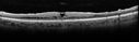
Severe Macular Puckervista 482 veces57 year old female with 20/200 vision and dense macular pucker. Vision improved to 20/50 within about 6 months of surgery.
|
|
| 5013 archivos en 51 página(s) |
 |
|
|
|
|
23 | |
|
|
|
|


