Most viewed - Macular Disease
|
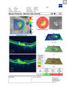
Angioid Streaks Scar and new CNVM439 views49 y/o WF with decreased vision x 1 month. Her OD is 20/400 and her OS is seeing 20/70. No cutaneous lesions or Ehlers-Danlos. The FP and FA show a predominant classic CNV (with fibrosis) OD and an Occult CNV OS with what appears to be a RAP lesion.
|
|

Angioid Streaks Scar and new CNVM439 views49 y/o WF with decreased vision x 1 month. Her OD is 20/400 and her OS is seeing 20/70. No cutaneous lesions or Ehlers-Danlos. The FP and FA show a predominant classic CNV (with fibrosis) OD and an Occult CNV OS with what appears to be a RAP lesion.
|
|

Punctate Inner Choroidopathy and Choroidal Neovascular Membrane439 views27 year old female with vision loss for 4 months. VA was 20/200. She had a CNVM which was extrafoveal with no subretinal fluid. Her vision improved over 6 months to 20/50 with Ranibizumab injections (she might have improved without treatment)
|
|

Concentric Geographic Atrophy - 76 year old man439 viewsGradual vision loss, worse at night. VA 20/40 OU.
|
|
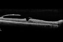
Macular Pucker - Spontaneously Partial Peel after 20 Years - OCT Video - Takes Time to Load438 views83-year-old woman decreased vision in the right eye for about a month. 20 years ago she was diagnosed with a macular pucker OD 20/50, OS 20/20.
|
|

Small vessel vasculitis causing cystoid macular edema - right eye - early frame fluorescein angiography 438 views36 year old female with no significant past medical history noted painless, progressive loss of vision over one month. BCVA was 20/100 in her right eye. Dilated fundus exam, OCT and FA identified small vessel vasculitis associated with cystoid macular edema. Left eye was also involved with less marked findings.
|
|

438 views
|
|

438 views
|
|

IPCV - idiopathic polypoidal choroidal vasculopathy - juxtapapillary CNVM with hemorrhage both eyes (also choroidal nevus right eye) - Subretinal hemorrhage both eyes438 views86 year old woman with shadow in vision OD and new hemmorhage OU. OCT shows progression of JP CNVM
|
|

Macular Telangiectasis with Symptomatic Blind Spots438 views55 year old man with non-neovascular MacTel. VA 20/20 right eye 20/50 left eye. The color images show gray areas temporally. The FAF shows decreased macular pigment. The OCT shows outer retinal atrophy.
|
|

Macular Telangiectasis with Symptomatic Blind Spots438 views55 year old man with non-neovascular MacTel. VA 20/20 right eye 20/50 left eye. The color images show gray areas temporally. The FAF shows decreased macular pigment. The OCT shows outer retinal atrophy.
|
|

MacTel - Juxtafoveal Telangiectasis438 views56 year old man with a lot of difficulty adjusting to vision loss
|
|

Pattern Dystrophy - Foveal Atrophy438 views73 year old female with vision loss - looks like macular hole
|
|
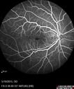
Pseudophakic Cystoid Macular Edema Both Eyes438 views81 year old man with vision loss about 4 months following ECCE. Did not respond to topical therapy but did fine with PST kenalog
|
|

Vitreomacular Traction -> Macular Hole -> Aborted Macular hole438 viewsProgression of VMT in both eyes over time
|
|

Central Serous Chorioretinopathy - Moderate438 views49 year old man with episodes in 2011, 2017, and 2018 - now with some paracentral vision loss in the right eye and subretinal fluid.
|
|
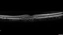
Choroidal Neovascular Membrane following Prior Central Serous Chorioretinopathy - OCT-A438 views67 year old female with CSR in the left eye first 20 years ago. She was managed with out therapy and has had a few episodes since. Now ther eis distortion in the left eye for a few days. She had a steroid shot in her shoulder 5 months ago.
VA 20/20 OD, 20/50 OS- ICG and OCT-A confirm CNVM in the left eye
|
|

Myopic CNVM in the left eye. New Classic subfoveal lesion438 views64 year old female. She had retinal detachment repair in the right eye in 1996 (25 years ago). The left eye has new distortion for a week or two.
VA 20/40 OU.
Left eye has a very small subfoveal CNVM
|
|

Myopic CNVM in the left eye. New Classic subfoveal lesion438 views64 year old female. She had retinal detachment repair in the right eye in 1996 (25 years ago). The left eye has new distortion for a week or two.
VA 20/40 OU.
Left eye has a very small subfoveal CNVM
|
|

Macular Pucker - Diabetic - Severe non-foveal Edema OS437 views74-year-old man has a macular pucker in the left eye and diabetic retinopathy.
VISUAL ACUITY: Vision OD is 20/25, OS is 20/40.
|
|

Pattern Dystrophy - Adult Vitelliform Dystrophy - Macular Degeneration437 views 78 Year Old Woman with vision has diminished in the right eye over the past couple of years, but the diminishment has been slow.
20/60+1 OD, 20/25 OS
|
|

Wet AMD OD and Lamellar Macular hole OS437 viewsWet AMD in right and better eye failed extend to 2 months but was able to stop therapy once he started on systemic Avastin for cancer. The last image shows the macula dry 9 months without local therapy. The left eye has a lamellar macular hole.
|
|

New wet AMD OS with subretinal hemorrhage and recent vision loss, Scar OD437 views
|
|

New wet AMD OS with subretinal hemorrhage and recent vision loss, Scar OD437 views
|
|

Cuticular Drusen - 26 year old female - Glomerulonephritis437 viewsVA 20/25 OU. Images are over 4 years
|
|

Dry AMD - Confluent Drusen Sparing Center of Macula which has Atrophy437 views65 year old woman, VA 20/40 OD; 20/80 OS. The center of the macula has few or no drusen with predominantly non-geographic atrophy
|
|

Chronic CSR with mild vision loss - Macular Atrophy Right Eye437 views55 year old man has not been to an eye doctor in years. VA 20/40 OD and 20/25 OS. OCT shows outer retinal atrophy and choroidal thickening. FAF shows guttering. FA shows no active lesions. ICG shows patchy mild leakage visible at about 5 minutes.
|
|

Vitreomacular Traction - Spontaneous Aborted - Both Eyes - 4 months437 views
|
|

Adult Pseudovitelliform Macular Dystrophy437 views89 year old female states dark smudge on ceiling during night time came and went. Decreased vision in the left eye
VA OD: Dcc20/32
VA OS: Dcc20/63
|
|

Basal Laminar Drusen - Cuticular Drusen - Equatorial Drusen - 70 Year Old Woman 436 views70 year old woman has had macular drusen since her thirties and she said her sister has a similar problem. Recently the vision in the left eye has substantially declined. OD 20/16, OS 10/200.
|
|

436 views
|
|

Choroidal Effusions and Serous retinal detachment with Systemic Testosterone and Cialis in 75 year old Man436 viewsFluorescein angiogram shows some staining of the disk - confocal system renders peripheral effusions black
|
|

Submacular Hemorrhage - wet AMD436 views84 year old man with 20/200 from submacular hemorrhage. Vision improved to 20/32 with only Avastin therapy (no surgical intervention). The initial FAF is interesting because parts of the blood hyper auto fluoresce.
|
|

Chronic CSR with mild vision loss - Macular Atrophy Right Eye436 views55 year old man has not been to an eye doctor in years. VA 20/40 OD and 20/25 OS. OCT shows outer retinal atrophy and choroidal thickening. FAF shows guttering. FA shows no active lesions. ICG shows patchy mild leakage visible at about 5 minutes.
|
|

Stage II Macular Hole - PPV without ILM peel436 views77 year old man with 20/40 VA on presentation. Macular hole did not abort and vision dropped to 20/63. Post PPV vision was 20/25 within 6 months
|
|

pucker-prepeel-MC436 views
|
|

Non-exudative wet AMD right eye (treatment naive quiescent)436 views82 year old man. ICG shows CNVM in the right eye. This stayed quiet for 3 years and then started leaking and responded well to Avastin.
|
|

Macular Telangiectasis and Vision loss following vitrectomy with membrane peel436 views76 year old man with MacTel who had membrane peel in the right eye 10 years ago with permanent vision loss.
VA OD: sc20/200-4 and VA OS: sc20/25-1
|
|

Macular Telangiectasis and Vision loss following vitrectomy with membrane peel436 views76 year old man with MacTel who had membrane peel in the right eye 10 years ago with permanent vision loss.
VA OD: sc20/200-4 and VA OS: sc20/25-1
|
|

Choroidal Neovascular Membrane following Prior Central Serous Chorioretinopathy - OCT-A436 views67 year old female with CSR in the left eye first 20 years ago. She was managed with out therapy and has had a few episodes since. Now ther eis distortion in the left eye for a few days. She had a steroid shot in her shoulder 5 months ago.
VA 20/20 OD, 20/50 OS- ICG and OCT-A confirm CNVM in the left eye
|
|

Vitreomacular traction with traction over retinal vessels in non-diabetic - Fluorescein leakage in areas of traction - Progressing vision loss left eye435 views80-year-old woman had vitrectomy surgery done in the right eye in 2007. Vision has been poor since despite a good anatomical result. Recently her vision in the left eye has been declining. The vision has declined in the left eye. Vision OD is 20/160, OS is 20/80
|
|
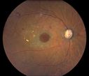
Macular Hole with Peripheral Laser Scars435 viewsDiabetic patient comes in with flashes and floaters in both eyes. Patient's VA is 20/400, right eye and 20/25, left eye. Fundus exam presents with large macular hole with diabetic changes in the retina. Laser scars in the peripheral.
|
|

Choroidal Effusions and Serous retinal detachment with Systemic Testosterone and Cialis in 75 year old Man435 viewsSD OCT shows choroidal thickening and subretinal fluid
|
|

Asymptomatic wet AMD - Subfoveal CNM both eyes435 views82 year old woman - 20/25 OU. Images show occult subfoveal CNVM both eyes best seen on late ICG. The left eye also has probable polypoidal choroidal vasculopathy. No treatment was recommended on the first visit - follow-up at 1 month.
|
|

Multifocal Bilateral Central Serous Retinopathy in African American Male435 views67 year old african american male with : Blurred Vision OD. Duration of Problem: 4-6 weeks. VA 20/32 OD 20/20 OS
|
|

Macular Hole - Asymptomatic435 views71 year old female did not notice 20/200 macular hole. Post surgery vision improved to 20/30
|
|

Macular Hole - Asymptomatic435 views71 year old female did not notice 20/200 macular hole. Post surgery vision improved to 20/30
|
|

MacTel - Juxtafoveal Telangiectasis435 views56 year old man with a lot of difficulty adjusting to vision loss
|
|

Pseudophakic Cystoid Macular Edema Both Eyes435 views81 year old man with vision loss about 4 months following ECCE. Did not respond to topical therapy but did fine with PST kenalog
|
|

Chronic Central Serous Chorioretinopathy435 views77 year old man was seen at uveitis clinic and tested positive to Anti-retinal antibody against 35 kDa and anti-optic nerve antibody. He did not have EDI OCT, FAF, or ICG. All testing was consistent with Chronic CSR. His immunosuppresives were stopped and he did fine for 3 years.
|
|

Chronic Central Serous Chorioretinopathy435 views77 year old man was seen at uveitis clinic and tested positive to Anti-retinal antibody against 35 kDa and anti-optic nerve antibody. He did not have EDI OCT, FAF, or ICG. All testing was consistent with Chronic CSR. His immunosuppresives were stopped and he did fine for 3 years.
|
|

Macular Telangiectasis - Very Mild435 views52 year old female with normal vision and suspicious OCT referred. Multimodal imaging shows very mild mactel (loss of macular pigment, ring on multicolor image and very mild OCT findings)
|
|

Rapid macular atrophy with anti-VEGF therapy for myopic CNVM435 views64 year old female with 20/32 vision and distortion in the left and better eye. She was treated with monthly lucentis and her vision declined to 20/100. Medicine was stopped and she improved a little to 20/80.
|
|

Acute Bilateral Central Serous Chorioretinopathy - Female - Smokestack435 views39 year old female She had no prior epsisodes of vision loss and then a week ago she noticed decreased vision in the right eye. She has not had any health problems and she is not pregnant. Phendimetrazine started 3 months ago. She is not on steroids. VA OD: sc20/63-1 NscJ16 VA OS: sc20/125-1 Dcc20/80 NscJ16 IOP: TP: OD:14 OS:14 Time:11:23 AM
|
|

Choroidal Neovascular Membrane following Prior Central Serous Chorioretinopathy - OCT-A435 views67 year old female with CSR in the left eye first 20 years ago. She was managed with out therapy and has had a few episodes since. Now ther eis distortion in the left eye for a few days. She had a steroid shot in her shoulder 5 months ago.
VA 20/20 OD, 20/50 OS- ICG and OCT-A confirm CNVM in the left eye
|
|

Myopic CNVM in the left eye. New Classic subfoveal lesion435 views64 year old female. She had retinal detachment repair in the right eye in 1996 (25 years ago). The left eye has new distortion for a week or two.
VA 20/40 OU.
Left eye has a very small subfoveal CNVM
|
|

Vitrectomy with membrane peel434 viewsIntraoperative images showing the peeling of an epiretinal membrane - macular pucker - with a 25 gauge forcepts.
|
|

Asymptomatic wet AMD - Subfoveal CNM both eyes434 views82 year old woman - 20/25 OU. Images show occult subfoveal CNVM both eyes best seen on late ICG. The left eye also has probable polypoidal choroidal vasculopathy. No treatment was recommended on the first visit - follow-up at 1 month.
|
|

Outer Retinal Tubulation434 views78 year old man chronic wet AMD - 20/200
|
|

MacTel - Juxtafoveal Telangiectasis434 views56 year old man with a lot of difficulty adjusting to vision loss
|
|

Angioid Streaks - CNVM OS with vision loss434 views48 year old soccer player. Prior trauma OS (with soccer ball). Vision loss for 3 weeks to 20/60 - with Lucentis vision improved to 20/30
|
|
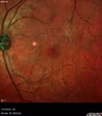
Vitreomacular Traction -> Macular Hole -> Aborted Macular hole434 viewsProgression of VMT in both eyes over time
|
|

Vitreomacular traction OU - Aborted in 3 months - Right eye initial visit434 views62 year old female with blurred vision in both eyes for 2 months intermittently. She was referred for macular edema.
A 20/32 OD, 20/25 OS
OCT showed VMT in one eye and edema in the other
3 months later in the last 2 OCT scans
VA 20/20 OD, 20/16 OS and VMT had released.
|
|

Chronic CSC Both Eyes - Recent vision loss Right Eye Treated with PDT434 views50 year old man The right eye has had vision loss for a few weeks. He had previous episodes seen by me in 2012 OS and 2008 OD. This was 2018. VA OS has been poor for several years.
VA OD: Dcc20/100
VA OS: Dcc20/200-3
The patient had PDT to leaky area superotemporal to the fovea 1/2 dose and within 3 months was back up to 20/25. The last two OCT scans show fluid absorbing
|
|

Central Serous Chorioretinopathy - Moderate434 views49 year old man with episodes in 2011, 2017, and 2018 - now with some paracentral vision loss in the right eye and subretinal fluid.
|
|

Macular Telangiectasis and Vision loss following vitrectomy with membrane peel434 views76 year old man with MacTel who had membrane peel in the right eye 10 years ago with permanent vision loss.
VA OD: sc20/200-4 and VA OS: sc20/25-1
|
|

Choroidal Neovascular Membrane following Prior Central Serous Chorioretinopathy - OCT-A434 views67 year old female with CSR in the left eye first 20 years ago. She was managed with out therapy and has had a few episodes since. Now ther eis distortion in the left eye for a few days. She had a steroid shot in her shoulder 5 months ago.
VA 20/20 OD, 20/50 OS- ICG and OCT-A confirm CNVM in the left eye
|
|

Myopic CNVM in the left eye. New Classic subfoveal lesion434 views64 year old female. She had retinal detachment repair in the right eye in 1996 (25 years ago). The left eye has new distortion for a week or two.
VA 20/40 OU.
Left eye has a very small subfoveal CNVM
|
|
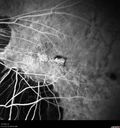
Myopic CNVM in the left eye. New Classic subfoveal lesion434 views64 year old female. She had retinal detachment repair in the right eye in 1996 (25 years ago). The left eye has new distortion for a week or two.
VA 20/40 OU.
Left eye has a very small subfoveal CNVM
|
|

Polypoidal Choroidal Vasculopathy Right Eye - Disciform Scar Left Eye433 views84-year-old woman has polypoidal choroidal vasculopathy in both eyes with macular degeneration. She has macular scars in both eyes and she remains with remarkably good vision in the right eye despite the scarring.
VISUAL ACUITY: OD 20/30, OS 1/200
Avastin Rx OD
|
|

Basal Laminar Drusen - Cuticular Drusen - Equatorial Drusen - 70 Year Old Woman 433 views70 year old woman has had macular drusen since her thirties and she said her sister has a similar problem. Recently the vision in the left eye has substantially declined. OD 20/16, OS 10/200.
|
|

Central Serous Retinopathy - Subretinal Fibrin433 views66 year old female with 20 year history of single episode of CSR. Now with vision loss for a month but improving - VA 20/63 OD; 20/32 OS
|
|

Submacular Hemorrhage - wet AMD433 views84 year old man with 20/200 from submacular hemorrhage. Vision improved to 20/32 with only Avastin therapy (no surgical intervention). The initial FAF is interesting because parts of the blood hyper auto fluoresce.
|
|

Non-exudative wet AMD right eye (treatment naive quiescent)433 views82 year old man. ICG shows CNVM in the right eye. This stayed quiet for 3 years and then started leaking and responded well to Avastin.
|
|

Adult Pseudovitelliform Macular Dystrophy433 views89 year old female states dark smudge on ceiling during night time came and went. Decreased vision in the left eye
VA OD: Dcc20/32
VA OS: Dcc20/63
|
|
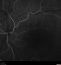
Cystoid Macular Edema and Macular Pucker433 viewshas been particular bad for 3 years. She has a macular pucker and severe CME.
VA 20/160 left eye
|
|

Myopic CNVM in the left eye. New Classic subfoveal lesion433 views64 year old female. She had retinal detachment repair in the right eye in 1996 (25 years ago). The left eye has new distortion for a week or two.
VA 20/40 OU.
Left eye has a very small subfoveal CNVM
|
|

Basal Laminar Drusen - Cuticular Drusen - Equatorial Drusen - 70 Year Old Woman 432 views70 year old woman has had macular drusen since her thirties and she said her sister has a similar problem. Recently the vision in the left eye has substantially declined. OD 20/16, OS 10/200.
|
|

432 views
|
|

Wet AMD OD and Lamellar Macular hole OS432 viewsWet AMD in right and better eye failed extend to 2 months but was able to stop therapy once he started on systemic Avastin for cancer. The last image shows the macula dry 9 months without local therapy. The left eye has a lamellar macular hole.
|
|
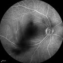
Cystoid Macular Edema 10 years after Cataract Surgery432 viewsNo overt uveitis but the patient had aching in the eyes and responded to topical anti-inflammatory drops with recurrence of the CME on discontinuation of topical therapy
|
|

Asymptomatic wet AMD - Subfoveal CNM both eyes432 views82 year old woman - 20/25 OU. Images show occult subfoveal CNVM both eyes best seen on late ICG. The left eye also has probable polypoidal choroidal vasculopathy. No treatment was recommended on the first visit - follow-up at 1 month.
|
|

Multifocal Bilateral Central Serous Retinopathy in African American Male432 views67 year old african american male with : Blurred Vision OD. Duration of Problem: 4-6 weeks. VA 20/32 OD 20/20 OS
|
|

Cystoid Macular Edema following Cataract surgery and vitrectomy for floaters432 views73 year old woman who had decreased vision 4 months following combined vitrectomy and cataract surgery. VA 20/63 improved in 1 month to 20/25 with ketorolac and prednisolone drops.
|
|
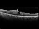
Macular Pucker 20/32 with increased distortion for a month432 views
|
|

Polypoidal Choroidal Vasculopathy Left Eye432 views74 year old man with vision loss to 20/100 over a few months. He improved to 20/20 with Lucentis. The color and ICG show the polyps.
|
|
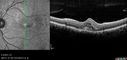
Angioid Streaks - CNVM OS with vision loss432 views48 year old soccer player. Prior trauma OS (with soccer ball). Vision loss for 3 weeks to 20/60 - with Lucentis vision improved to 20/30
|
|

Non-exudative wet AMD right eye (treatment naive quiescent)432 views82 year old man. ICG shows CNVM in the right eye. This stayed quiet for 3 years and then started leaking and responded well to Avastin.
|
|

Polypoidal Choroidal Vasculopathy - Right Eye432 views83 year old female presented in June 2013 with vision loss for 3 months. She had 2 injections elsewhere with no improvement (avastin). The first images show a peripapillary CNVM with polyps. I treated her once with PDT and she stayed dry with good vision for 5 years (2019). VA on presentation 20/80 OD. Improved to 20/40
|
|

Macular Telangiectasia - Mild432 views56 year old female with good vision and mild macular telangiectasia. VA 20/40 OD 20/16 OS
|
|

Choroidal Neovascular Membrane following Prior Central Serous Chorioretinopathy - OCT-A432 views67 year old female with CSR in the left eye first 20 years ago. She was managed with out therapy and has had a few episodes since. Now ther eis distortion in the left eye for a few days. She had a steroid shot in her shoulder 5 months ago.
VA 20/20 OD, 20/50 OS- ICG and OCT-A confirm CNVM in the left eye
|
|

431 views
|
|

431 views
|
|

431 views
|
|

Myopic Traction Maculopathy - Long Term Follow-up431 views88-92 year old woman with about 20/40 vision and myopic tractional maculopathy. Her vision fluctuates a little. The serial OCT's show no progression.
|
|

Myopic Traction Maculopathy - Long Term Follow-up431 views88-92 year old woman with about 20/40 vision and myopic tractional maculopathy. Her vision fluctuates a little. The serial OCT's show no progression.
|
|

Multifocal Bilateral Central Serous Retinopathy in African American Male431 views67 year old african american male with : Blurred Vision OD. Duration of Problem: 4-6 weeks. VA 20/32 OD 20/20 OS
|
|

Central Serous Retinopathy - Subretinal Fibrin431 views66 year old female with 20 year history of single episode of CSR. Now with vision loss for a month but improving - VA 20/63 OD; 20/32 OS
|
|

Dry AMD - Confluent Drusen Sparing Center of Macula which has Atrophy431 views65 year old woman, VA 20/40 OD; 20/80 OS. The center of the macula has few or no drusen with predominantly non-geographic atrophy
|
|

Submacular Hemorrhage - wet AMD431 views84 year old man with 20/200 from submacular hemorrhage. Vision improved to 20/32 with only Avastin therapy (no surgical intervention). The initial FAF is interesting because parts of the blood hyper auto fluoresce.
|
|
| 5013 files on 51 page(s) |
 |
|
|
|
|
30 | |
|
|
|
|


