Most viewed - Macular Disease
|

Submacular Hemorrhage - wet AMD425 views84 year old man with 20/200 from submacular hemorrhage. Vision improved to 20/32 with only Avastin therapy (no surgical intervention). The initial FAF is interesting because parts of the blood hyper auto fluoresce.
|
|

Polypoidal Choroidal Vasculopathy Left Eye425 views74 year old man with vision loss to 20/100 over a few months. He improved to 20/20 with Lucentis. The color and ICG show the polyps.
|
|

Polypoidal Choroidal Vasculopathy Left Eye425 views74 year old man with vision loss to 20/100 over a few months. He improved to 20/20 with Lucentis. The color and ICG show the polyps.
|
|

Juxtapapillary CNVM - Hemorrhage425 views83 year old female VA 20/200 improved to 20/32 within 2 months with Lucentis
|
|

Vitreomacular Traction -> Macular Hole -> Aborted Macular hole425 viewsProgression of VMT in both eyes over time
|
|

Non-exudative wet AMD right eye (treatment naive quiescent)425 views82 year old man. ICG shows CNVM in the right eye. This stayed quiet for 3 years and then started leaking and responded well to Avastin.
|
|

425 views
|
|
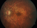
Concentric Geographic Atrophy - 76 year old man425 viewsGradual vision loss, worse at night. VA 20/40 OU.
|
|

Myopic CNVM in the left eye. New Classic subfoveal lesion425 views64 year old female. She had retinal detachment repair in the right eye in 1996 (25 years ago). The left eye has new distortion for a week or two.
VA 20/40 OU.
Left eye has a very small subfoveal CNVM
|
|

Vitreomacular traction with traction over retinal vessels in non-diabetic - Fluorescein leakage in areas of traction - Progressing vision loss left eye424 views80-year-old woman had vitrectomy surgery done in the right eye in 2007. Vision has been poor since despite a good anatomical result. Recently her vision in the left eye has been declining. The vision has declined in the left eye. Vision OD is 20/160, OS is 20/80
|
|

Wet AMD OD and Lamellar Macular hole OS424 viewsWet AMD in right and better eye failed extend to 2 months but was able to stop therapy once he started on systemic Avastin for cancer. The last image shows the macula dry 9 months without local therapy. The left eye has a lamellar macular hole.
|
|

Multifocal Bilateral Central Serous Retinopathy in African American Male424 views67 year old african american male with : Blurred Vision OD. Duration of Problem: 4-6 weeks. VA 20/32 OD 20/20 OS
|
|

Submacular Hemorrhage - wet AMD424 views84 year old man with 20/200 from submacular hemorrhage. Vision improved to 20/32 with only Avastin therapy (no surgical intervention). The initial FAF is interesting because parts of the blood hyper auto fluoresce.
|
|

Outer Retinal Tubulation424 views78 year old man chronic wet AMD - 20/200
|
|

Concentric Geographic Atrophy - 76 year old man424 viewsGradual vision loss, worse at night. VA 20/40 OU.
|
|

Concentric Geographic Atrophy - 76 year old man424 viewsGradual vision loss, worse at night. VA 20/40 OU.
|
|

Macular Telangiectasia - Mild424 views56 year old female with good vision and mild macular telangiectasia. VA 20/40 OD 20/16 OS
|
|

Mild Central Serous Chorioretinopathy - Flonase related424 views58 year old man with vision loss in the left eye for 8 days. This has never happened before. He started taking flonase about a month ago. He stopped the flonase and the vision improved in less than a month. Initial vision was 20/20 OD, 20/40 OS
|
|

Hydroxychloroquine retinopathy - Plaquenil - Moderate424 views51 year old female on with trouble driving at night for a year. No eye examination for several years. 2009 (11 years ago) Plaquenil 300 then up to 400 a few years ago now down to 300 - 5'2" 135 lbs
VA 20/20 OD, 20/25 OS
|
|

Macular Telangiectasia (MacTel) and Pattern Dystrophy424 views71 year old man with diabetes for 5 years and blurred vision in the left eye. VA 20/25 OD, 20/50 OS. OCT OS shows MacTel
|
|

Macular Telangiectasia (MacTel) and Pattern Dystrophy424 views71 year old man with diabetes for 5 years and blurred vision in the left eye. VA 20/25 OD, 20/50 OS. OCT OS shows MacTel
|
|

hydroxychloroquine (Plaquenil) Toxicity - Taiwanese - 3 years post stopping424 views59 year old female on 400 mg plaquenil for 22 years. 3 years ago, when stopping plaquenil, VA 20/50 both eyes. Plaquenil was stopped. The vision in the left eye continued to deteriorate to 3/200. 5'2" 127 lb (6.6 mg/kg body weight) -- Vision in these images is 20/80 OD, 5/200 OS - Vision loss now seems stable.
|
|

Asymptomatic wet AMD - Subfoveal CNM both eyes423 views82 year old woman - 20/25 OU. Images show occult subfoveal CNVM both eyes best seen on late ICG. The left eye also has probable polypoidal choroidal vasculopathy. No treatment was recommended on the first visit - follow-up at 1 month.
|
|

Cystoid Macular Edema following Cataract surgery and vitrectomy for floaters423 views73 year old woman who had decreased vision 4 months following combined vitrectomy and cataract surgery. VA 20/63 improved in 1 month to 20/25 with ketorolac and prednisolone drops.
|
|

Outer Retinal Tubulation423 views78 year old man chronic wet AMD - 20/200
|
|

Chronic CSC Both Eyes - Recent vision loss Right Eye Treated with PDT423 views50 year old man The right eye has had vision loss for a few weeks. He had previous episodes seen by me in 2012 OS and 2008 OD. This was 2018. VA OS has been poor for several years.
VA OD: Dcc20/100
VA OS: Dcc20/200-3
The patient had PDT to leaky area superotemporal to the fovea 1/2 dose and within 3 months was back up to 20/25. The last two OCT scans show fluid absorbing
|
|

Macular Telangiectasia - Mild423 views56 year old female with good vision and mild macular telangiectasia. VA 20/40 OD 20/16 OS
|
|
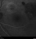
Polypiodal Choroidal Vasculopathy - Temporal Macula - Left eye423 views82 year old man with asymptomatic PCV in the left eye. Vision 20/50 OD, 20/32 OS did well with Lucentis
|
|

Myopic CNVM in the left eye. New Classic subfoveal lesion423 views64 year old female. She had retinal detachment repair in the right eye in 1996 (25 years ago). The left eye has new distortion for a week or two.
VA 20/40 OU.
Left eye has a very small subfoveal CNVM
|
|

Large and Extensive Macular Drusen423 views64 year old female with 20/25 vision taking vitamins in for a checkup.
|
|

Red Free Macular Hole 422 viewsFull Thickness Macular Hole
|
|
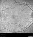
Macular Telangiectasis (MacTel)422 views
|
|

Pattern Dystrophy - Adult Vitellifrom (Best)422 views71 year old female - lesions nasal to the fovea in both eyes. (20/40 OU)
|
|

Polypoidal Choroidal Vasculopathy - Right Eye422 views83 year old female presented in June 2013 with vision loss for 3 months. She had 2 injections elsewhere with no improvement (avastin). The first images show a peripapillary CNVM with polyps. I treated her once with PDT and she stayed dry with good vision for 5 years (2019). VA on presentation 20/80 OD. Improved to 20/40
|
|

Rapid macular atrophy with anti-VEGF therapy for myopic CNVM422 views64 year old female with 20/32 vision and distortion in the left and better eye. She was treated with monthly lucentis and her vision declined to 20/100. Medicine was stopped and she improved a little to 20/80.
|
|

Rapid macular atrophy with anti-VEGF therapy for myopic CNVM422 views64 year old female with 20/32 vision and distortion in the left and better eye. She was treated with monthly lucentis and her vision declined to 20/100. Medicine was stopped and she improved a little to 20/80.
|
|

Choroidal Neovascular Membrane following Prior Central Serous Chorioretinopathy - OCT-A422 views67 year old female with CSR in the left eye first 20 years ago. She was managed with out therapy and has had a few episodes since. Now ther eis distortion in the left eye for a few days. She had a steroid shot in her shoulder 5 months ago.
VA 20/20 OD, 20/50 OS- ICG and OCT-A confirm CNVM in the left eye
|
|

Hydroxychloroquine retinopathy - Plaquenil - Moderate - Myelinated nerve fiber layer 422 views51 year old female on with trouble driving at night for a year. No eye examination for several years. 2009 (11 years ago) Plaquenil 300 then up to 400 a few years ago now down to 300 - 5'2" 135 lbs
VA 20/20 OD, 20/25 OS
|
|

Polypoidal Choroidal Vasculopathy Right Eye - Disciform Scar Left Eye421 views84-year-old woman has polypoidal choroidal vasculopathy in both eyes with macular degeneration. She has macular scars in both eyes and she remains with remarkably good vision in the right eye despite the scarring.
VISUAL ACUITY: OD 20/30, OS 1/200
Avastin Rx OD
|
|

421 views75 year old male seen for ARMD. Pt developed massive hemorrhage with lipid and macular edema.
|
|

New wet AMD OS with subretinal hemorrhage and recent vision loss, Scar OD421 views
|
|
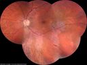
Normal Vision - Chronic Central Serous Chorioretinopathy421 views67 year old man - never had any symptoms - 20/16 vision in each eye. Lesions spare the fovea
|
|
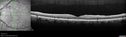
Spontaneously Peeled Macular Pucker 20/20421 views
|
|

Pattern Dystrophy - Foveal Atrophy421 views73 year old female with vision loss - looks like macular hole
|
|
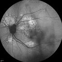
Chronic Central Serous Chorioretinopathy421 views77 year old man was seen at uveitis clinic and tested positive to Anti-retinal antibody against 35 kDa and anti-optic nerve antibody. He did not have EDI OCT, FAF, or ICG. All testing was consistent with Chronic CSR. His immunosuppresives were stopped and he did fine for 3 years.
|
|
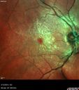
Vitreomacular Traction -> Macular Hole -> Aborted Macular hole421 viewsProgression of VMT in both eyes over time
|
|

Macular Telangiectasis - Very Mild421 views52 year old female with normal vision and suspicious OCT referred. Multimodal imaging shows very mild mactel (loss of macular pigment, ring on multicolor image and very mild OCT findings)
|
|
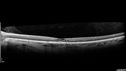
Macular Telangiectasis - Very Mild421 views52 year old female with normal vision and suspicious OCT referred. Multimodal imaging shows very mild mactel (loss of macular pigment, ring on multicolor image and very mild OCT findings)
|
|

Macular Telangiectasis - Mild - Retinal Crystals421 views58 year old man with long term mild vision loss. Considering Cataract surgery. VA 20/50 OD and 20/63 OS - Moderate cataract are present
|
|

Macular Telangiectasis - Mild - Retinal Crystals421 views58 year old man with long term mild vision loss. Considering Cataract surgery. VA 20/50 OD and 20/63 OS - Moderate cataract are present
|
|
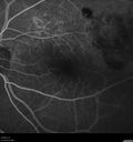
Polypiodal Choroidal Vasculopathy - Temporal Macula - Left eye421 views82 year old man with asymptomatic PCV in the left eye. Vision 20/50 OD, 20/32 OS did well with Lucentis
|
|

Lamellar Macular Hole - Evoluation over 5 years421 views65 year old man with Sturge weber and a choroidal hemangioma in the left eye. (See other images under sturge weber on this website). Has ongoing problem with his left eye. Now with Blurred vision in the right eye gradually worse the last few weeks. He has had a vitrectomy in the right eye 4 years ago for floaters. Laser for tears and cryo in the right eye 5 years ago. He is on Dorzolamide and Lumigan for glaucoma.
VA 20/32 OD, 20/40 OS
IOP 23 OD, 19 OS
|
|

Central Serous Retinopathy 41 year old woman with Vision Loss for One Year420 views41-year-old woman has chronic central serous retinopathy in the right eye with decreased vision in that eye for over a year. Her vision has been waxing and waning and recently is getting worse. Her vision in the left eye is fine.
VISUAL ACUITY: OD 20/40
|
|

Choroidal Effusions and Serous retinal detachment with Systemic Testosterone and Cialis in 75 year old Man420 viewsFluorescein angiogram shows some staining of the disk - confocal system renders peripheral effusions black
|
|

Wet AMD OD and Lamellar Macular hole OS420 viewsWet AMD in right and better eye failed extend to 2 months but was able to stop therapy once he started on systemic Avastin for cancer. The last image shows the macula dry 9 months without local therapy. The left eye has a lamellar macular hole.
|
|

Dry AMD - Confluent Drusen Sparing Center of Macula which has Atrophy420 views65 year old woman, VA 20/40 OD; 20/80 OS. The center of the macula has few or no drusen with predominantly non-geographic atrophy
|
|

Polypoidal Choroidal Vasculopathy Left Eye420 views74 year old man with vision loss to 20/100 over a few months. He improved to 20/20 with Lucentis. The color and ICG show the polyps.
|
|

Pattern Dystrophy - Foveal Atrophy420 views73 year old female with vision loss - looks like macular hole
|
|

Angioid Streaks - CNVM OS with vision loss420 views48 year old soccer player. Prior trauma OS (with soccer ball). Vision loss for 3 weeks to 20/60 - with Lucentis vision improved to 20/30
|
|

Non-exudative wet AMD right eye (treatment naive quiescent)420 views82 year old man. ICG shows CNVM in the right eye. This stayed quiet for 3 years and then started leaking and responded well to Avastin.
|
|

Polypoidal Choroidal Vasculopathy - Right Eye420 views83 year old female presented in June 2013 with vision loss for 3 months. She had 2 injections elsewhere with no improvement (avastin). The first images show a peripapillary CNVM with polyps. I treated her once with PDT and she stayed dry with good vision for 5 years (2019). VA on presentation 20/80 OD. Improved to 20/40
|
|

Acute Bilateral Central Serous Chorioretinopathy - Female - Smokestack420 views39 year old female She had no prior epsisodes of vision loss and then a week ago she noticed decreased vision in the right eye. She has not had any health problems and she is not pregnant. Phendimetrazine started 3 months ago. She is not on steroids. VA OD: sc20/63-1 NscJ16 VA OS: sc20/125-1 Dcc20/80 NscJ16 IOP: TP: OD:14 OS:14 Time:11:23 AM
|
|

Acute Bilateral Central Serous Chorioretinopathy - Female - Smokestack420 views39 year old female She had no prior epsisodes of vision loss and then a week ago she noticed decreased vision in the right eye. She has not had any health problems and she is not pregnant. Phendimetrazine started 3 months ago. She is not on steroids. VA OD: sc20/63-1 NscJ16 VA OS: sc20/125-1 Dcc20/80 NscJ16 IOP: TP: OD:14 OS:14 Time:11:23 AM
|
|

Punctate Inner Choroidopathy and Choroidal Neovascular Membrane420 views27 year old female with vision loss for 4 months. VA was 20/200. She had a CNVM which was extrafoveal with no subretinal fluid. Her vision improved over 6 months to 20/50 with Ranibizumab injections (she might have improved without treatment)
|
|

Punctate Inner Choroidopathy and Choroidal Neovascular Membrane420 views27 year old female with vision loss for 4 months. VA was 20/200. She had a CNVM which was extrafoveal with no subretinal fluid. Her vision improved over 6 months to 20/50 with Ranibizumab injections (she might have improved without treatment)
|
|

Myopic CNVM in the left eye. New Classic subfoveal lesion420 views64 year old female. She had retinal detachment repair in the right eye in 1996 (25 years ago). The left eye has new distortion for a week or two.
VA 20/40 OU.
Left eye has a very small subfoveal CNVM
|
|

Macular Telangiectasi - Stage V - CNVM OU420 views60 year old female who has been treated with injections for 5 years starting in the left eye. She had no treatment 6 months prior to this visit and her vision has been getting worse. Her vision is 20/100 OD, 20/32 OS. Vision improved with intravitreal Avastin to 20/30 OU.
|
|

Severe Geographic Atrophy from Dry Age-Related Macular Degeneration420 views90 year old female with severe vision loss over the last few years.
VA 3/200 OD
1/200 OS
|
|

Mild Central Serous Chorioretinopathy - Flonase related420 views58 year old man with vision loss in the left eye for 8 days. This has never happened before. He started taking flonase about a month ago. He stopped the flonase and the vision improved in less than a month. Initial vision was 20/20 OD, 20/40 OS
|
|

Large and Extensive Macular Drusen420 views64 year old female with 20/25 vision taking vitamins in for a checkup.
|
|

Vitrectomy with membrane peel419 viewsIntraoperative images showing the peeling of an epiretinal membrane - macular pucker - with a 25 gauge forcepts.
|
|

Dry AMD - Confluent Drusen Sparing Center of Macula which has Atrophy419 views65 year old woman, VA 20/40 OD; 20/80 OS. The center of the macula has few or no drusen with predominantly non-geographic atrophy
|
|

Chronic Central Serous Chorioretinopathy419 views77 year old man was seen at uveitis clinic and tested positive to Anti-retinal antibody against 35 kDa and anti-optic nerve antibody. He did not have EDI OCT, FAF, or ICG. All testing was consistent with Chronic CSR. His immunosuppresives were stopped and he did fine for 3 years.
|
|
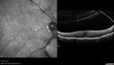
Vitreomacular Traction -> Macular Hole -> Aborted Macular hole419 viewsProgression of VMT in both eyes over time
|
|
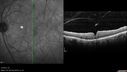
Vitreomacular Traction -> Macular Hole -> Aborted Macular hole419 viewsProgression of VMT in both eyes over time
|
|

Non-exudative wet AMD right eye (treatment naive quiescent)419 views82 year old man. ICG shows CNVM in the right eye. This stayed quiet for 3 years and then started leaking and responded well to Avastin.
|
|

Macular Telangiectasis - Very Mild419 views52 year old female with normal vision and suspicious OCT referred. Multimodal imaging shows very mild mactel (loss of macular pigment, ring on multicolor image and very mild OCT findings)
|
|

Macular Telangiectasis - Very Mild419 views52 year old female with normal vision and suspicious OCT referred. Multimodal imaging shows very mild mactel (loss of macular pigment, ring on multicolor image and very mild OCT findings)
|
|

Choroidal Neovascular Membrane following Prior Central Serous Chorioretinopathy - OCT-A419 views67 year old female with CSR in the left eye first 20 years ago. She was managed with out therapy and has had a few episodes since. Now ther eis distortion in the left eye for a few days. She had a steroid shot in her shoulder 5 months ago.
VA 20/20 OD, 20/50 OS- ICG and OCT-A confirm CNVM in the left eye
|
|

Choroidal Neovascular Membrane following Prior Central Serous Chorioretinopathy - OCT-A419 views67 year old female with CSR in the left eye first 20 years ago. She was managed with out therapy and has had a few episodes since. Now ther eis distortion in the left eye for a few days. She had a steroid shot in her shoulder 5 months ago.
VA 20/20 OD, 20/50 OS- ICG and OCT-A confirm CNVM in the left eye
|
|

Intermediate Dry Age-Related Macular Degeneration - Soft Drusen419 views70-year-old Left eye worse than the right. Both eyes see distortion in the amsler grid for a few months
HTN, High Lipids, Osteoporosis
Meds: Lutein once daily, Calcium, Fish oil, Red Yeast Rice, CoQ10, Magnesium, MVI, Xanax
VA 20/16 OU
2+NS cataract
|
|

Chronic Central Serous Retinopathy - Gutter in Both Eyes - Treated with PDT - Visudyne418 views69-year-old man with blurred vision in the right eye for about three weeks. He had a similar episode about twenty years ago which lasted about six weeks. He is not taking any steroids. He takes Lupron injections for his prostate cancer every three months.
VISUAL ACUITY: Vision OD is 20/80
FU: Treated with PDT after 6 months of observation with success. Treatment was done to area between nerve and fovea at 1/2 fluence.
|
|

Vitreomacular traction with traction over retinal vessels in non-diabetic - Fluorescein leakage in areas of traction - Progressing vision loss left eye418 views80-year-old woman had vitrectomy surgery done in the right eye in 2007. Vision has been poor since despite a good anatomical result. Recently her vision in the left eye has been declining. The vision has declined in the left eye. Vision OD is 20/160, OS is 20/80
|
|

Vitreomacular traction with traction over retinal vessels in non-diabetic - Fluorescein leakage in areas of traction - Progressing vision loss left eye418 views80-year-old woman had vitrectomy surgery done in the right eye in 2007. Vision has been poor since despite a good anatomical result. Recently her vision in the left eye has been declining. The vision has declined in the left eye. Vision OD is 20/160, OS is 20/80
|
|

418 views
|
|
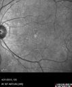
Adult Vitelliform Macular Dystrophy - Pattern Dystrophy418 views
|
|

Macular Telangiectasis with Symptomatic Blind Spots418 views55 year old man with non-neovascular MacTel. VA 20/20 right eye 20/50 left eye. The color images show gray areas temporally. The FAF shows decreased macular pigment. The OCT shows outer retinal atrophy.
|
|

Stage 4 Macular Hole418 views57 year old female with 20/160 vision and stage 4 macular hole
|
|

Outer Retinal Tubulation418 views78 year old man chronic wet AMD - 20/200
|
|

Pattern Dystrophy - Foveal Atrophy418 views73 year old female with vision loss - looks like macular hole
|
|

Non-central vitreomacular traction418 views
|
|

Central Serous Chorioretinopathy - Moderate418 views49 year old man with episodes in 2011, 2017, and 2018 - now with some paracentral vision loss in the right eye and subretinal fluid.
|
|
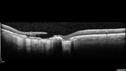
Macular Scars - Laser (1980s) Ocular Histoplasmosis418 views70 year old man who had laser at Wills, Wilmer and the Mayo clinic before better therapies were available for ocular histoplasmosis related CNVM
|
|
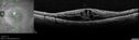
Cystoid Macular Edema and Macular Pucker418 viewshas been particular bad for 3 years. She has a macular pucker and severe CME.
VA 20/160 left eye
|
|

Choroidal Neovascular Membrane following Prior Central Serous Chorioretinopathy - OCT-A418 views67 year old female with CSR in the left eye first 20 years ago. She was managed with out therapy and has had a few episodes since. Now ther eis distortion in the left eye for a few days. She had a steroid shot in her shoulder 5 months ago.
VA 20/20 OD, 20/50 OS- ICG and OCT-A confirm CNVM in the left eye
|
|

418 views
|
|

Vitreomacular traction with traction over retinal vessels in non-diabetic - Fluorescein leakage in areas of traction - Progressing vision loss left eye417 views80-year-old woman had vitrectomy surgery done in the right eye in 2007. Vision has been poor since despite a good anatomical result. Recently her vision in the left eye has been declining. The vision has declined in the left eye. Vision OD is 20/160, OS is 20/80
|
|
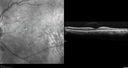
Normal Vision - Chronic Central Serous Chorioretinopathy417 views67 year old man - never had any symptoms - 20/16 vision in each eye. Lesions spare the fovea
|
|
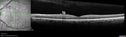
Spontaneously Peeled Macular Pucker 20/20417 views
|
|

Outer Retinal Tubulation417 views78 year old man chronic wet AMD - 20/200
|
|
| 5013 files on 51 page(s) |
 |
|
|
|
|
32 | |
|
|
|
|


