Most viewed - Macular Disease
|

Wet AMD 62 year old male smoker 716 views62-year-old man has wet age-related macular degeneration in the right eye. He has been undergoing treatment with Lucentis for that. Then his left eye developed hazy vision a few weeks ago while he was overseas and now his vision is hazy in both eyes. OD 20/40, OS 20/30. FA shows a classic subfoveal choroidal neovascular membrane in the left eye, which is leaking and in the right eye, which is dry. He is going to stop smoking today. FU: VA 20/30 OU with 2 years of every 6 weeks bilateral avastin!
|
|

Angioid Streaks - Peau D'Orange - Pseudoxanthoma Elasticum - Asymptomatic Extrafoveal Choroidal Neovascular Membrane716 views60-year-old woman has pseudoxanthoma elasticum since 2008. Her sister has the same problem and lost vision in both eyes. She had a problem with blood vessel growth in the left eye for which she had injections of Avastin in August, September, and October of 2011 (5 months ago). Subsequent to that, the macula has been dry and you have just been watching her carefully. She is in Florida for a short time but will ultimately be spending nine months out of the year here. Both of her eyes have good vision at
|
|

Acute Central Serous Retinopathy - CSR - Steroid Induced (probably) - Good Vision - ICG Affected Eye - Choroidal Leakage716 views42-year-old man was seen in the office on October 5, 2011. He had noticed starting in August after a course of antibiotic and steroids, that he developed new spots in his vision in the right eye. He may have had an episode like this sometime in the past. He did take steroids a few years ago and his vision did change at that time, but then returned.
VISUAL ACUITY: OD 20/32, OS 20/32. The OCT scan of the right eye confirms subretinal fluid.
|
|

Cartwheel CNVM - Classic - wet AMD - evidence of possible old CSR - FA shows cartwheel shaped CNVM716 views75 y.o Decreased Vision right eye for one year. VA OD: sc20/125+2 OS 20/25. Autofluorescence and SD OCT show evidence of old serous retinal detachment superior to the fovea suggesting a history of CSR. The macular lesion on FA shows a classic cartwheel appearance
|
|

Plaquenil Toxicity - Bulls Eye Maculopathy715 views70-year-old woman with systemic Lupus erythematosus and clotting problems. She was on the Plaquenil for about eight years and then off the Plaquenil for the last eight years because she developed macular toxicity. Although her vision was hazy, it was stable. Recent deceased vision left eye: OD 20/60, OS 20/100. IOP: OD 18, OS 19.
|
|
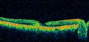
Group 2a Juxtafoveal Telangiectasis - MacTel - Idiopathic Juxtafoveal Macular Telangiectasia - Macular Hole715 views80-year-old man has juxtafoveal retinal telangiectasis in both eyes. He also has a true full-thickness macular hole in the left eye. The vision OD is 20/40, OS is 20/160
Group 2a Juxtafoveal Retinal Telangiectasis (telangiectasia), MacTel
|
|
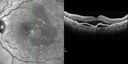
Polypoidal Choroidal Vasculopathy with Pulsating ICG polyp left eye715 views
|
|

Basal Laminar Drusen - Cuticular Drusen - Equatorial Drusen - 70 Year Old Woman Fundus Photo - Geographic Atrophy714 views70 year old woman has had macular drusen since her thirties and she said her sister has a similar problem. Recently the vision in the left eye has substantially declined. OD 20/16, OS 10/200.
|
|

HD OCT of Macular Hole with Operculum714 views
|
|

Cuticular Drusen (Basal Laminar Drusen) and Bilateral Chronic Subfoveal Fluid (Serous Retinal Detachment) unresponsive to monthly Avastin - ICG OS713 views66-year-old man with bilateral subfoveal fluid unresponsive to one year of monthly avastin therapy.
OD 20/160, Pinhole 20/80. OS 20/63, Pinhole 20/50. OCT scan shows subretinal fluid with normal choroidal thickness. Fluorescein angiogram shows cuticular/basal laminar drusen. ICG shows no foci of choriocapillaris leakage.
|
|

Cartwheel CNVM - Classic - wet AMD - evidence of possible old CSR - FA shows cartwheel shaped CNVM713 views75 y.o Decreased Vision right eye for one year. VA OD: sc20/125+2 OS 20/25. Autofluorescence and SD OCT show evidence of old serous retinal detachment superior to the fovea suggesting a history of CSR. The macular lesion on FA shows a classic cartwheel appearance
|
|
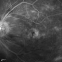
Macular Edema and dry AMD - excellent example of why FA and ICG are necessary in management of wet AMD713 viewsThis 72 year old woman has an OCT that looks like wet AMD. Only her FA and ICG show that she has inflammatory macular edema. She had no clinical signs or symptoms of uveitis. Nevertheless, the macular edema responded to subtenon's kenalog.
|
|

Basal Laminar Drusen - Cuticular Drusen - 19 Year Old with Lupus Nephritis712 views19-year-old. She has a history of developing severe systemic lupus erythematosus in 2001. She was in renal failure and had biopsy proven Class 2 lupus nephritis. She is currently on Plaquenil 400 mg for six years, as well as Prednisolone orally, every other day. OD 20/20, OS 20/25.
|
|

Cystoid Macular Edema - Xalatan - ECCE was done 20 years previously712 views71 Year old woman who noticed about a month ago the central vision in the right eye fogged up. OD 20/200 POSSIBLE XALATAN INDUCED CYSTOID MACULAR EDEMA
|
|
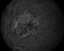
Angioid Streaks and Treated CNVM711 viewsMaculopathy associated to angioid streaks in a 45 years old patient, man,
treated for related CNV
|
|

Hyperpigmentation of the Retinal Pigment Epithelium in Dry AMD709 views87-year-old woman has wet age-related macular degeneration in the left eye. Vision OD is 20/25, OS is 20/400
|
|
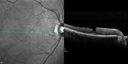
Psuedomacular Hole - Epiretinal Membrane - Vision unchanged 20/40 for 6 months709 views
|
|
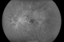
Myopic Macular Degeneration - Dry 708 views55-year-old woman has myopic macular degeneration in both eyes with vision and an Amsler grid change starting a few days ago in the left eye.
VISUAL ACUITY: OD: 20/60. PH: 20/25. OS: 20/200. PH: 20/30.
|
|

708 views
|
|

Concentric Geograophic Atrophy - Age-related Macular Degeneration - Charles Bonnet Syndrome - Bulls Eye708 views88-year-old woman has age-related with vision gradually declining over the last year. She sees spots in her vision, like red spots and dark spots and she even notices during the daytime, for example when she was looking at her minister, it looked like there were little flowering pots coming out of his head.
VISUAL ACUITY: OD 20/200, OS 20/400.
|
|

Choroidal Neovascular Membrane - Optic Disc Drusen - Hemorrhage (Coumadin) 708 views82-year-old man with decreased vision in the left eye over about the last six to 12 months. He is also on Coumadin. OD 20/20, OS 20/80
|
|

Geographic Atrophy 20/160 Vision708 views84-year-old woman has age-related macular degeneration in both eyes with foveal atrophy. She also has a central retinal vein occlusion
in the left eye for which she had intravitreal Kenalog and finally chorioretinal anastomosis laser a year
ago. VISUAL ACUITY: Vision OD is 20/160, OS is 7/200.
|
|

New Subfoveal Classic CNVM OS - Wet AMD - Macular Scar Congenital Toxoplasmosis Right Eye 708 views77-year-old woman as had poor vision in the right eye since birth from congenital toxoplasmosis of her macula in the right eye. Two to three weeks ago when she noticed vision loss in the left eye. VISUAL ACUITY: OD 2/200, OS 20/200.
|
|

Juxtafoveal Telangiectasis - MacTel - Group 2a - Stage IV - Pigment Plaque 89 Year Old Man (Also Nevus)708 views89-year-old man has stage IV juxtafoveal retinal telangiectasis in both eyes with background diabetic retinopathy. His vision is stable since he was here last which is about 6 months ago. OD 20/50, OS 20/70.
|
|

Small Retinal Pigment Epithelial Rip and Poor Vision (20/400)707 views80-year-old woman vision in the right eye has substantially declined.
OD: 20/400; OS: 20/50
|
|

Hyperpigmentation of Retinal Pigment Epithlium in Dry AMD (hyperplasia)707 views87-year-old woman has wet age-related macular degeneration in the left eye. She had treatment up north but there has been a gap in therapy and the vision in the left eye is poor. Vision OD is 20/25, OS is 20/400
|
|

Basal Laminar Drusen - Cuticular Drusen - Equatorial Drusen - 70 Year Old Woman - Fluorescein Angiogram Montage Left Eye707 views70 year old woman has had macular drusen since her thirties and she said her sister has a similar problem. Recently the vision in the left eye has substantially declined. OD 20/16, OS 10/200.
|
|

Plaquenil Toxicity - Bulls Eye Maculopathy706 views70-year-old woman with systemic Lupus erythematosus and clotting problems. She was on the Plaquenil for about eight years and then off the Plaquenil for the last eight years because she developed macular toxicity. Although her vision was hazy, it was stable. Recent deceased vision left eye: OD 20/60, OS 20/100. IOP: OD 18, OS 19.
|
|

Plaquenil Toxicity - Bulls Eye Maculopathy706 views70-year-old woman with systemic Lupus erythematosus and clotting problems. She was on the Plaquenil for about eight years and then off the Plaquenil for the last eight years because she developed macular toxicity. Although her vision was hazy, it was stable. Recent deceased vision left eye: OD 20/60, OS 20/100. IOP: OD 18, OS 19.
|
|

Drusen Maculopathy - Macular Degeneration (Dry) - 50 year old African-American Woman706 views50-year-old woman has drusen and maculopathy. VISUAL ACUITY: OD 20/30, OS 20/25.
|
|

Juxtafoveal Telangiectasis - Stage 5 Left Eye - New Blood and Neovascularization MacTel FAF706 views60-year-old man has juxtafoveal retinal telangiectasis recent vision loss OD 20/50, OS 20/70 : Hemorrhage OS and new SRNVM
|
|

Chronic Pseudophakic Cystoid Macular Edema - Haptic Migrated into Anterior Chamber Through Peripheral Iridotomy705 views57-year-old man with congenital nystagmus. He had cataract surgery done 30 years ago and has decreased vision left eye for 6 months. OD is 20/50, OS is 20/100
|
|

Reticular Macular Disease (Dry Macular Degeneration) - Infrared reflectance705 views85 year old woman with 20/25 vision OU and reticular macular disease
|
|

Macular Scars - Laser (1980s) Ocular Histoplasmosis704 views70 year old man who had laser at Wills, Wilmer and the Mayo clinic before better therapies were available for ocular histoplasmosis related CNVM
|
|
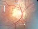
Angioid Streaks - Pseudoxanthoma Elasticum - Supressed CNVM704 views54-year-old woman had angioid streaks and macular degeneration in both eyes. She has had intravitreal Avastin in the right eye about every three months and her vision in that eye is good.
VISUAL ACUITY: Vision OD is 20/30, OS is 2/200
|
|

Acute Central Serous Retinopathy - CSR - Steroid Induced (probably) - Good Vision 704 views42-year-old man was seen in the office on October 5, 2011. He had noticed starting in August after a course of antibiotic and steroids, that he developed new spots in his vision in the right eye. He may have had an episode like this sometime in the past. He did take steroids a few years ago and his vision did change at that time, but then returned.
VISUAL ACUITY: OD 20/32, OS 20/32. The OCT scan of the right eye confirms subretinal fluid.
|
|

Wet AMD - PED with exudate 20/400 87 Year old703 views87-year-old woman has wet age-related macular degeneration in the left eye. Vision OD is 20/25, OS is 20/400
|
|

Cuticular Drusen (Basal Laminar Drusen) and Bilateral Chronic Subfoveal Fluid (Serous Retinal Detachment) unresponsive to monthly Avastin - Late FA OS703 views66-year-old man with bilateral subfoveal fluid unresponsive to one year of monthly avastin therapy.
OD 20/160, Pinhole 20/80. OS 20/63, Pinhole 20/50. OCT scan shows subretinal fluid with normal choroidal thickness. Fluorescein angiogram shows cuticular/basal laminar drusen. ICG shows no foci of choriocapillaris leakage.
|
|

Idiopathic Cystoid Macular Edema - CME - Responded to Subtenons Kenalog - Early Fluorescein Angiogram Showing Dilated Capillaries702 views49-year-old man had a flu two weeks ago the right eye has been throbbing and his vision in the right eye has been foggy. OD 20/80, OS 20/40. IOP: 14 OU.
|
|

Patterned Pigment Dystrophy of the Fovea 79 YO Woman VA 20/20 Right 20/30 Left702 views
|
|

Plaquenil Toxicity - Bull's Eye Maculopathy - 25 Years Therapy - OCT Posterior Pole MAP702 views73-year-old woman has been on Plaquenil for twenty five years. Plaquenil has now been stopped for 6 months. VA continues to decline as does visual function - 20/80 OD , 20/40 OS
|
|

Idiopathic Polypoidal Choroidal Vasculopathy - False Color702 views
|
|

Idiopathic Cystoid Macular Edema - CME - Responded to Subtenons Kenalog - Red Free Photos Shows Foveal Cysts701 views49-year-old man had a flu two weeks ago the right eye has been throbbing and his vision in the right eye has been foggy. OD 20/80, OS 20/40. IOP: 14 OU.
|
|

Pigment Epithelial Detachment with Subretinal Fluid and Exudate 20/400 Vision701 views87-year-old woman has wet age-related macular degeneration in the left eye. She had treatment up north but there has been a gap in therapy and the vision in the left eye is poor. Vision OD is 20/25, OS is 20/400
|
|

Wet AMD - New Small Hemorrhage Both Eyes - Mature CNVM visible on Early Indocyanine Green Angiogram701 views
|
|

Presumed Ocular Histoplasmosis with new CNVM in right and better eye - VA 20/80 OD; 20/800 OS701 viewsPeripheral punched out small scars typical for OHS
|
|
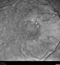
Multifocal Pattern Dystrophy701 views51 year old with normal vision - images look a lot like fundus flavimaculatus
|
|

Myopic Choroidal Neovascular Membrane 54 year old woman 20/20700 views54-year-old woman has had problems in her vision in the right eye for about the last three months. Inferotemporally she sees a gray area, which is distorted. OD is 20/20, OS is 20/20
|
|
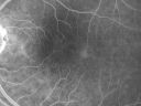
Group 2a Juxtafoveal Telangiectasis - MacTel - Idiopathic Juxtafoveal Macular Telangiectasia - Macular Hole700 views80-year-old man has juxtafoveal retinal telangiectasis in both eyes. He also has a true full-thickness macular hole in the left eye. The vision OD is 20/40, OS is 20/160
Group 2a Juxtafoveal Retinal Telangiectasis (telangiectasia), MacTel
|
|

Acute Central Serous Retinopathy - CSR - Steroid Induced (probably) - Good Vision - FA Affected Eye700 views42-year-old man was seen in the office on October 5, 2011. He had noticed starting in August after a course of antibiotic and steroids, that he developed new spots in his vision in the right eye. He may have had an episode like this sometime in the past. He did take steroids a few years ago and his vision did change at that time, but then returned.
VISUAL ACUITY: OD 20/32, OS 20/32. The OCT scan of the right eye confirms subretinal fluid.
|
|

Reticular Macular Disease (Pseudo-drusen) Both Eyes - Occult CNVM and Macular Edema Right Eye 700 viewsSpectral Domain Optical Coherence Tomography
|
|

Basal Laminar Drusen - Cuticular Drusen - 19 Year Old with Lupus Nephritis699 views19-year-old. She has a history of developing severe systemic lupus erythematosus in 2001. She was in renal failure and had biopsy proven Class 2 lupus nephritis. She is currently on Plaquenil 400 mg for six years, as well as Prednisolone orally, every other day. OD 20/20, OS 20/25.
|
|

Plaquenil Toxicity - Bulls Eye Maculopathy699 views70-year-old woman with systemic Lupus erythematosus and clotting problems. She was on the Plaquenil for about eight years and then off the Plaquenil for the last eight years because she developed macular toxicity. Although her vision was hazy, it was stable. Recent deceased vision left eye: OD 20/60, OS 20/100. IOP: OD 18, OS 19.
|
|

Plaquenil Toxicity - Bulls Eye Maculopathy699 views70-year-old woman with systemic Lupus erythematosus and clotting problems. She was on the Plaquenil for about eight years and then off the Plaquenil for the last eight years because she developed macular toxicity. Although her vision was hazy, it was stable. Recent deceased vision left eye: OD 20/60, OS 20/100. IOP: OD 18, OS 19.
|
|

Wet AMD - PED with exudate 20/400 87 Year old699 views87-year-old woman has wet age-related macular degeneration in the left eye. Vision OD is 20/25, OS is 20/400
|
|

Chronic Pseudophakic Cystoid Macular Edema - Haptic Migrated into Anterior Chamber Through Peripheral Iridotomy699 views57-year-old man with congenital nystagmus. He had cataract surgery done 30 years ago and has decreased vision left eye for 6 months. OD is 20/50, OS is 20/100
|
|

Macular Edema and dry AMD - excellent example of why FA and ICG are necessary in management of wet AMD699 viewsThis 72 year old woman has an OCT that looks like wet AMD. Only her FA and ICG show that she has inflammatory macular edema. She had no clinical signs or symptoms of uveitis. Nevertheless, the macular edema responded to subtenon's kenalog.
|
|

Wet AMD - PED with exudate 20/400 87 Year old698 views87-year-old woman has wet age-related macular degeneration in the left eye. Vision OD is 20/25, OS is 20/400
|
|

PXE with Angioid Streaks698 viewsPXE with angioid streaks OD (12/10/2010)
|
|

Chronic Central Serous Retinopathy 1 month after Photodynamic Therapy (Visudyne)698 viewsSerous Retinal Detachment is Gone (Absorbed)
|
|

Polypoidal Choroidal Vasculopathy with Pulsating ICG polyp left eye698 views
|
|

Myopic CNVM - Wet - rx Avastin for 1 year697 views55-year-old woman OD is 20/40, OS is 20/20. There is a hyperpigmented disc diameter choroidal neovascular membrane with fluid touching the fovea.
|
|

Polypoidal Choroidal Vasculopathy with Pulsating ICG polyp left eye697 views
|
|

Geographic Atrophy 20/160 Vision696 views84-year-old woman has age-related macular degeneration in both eyes with foveal atrophy. She also has a central retinal vein occlusion
in the left eye for which she had intravitreal Kenalog and finally chorioretinal anastomosis laser a year
ago. VISUAL ACUITY: Vision OD is 20/160, OS is 7/200.84-year-old woman has age-related macular degeneration in both eyes with foveal atrophy. She also has a central retinal vein occlusion
in the left eye for which she had intravitreal Kenalog and finally chorio
|
|

Wet AMD - New Small Hemorrhage Both Eyes - Mature CNVM visible on Early Indocyanine Green Angiogram696 views
|
|

Wet AMD - New Small Hemorrhage Both Eyes - Mature CNVM visible on Early Indocyanine Green Angiogram696 views
|
|
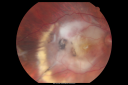
Disciform Macular Scar Both Eyes from Wet Macular Degeneration695 views86-year-old woman has disciform scars OD: 20/400; OS: 20/100
|
|

Angioid Streaks - New Extrafoveal Choroidal Neovascular Membrane Right Eye695 views61-year-old woman twelve days ago, saw a spot to the right of her central vision. OD is 20/25, OS is 20/25
Treated with one year of avastin with good result and no vision loss.
|
|

Vision Loss, Blood, and Cartwheel CNVM OD with no fluid on OCT Scan695 views86-year-old man has had changing vision. He says his vision has been gradually declining since I saw him in June. You saw a hemorrhage in the eye and suggested he comes in here for an evaluation. He is on Combigan and Travatan in the right eye for his glaucoma.
VISUAL ACUITY: Vision OD is 6/200 OS is 8/200 The macula in the right eye is wet even though interestingly the OCT does not show subretinal fluid.
|
|
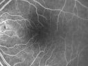
Group 2a Juxtafoveal Telangiectasis - MacTel - Idiopathic Juxtafoveal Macular Telangiectasia - Macular Hole695 views80-year-old man has juxtafoveal retinal telangiectasis in both eyes. He also has a true full-thickness macular hole in the left eye. The vision OD is 20/40, OS is 20/160
Group 2a Juxtafoveal Retinal Telangiectasis (telangiectasia), MacTel
|
|

Pattern Dystrophy - Adult Vitelliform Dystrophy - Macular Degeneration695 views 78 Year Old Woman with vision has diminished in the right eye over the past couple of years, but the diminishment has been slow.
20/60+1 OD, 20/25 OS
|
|

Lamellar Hole Epiretinal Proliferation - LHEP695 views
|
|

Pattern Dystrophy - Adult Vitelliform - 95 Year Old Man with Good Vision694 views95-year-old man in excellent health and he doesn’t notice a vision problem. OD 20/32, OS 20/80
|
|

Cuticular Drusen (Basal Laminar Drusen) and Bilateral Chronic Subfoveal Fluid (Serous Retinal Detachment) unresponsive to monthly Avastin Early FA OD694 views66-year-old man with bilateral subfoveal fluid unresponsive to one year of monthly avastin therapy.
OD 20/160, Pinhole 20/80. OS 20/63, Pinhole 20/50. OCT scan shows subretinal fluid with normal choroidal thickness. Fluorescein angiogram shows cuticular/basal laminar drusen. ICG shows no foci of choriocapillaris leakage.
|
|
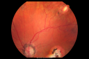
Macular Scars - Laser (1980s) Ocular Histoplasmosis693 views70 year old man who had laser at Wills, Wilmer and the Mayo clinic before better therapies were available for ocular histoplasmosis related CNVM
|
|

Reticular Macular Disease (Pseudo-drusen) Both Eyes - Occult CNVM and Macular Edema Right Eye 693 viewsSpectral Domain Optical Coherence Tomography
|
|
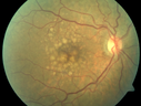
Polypoidal Choroidal Vasculopathy with Pulsating ICG polyp left eye693 views
|
|

Dry AMD - Confluent Drusen Sparing Center of Macula which has Atrophy693 views65 year old woman, VA 20/40 OD; 20/80 OS. The center of the macula has few or no drusen with predominantly non-geographic atrophy
|
|

Non-Foveal Geographic Atrophy Both Eyes692 views81-year-old man has age-related macular degeneration in both eyes. Vision OD is 20/40, OS is 20/80.
|
|

Angioid Streaks - Peau D'Orange - Pseudoxanthoma Elasticum - Asymptomatic Extrafoveal Choroidal Neovascular Membrane692 views60-year-old woman has pseudoxanthoma elasticum since 2008. Her sister has the same problem and lost vision in both eyes. She had a problem with blood vessel growth in the left eye for which she had injections of Avastin in August, September, and October of 2011 (5 months ago). Subsequent to that, the macula has been dry and you have just been watching her carefully. She is in Florida for a short time but will ultimately be spending nine months out of the year here. Both of her eyes have good vision at
|
|

Lumigan (Bimatoprost) Cystoid Macular Edema - Eye Better 1 month after Stopping Lumigan692 views 82-year-old man had cataract surgery in 1993. He has had glaucoma for seven or eight years for a long time. He is taking Lumigan only in the left eye. About a year ago he started in the right eye and then starting just about a few months ago he has noticed intermittent blurring in the vision in the right eye. Vision 20/25 OD, 20/20 OS
|
|

Wet AMD - New Small Hemorrhage Both Eyes - Mature CNVM visible on Early Indocyanine Green Angiogram692 views
|
|

Central Serous Retinopathy - Subretinal Fibrin692 views66 year old female with 20 year history of single episode of CSR. Now with vision loss for a month but improving - VA 20/63 OD; 20/32 OS
|
|

Plaquenil (Hydroxycholorquine) Toxicity - Bull's Eye Maculopathy - 73 year old womand 25 Years therapy - 64 inches tall - 110 lbs691 views73-year-old woman has been on Plaquenil for twenty five years. She noticed loss of paracentral vision a few months ago. VISUAL ACUITY: OD 20/70, OS 20/30. Her color vision was 0 out 10 correct in both eyes and she also missed the controls.
|
|

Plaquenil Toxicity - Bull's Eye Maculopathy - 25 Years Therapy - Infra-red Image691 views73-year-old woman has been on Plaquenil for twenty five years. Plaquenil has now been stopped for 6 months. VA continues to decline as does visual function - 20/80 OD , 20/40 OS
|
|

Juxtafoveal Telangiectasis - Stage 5 Left Eye - New Blood and Neovascularization MacTel 691 views60-year-old man has juxtafoveal retinal telangiectasis recent vision loss OD 20/50, OS 20/70 : Hemorrhage OS and new SRNVM
|
|

Outer Retinal Holes - Probably Solar Retinopathy (history is negative)691 views
|
|

Patterned Pigment Dystrophy of the Fovea 79 YO Woman VA 20/20 Right 20/30 Left690 views
|
|

Macular Pucker Pre-OP 20/200 690 views
|
|

Acute Central Serous Retinopathy - CSR - Steroid Induced (probably) - Good Vision 690 views42-year-old man was seen in the office on October 5, 2011. He had noticed starting in August after a course of antibiotic and steroids, that he developed new spots in his vision in the right eye. He may have had an episode like this sometime in the past. He did take steroids a few years ago and his vision did change at that time, but then returned.
VISUAL ACUITY: OD 20/32, OS 20/32. The OCT scan of the right eye confirms subretinal fluid.
|
|

Chronic Central Serous Retinopathy - Indocyanine Green Angiogram 690 views
|
|

689 views
|
|

Basal Laminar (cuticular) Drusen both eyes 60 Year Old Woman689 views60-year-old woman with minimal vision loss. Vision OD is 20/30 OS 20/40
|
|

Outer Retinal Holes - Probably Solar Retinopathy (history is negative)689 views
|
|

Myopic Choroidal Neovascular Membrane 54 year old woman 20/20688 views54-year-old woman has had problems in her vision in the right eye for about the last three months. Inferotemporally she sees a gray area, which is distorted. OD is 20/20, OS is 20/20
|
|
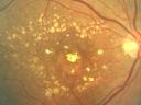
688 views
|
|

Angioid Streaks - Pseudoxanthoma Elasticum - Supressed CNVM688 views54-year-old woman had angioid streaks and macular degeneration in both eyes. She has had intravitreal Avastin in the right eye about every three months and her vision in that eye is good.
VISUAL ACUITY: Vision OD is 20/30, OS is 2/200
|
|

Multifocal Bilateral Central Serous Retinopathy in African American Male688 views67 year old african american male with : Blurred Vision OD. Duration of Problem: 4-6 weeks. VA 20/32 OD 20/20 OS
|
|
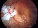
Myopic CNVM - Wet - rx Avastin for 1 year687 views55-year-old woman OD is 20/40, OS is 20/20. There is a hyperpigmented disc diameter choroidal neovascular membrane with fluid touching the fovea.
|
|

Myopic Traction Maculopathy - Vitreoretinal Traction - Possible Occult Neovascular Membrane687 views58-year-old woman was seen in the office on June 30, 2011. In 2002, her vision was 20/30. Subsequent to that she had her left eye made her distance eye, which was dominate. She had LASIK surgery on the right eye to make that her near eye and she was getting along well until about a year ago when she noticed substantial vision loss in the left eye only. VISUAL ACUITY: The near eye is 20/200, but she reads J2. OS 20/100- and J16.
|
|
| 5015 files on 51 page(s) |
 |
|
|
|
|
7 | |
|
|
|
|


