Most viewed - Retinal Vascular Disease
|

Nonproliferative Diabetic Retinopathy8351 views65-year old female with diabetes. Has had cataract surgery in the left eye with VA 20/25. She has had laser in the past. Fundus examination shows microaneurysms with retinal hemorrahages and exudates in the left eye.
|
|

Nonproliferative Diabetic Retinopathy7676 views65-year old female with diabetes. Has had cataract surgery in the left eye with VA 20/25. She has had laser in the past. Fundus examination shows microaneurysms with retinal hemorrahages and exudates in the left eye.
|
|

Nonproliferative Diabetic Retinopathy6792 views65-year old female with diabetes. Has had cataract surgery in the left eye with VA 20/25. She has had laser in the past. Fundus examination shows microaneurysms with retinal hemorrahages and exudates in the left eye.
|
|

Nonproliferative Diabetic Retinopathy5940 views65-year old female with diabetes. Has had cataract surgery in the left eye with VA 20/25. She has had laser in the past. Fundus examination shows microaneurysms with retinal hemorrahages and exudates in the left eye.
|
|

Nonproliferative Diabetic Retinopathy5851 views65-year old female with diabetes. Has had cataract surgery in the left eye with VA 20/25. She has had laser in the past. Fundus examination shows microaneurysms with retinal hemorrahages and exudates in the left eye.
|
|

Juvenile Onset Diabetic - Proliferative Diabetic retinopathy with NVE and NVD4706 viewsProliferative Diabetic Retinopathy with Neovascularization of the Disc and Neovascularization Elsewhere.
James L. Perron, C.R.A.
|
|
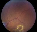
Nonproliferative Diabetic Retinopathy4519 views65-year old female with diabetes. Has had cataract surgery in the left eye with VA 20/25. She has had laser in the past. Fundus examination shows microaneurysms with retinal hemorrahages and exudates in the left eye.
|
|

Purtscher's Retinopathy - Motor Vehicle Accident4418 viewsNerve Fiber Layer Inflammation and retinal hemorrhages due to severe trauma. Patient was a teenage boy that got into a car accident.
|
|

Proliferative Diabetic Retinopathy with Macular Hole3184 viewsPatitent with diabetes diagnosed 12 years ago and low visual acuity.
|
|
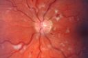
Non-Ischemic Central Retinal Vein Occlusion in Young Man (30 years) - Many Cotton Wool Spots3056 views30-year-old man with a history of a central retinal vein occlusion for which he was first examined on 2 months ago and his vision then was 4/200. He responded nicely to two Avastin treatments. OD is 20/20, OS is 20/60
|
|

Nonproliferative Diabetic Retinopathy2920 views65-year old female with diabetes. Has had cataract surgery in the left eye with VA 20/25. She has had laser in the past. Fundus examination shows microaneurysms with retinal hemorrahages and exudates in the left eye.
|
|

Prominent Posterior Hyaloid with Background Diabetic Retinopathy2859 viewsDiabetic patient comes in for follow up for her Background Diabetic Retinopathy and glaucoma. VA is 20/30. left eye. Fundus exam shows posterior Hyaloid with hemorrhage inferiorily.
|
|

Asymptomatic Hollenhorst Plaque - Cholesterol Embolis2697 views71-year-old woman is not noticing any vision change. She does take pressure-lowering drops for glaucoma, both Timolol and Xalatan.
VISUAL ACUITY: Vision OD is 20/30, OS is 20/16
|
|

Retinal Vasculitis due to Lupus (color)2529 viewsSystemic Lupus Erythematosis - Vasculitis
|
|

Prominent Posterior Hyaloid with Background Diabetic Retinopathy2527 viewsDiabetic patient comes in for follow up for her Background Diabetic Retinopathy and glaucoma. VA is 20/30. left eye. Fundus exam shows posterior Hyaloid with hemorrhage inferiorily.
|
|

Non-Ischemic Central Retinal Vein Occlusion in Young Man (30 years) - Many Cotton Wool Spots2416 views30-year-old man with a history of a central retinal vein occlusion for which he was first examined on 2 months ago and his vision then was 4/200. He responded nicely to two Avastin treatments. OD is 20/20, OS is 20/60
|
|

Nonproliferative Diabetic Retinopathy2407 views65-year old female with diabetes. Has had cataract surgery in the left eye with VA 20/25. She has had laser in the past. Fundus examination shows microaneurysms with retinal hemorrahages and exudates in the left eye.
|
|

Malignant Hypertension - Cotton Wool Spots - Elschnig Spots - Optic Nerve Edema 2190 viewsElschnig Spots
|
|

Diabetic Macular Edema and Hypertensive Retinopathy - Circinate Exudate (Ring Exudate) 2172 views55-year-old woman diabetic for fifteen years and high blood pressure, especially over the last few months. She has had problems with nosebleeds, headaches, and there has been some difficulty bringing her blood pressure down. She said now the blood pressure is under control but it was running 220/120 mmHg for some time. Her vision in the right eye has been poor for four weeks with a spot in the central vision and both eyes have been blurred.
VISUAL ACUITY: Vision OD is 20/160, OS is 20/60
|
|
Malignant Hypertension - Cotton Wool Spots - Elschnig Spots - Optic Nerve Edema 2051 viewsElschnig Spots
|
|
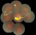
coats disease; exudative retinitis; retinal telangiectasis; leber multiple miliary aneurysm disease2017 viewsclassic coats
|
|
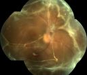
TRD w/ fibrovascular proliferation and sub retinal PVR OD1927 views41 year old african american female with tractional retinal detachment, fibrovascular proliferation, old vitreous hemorrhage, disc neovascularization, and proliferative vitreoretinopathy. Pt has a vision of 20/200
|
|

Background Diabetic Retinopathy1829 viewsPatient with diabetes for over 11-years comes in with blurred vision. Blood sugar control is very poor. VA 20/50- right eye, 20/25-left eye. Fundus exam shows hard exudates with Circinate Rings and edema. Micro-aneurysms and retinal hemorrhage are present in both eyes. Patient will come back for laser treatment.
|
|

Retinal Arterial Macroaneurysm - Recurrent Hemorrhage 6 months post laser1822 views6 months post laser: Her vision had improved, but then three weeks ago it worsened again. She is not on any blood thinners. She wasn’t doing any heavy lifting or straining.
VISUAL ACUITY: OD 3/200
|
|
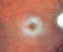
Central Retinal Artery Occlusion - 2 months old by history1820 views77-year-old man was doing fine. His left eye was a little worse than the right eye, and then in the 2 months ago he had sudden severe vision loss in the right eye. Vision OD is 3/200; OS is 20/60. IOP: OD 30, OS 15.
OD: Vertical C/D ratio is 0.4. The macula is white from ischemia. The arteries are narrow.
Photos confirm clinical findings.
OCT SCAN: OCT scan of the right eye shows retinal thickening, consistent with macular edema associated with a central retinal artery occlusion. The left eye has severe inferior retinal thinning, consistent with an old hemicentral retinal artery occlusion in the left eye. The fovea in the left eye is a little bit thin but not too bad.
IMPRESSION: 1. CENTRAL RETINAL ARTERY OCCLUSION RIGHT EYE
2 OLD HEMICENTRAL RETINAL ARTERY OCCLUSION LEFT EYE
3 DIABETES WITHOUT RETINOPATHY
4 ELEVATED INTRAOCULAR PRESSURE RIGHT EYE STATUS POST INJECTION RIGHT EYE A WEEK AGO
DISCUSSION: I explained to the patient that the right eye unfortunately has had a central retinal artery and unfortunately we do not have anything to treat that. His vision will probably improve some over time. He needs to be watched though. It is possible he is developing rubeotic glaucoma, although it is probable the intraocular pressure is high from the injection last week. I asked him to restart the Alphagan, to stop the other two drops, and to return here in three weeks to make sure his pressure is more acceptable.
At this point, having lost almost all the retinal circulation in the right eye and half of the retinal circulation in the left eye, he only has half a retina in the left eye he is working off of. I told him it is very important he keep seeing you and anything that can be done to keep his blood pressure low and his cholesterol reasonable would be helpful to what remains of his retinal circulation.
|
|

Proliferative Diabetic Retinopathy - Preretinal Hemorrhage1799 viewsPreretinal Hemorrhage secondary to Proliferative Diabetic Retinopathy (PDR)
James L. Perron, C.R.A.
|
|

Central Retinal Artery Occlusion1759 views60 year old male with LP vision due to extensive blood flow loss.
|
|

Aplastic anemia1755 views29 y.o.m. with aplastic anemia OD VA 20/25.
|
|

Diabetic Macular Edema and Hypertensive Retinopathy - Circinate Exudate (Ring Exudate) 1719 views55-year-old woman diabetic for fifteen years and high blood pressure, especially over the last few months. She has had problems with nosebleeds, headaches, and there has been some difficulty bringing her blood pressure down. She said now the blood pressure is under control but it was running 220/120 mmHg for some time. Her vision in the right eye has been poor for four weeks with a spot in the central vision and both eyes have been blurred.
VISUAL ACUITY: Vision OD is 20/160, OS is 20/60
|
|

Malignant Hypertension - Cotton Wool Spots - Elschnig Spots - Optic Nerve Edema 1693 views
|
|

Diabetic Macular Edema and Hypertensive Retinopathy - Circinate Exudate (Ring Exudate) 1688 views55-year-old woman diabetic for fifteen years and high blood pressure, especially over the last few months. She has had problems with nosebleeds, headaches, and there has been some difficulty bringing her blood pressure down. She said now the blood pressure is under control but it was running 220/120 mmHg for some time. Her vision in the right eye has been poor for four weeks with a spot in the central vision and both eyes have been blurred.
VISUAL ACUITY: Vision OD is 20/160, OS is 20/60
|
|
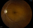
Retinal Artery Occlusion1688 viewsPatient comes in for eval on artery occlusions in both eyes. VA is 20/400, right eye and NLP, left eye. Fundus photos show paniretinal scars in the right eye with arterial narrowing and the left eye has arterial narrowing as well.
|
|

Clinically Significant Diabetic Macular Edema both Eyes with Exudates1685 views56-year-old woman with diabetes for twelve years. She has noticed blurring of the vision in the right eye for about six months. OD is 20/70, OS is 20/25.
|
|

Malignant Hypertension - Cotton Wool Spots - Elschnig Spots - Optic Nerve Edema 1651 viewsElschnig Spots are pigment spots in periphery
|
|

Inferotemporal and Inferonasal BRAO1639 viewsBranch Retinal Artery Occlusions - Multiple
|
|

Malignant Hypertension - Cotton Wool Spots - Elschnig Spots - Optic Nerve Edema 1626 views
|
|

Subhyloid Hem with PDR1624 views
|
|

Proliferative Diabetic Retinopathy both Eyes Neovascularization of the Disc1608 views63-year-old woman has been diabetic for twenty one years who sees new floaters in the right eye. OD 20/40, OS 20/40.
|
|

Hemi-retinal Vein Occlusion1598 views81 year old female seen in 2009 for a Hemi-retinal vein Occlusion OS with vision of 20/25.
|
|
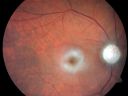
Central Retinal Artery Occlusion - 2 months old by history1594 views77-year-old man was doing fine. His left eye was a little worse than the right eye, and then in the 2 months ago he had sudden severe vision loss in the right eye. Vision OD is 3/200; OS is 20/60. IOP: OD 30, OS 15.
OD: Vertical C/D ratio is 0.4. The macula is white from ischemia. The arteries are narrow.
Photos confirm clinical findings.
OCT SCAN: OCT scan of the right eye shows retinal thickening, consistent with macular edema associated with a central retinal artery occlusion. The left eye has severe inferior retinal thinning, consistent with an old hemicentral retinal artery occlusion in the left eye. The fovea in the left eye is a little bit thin but not too bad.
IMPRESSION: 1. CENTRAL RETINAL ARTERY OCCLUSION RIGHT EYE
2 OLD HEMICENTRAL RETINAL ARTERY OCCLUSION LEFT EYE
3 DIABETES WITHOUT RETINOPATHY
4 ELEVATED INTRAOCULAR PRESSURE RIGHT EYE STATUS POST INJECTION RIGHT EYE A WEEK AGO
DISCUSSION: I explained to the patient that the right eye unfortunately has had a central retinal artery and unfortunately we do not have anything to treat that. His vision will probably improve some over time. He needs to be watched though. It is possible he is developing rubeotic glaucoma, although it is probable the intraocular pressure is high from the injection last week. I asked him to restart the Alphagan, to stop the other two drops, and to return here in three weeks to make sure his pressure is more acceptable.
At this point, having lost almost all the retinal circulation in the right eye and half of the retinal circulation in the left eye, he only has half a retina in the left eye he is working off of. I told him it is very important he keep seeing you and anything that can be done to keep his blood pressure low and his cholesterol reasonable would be helpful to what remains of his retinal circulation.
|
|

Proliferative Diabetic Retinopathy - Vitreous Hemorrhage and Tractional Retinal Detachment Left Eye1526 views52-year-old man has been diabetic for nineteen years developed substantial vision loss over the last month or two. OD 20/70, OS 20/200.
|
|

Central Retinal Artery Occlusion with Cilioretinal Sparing - Acute - SD-OCT Line Scan Inner retinal edema1523 views 77-year-old man was doing fine until midnight of last night when he noticed sudden severe vision loss in the left eye. VISUAL ACUITY: OD 20/20, OS 2/200.
|
|

Proliferative Diabetic Retinopathy - Moderate - mild NVD with NVE 1510 viewsVenous beading, vascular loops and NVE are visible on photos
|
|

Eales Disease1503 views
|
|

Macular Branch Retinal Vein Occlusions (2) Left Eye - No Macular Edema (yet) 1482 views77-year-old woman with two macular branch retinal vein occlusions in the left eye OD 20/25
|
|

Capillary Non-Perfusion - Severe Non-Proliferative (Background) Diabetic Retinopathy - Type I - 20 Year Old Man Diabetic 16 years1481 views20 year old diabetic with poor vision for the past three or four years. He has been diabetic for sixteen years and hemoglobin A1C of 10.0. 20/25 OD, 20/30 OS.
|
|

Diabetic Retinopathy1457 viewsAnnual diabetic exam. VA 20/30, right eye and 20/50, left eye. Fundus photos shows hemorrhages with cotton wool spots. Exudates are visible around the macula in the right eye. Scattered hemorrhages in the left eye.
|
|

Malignant Hypertension - Cotton Wool Spots - Elschnig Spots - Optic Nerve Edema 1453 views
|
|

Diabetic Macular Edema and Hypertensive Retinopathy - Circinate Exudate (Ring Exudate) 1452 views55-year-old woman diabetic for fifteen years and high blood pressure, especially over the last few months. She has had problems with nosebleeds, headaches, and there has been some difficulty bringing her blood pressure down. She said now the blood pressure is under control but it was running 220/120 mmHg for some time. Her vision in the right eye has been poor for four weeks with a spot in the central vision and both eyes have been blurred.
VISUAL ACUITY: Vision OD is 20/160, OS is 20/60
|
|

Hemi Central Retinal Vein Occlusion 35 year old non-compliant man1444 views35-year-old man hazy vision in his left eye for about the last three days. OD 20/20, OS 20/30
|
|

Malignant Hypertension - Cotton Wool Spots - Elschnig Spots - Optic Nerve Edema 1424 views
|
|
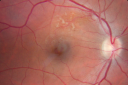
Twig Branch Retinal Vein Occlusion - Recurrent Macular Edema with extended interval Lucentis Therapy after first 6 monthly Injections1407 views39-year-old man has a macular branch retinal vein occlusion with macular edema. He had an excellent response to Lucentis for his first 6 treatments. He was last here 6 weeks ago and his vision seems OK to him. OD 20/40.
|
|

Post- treatment1394 views
|
|

Diabetic Macular Edema Left Eye - Post Laser1388 views82-year-old woman diabetic for many years, last eye exam 5 years ago with gradual vision loss. OD 20/60, OS 20/70. IOP: OD 16, OS 17.
|
|

Capillary Non-Perfusion - Severe Non-Proliferative (Background) Diabetic Retinopathy - Type I - 20 Year Old Man Diabetic 16 years1377 views20 year old diabetic with poor vision for the past three or four years. He has been diabetic for sixteen years and hemoglobin A1C of 10.0. 20/25 OD, 20/30 OS.
|
|

Post- treatment1368 views
|
|

Sickle Cell1358 views
|
|

Asymptomatic Hollenhorst Plaque - Cholesterol Embolis1357 views71-year-old woman is not noticing any vision change. She does take pressure-lowering drops for glaucoma, both Timolol and Xalatan.
VISUAL ACUITY: Vision OD is 20/30, OS is 20/16
|
|
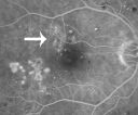
Focal Laser for diabetic macular edema1345 views66 year old female with diabetes for 45 years. VA 20/20 OU. Extrafoveal edema in the left eye treated with light focal laser.
|
|

Malignant Hypertension - Cotton Wool Spots - Elschnig Spots - Optic Nerve Edema 1344 viewsElschnig Spots are pigment spots in periphery
|
|

Central Retinal Vein Occlusion1338 viewsPatient comes in with mild blurred vision in the right eye. Fundus exam shows CRVO with scattered retinal hemorrhages in the right eye.
|
|

Proliferative Diabetic Retinopathy1334 viewsFundus photography shows severe fibrosis and arterial narrowing. Peripheral laser scars in both eyes. VA is 20/40, right eye and 20/50, left eye.
|
|

Non-Ischemic Central Retinal Vein Occlusion in Young Man (30 years) - Many Cotton Wool Spots1330 views30-year-old man with a history of a central retinal vein occlusion for which he was first examined on 2 months ago and his vision then was 4/200. He responded nicely to two Avastin treatments. OD is 20/20, OS is 20/60
|
|

Bilateral Retinal Arteriol Occlusions - Possible Susac Syndrome 1329 views80-year-old woman one month ago had vision loss and a vascular occlusion in the right eye. Vision loss occurred in the left eye about 9 years ago with cotton wool spots. Patients has a 30 year history of tinnitis.
|
|
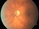
Microaneurysms from Ocular Ischemic Syndrome (non-diabetic) Right Eye - Left eye CRAO 1 year ago1325 views66-year-old woman lost vision in the left eye from a central retinal artery occlusion 1 year ago. Vision OD is fine. OD 20/25, OS 20/50
|
|

Hypertensive Choroidopathy - Pheochromocytoma - Chronic Very High Blood Pressure 49 YO Male1320 views49-year-old man chronic very high blood pressure sometimes the blood pressure will go as high as 260/160, sometimes it is 200/120 and sometimes it is 160/80. He has a pheochromocytoma on the adrenal gland and he has had vision loss in the right eye starting between three and four years ago.
VISUAL ACUITY: OD 20/60, OS 20/25.
|
|

Malignant Hypertension - Cotton Wool Spots - Elschnig Spots - Optic Nerve Edema 1319 views
|
|

Angiogram of PDR1313 views
|
|

Vitreo-Traction due to Diabetic Retinopathy1302 viewsPatient comes in for follow up on her diabetes. VA is 20/30 in the right eye. Fundus photos shows thick traction nasally to the optic nerve forming a ring around the macula. Will follow up in 6-months.
|
|

PDR OS with NVE1296 views57-year-old man has diabetic retinopathy in both eyes.
Diabetic for 14 years with HgB A1C often over 10.
VISUAL ACUITY: OD 20/30, OS 20/40. PDR OS BDR OD
|
|

Mild Clinically Significant Diabetic Macular Edema1293 views58-year-old woman diabetic for 20 years who has diabetic macular edema in both eyes. She has had waxing and waning edema in the past and I thought it might improve on its own but it did not. She notices her vision is still a little hazy.
VISUAL ACUITY: Vision OD is 20/30, OS is 20/25.
|
|

Three years after Injury - Optic Atrophy - Vision is 1/200 in each eye1290 views
|
|

Asymptomatic Hollenhorst Plaque - Cholesterol Embolis1289 views71-year-old woman is not noticing any vision change. She does take pressure-lowering drops for glaucoma, both Timolol and Xalatan.
VISUAL ACUITY: Vision OD is 20/30, OS is 20/16
|
|

Prominent Posterior Hyaloid with Background Diabetic Retinopathy1284 viewsPatient comes in for follow up on her Diabetic Retinopathy and glaucoma. Patient's VA was 20/30 in the left eye. Fundus exam presents a Posterior Hyaloid with hemorrhage inferiorily. Patient will be seen again in 6-months for follow up.
|
|
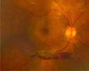
PDR with subhyloid hem1283 viewsPDR with subhyloid hem
|
|

HTN Retinopathy w/ cotton wool spots and hemorrhages1276 views23 year old african american male w/ visual acuity of 20/20
|
|
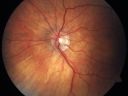
Cilioretinal artery occlusion and Ophthalmic Artery Occlusion with Relative Sparing of the Central Retinal Artery1268 views80-year-old woman who says she was looking in the mirror, covered her right eye and noticed she could not see well out of the left eye. That just happened yesterday. Her last eye check was a few years ago. She also had a carotid endarterectomy on the right side in 2006 shortly after coronary bypass surgery. OD is 20/40, OS is 20/200.
|
|

Ruptured Retinal Arterial Macroaneurysm - Submacular Hemorrhage - Vision 20/400 never recovered - Hypertension 205/941263 views
|
|
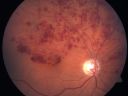
Hemicentral Retinal Vein Occlusion Vision 20/60 1257 views84-year-old man has a hemicentral retinal vein occlusion in the right eye. His vision has declined since then.
VISUAL ACUITY: Vision OD is 20/60.
|
|

Diabetic Retinopathy with CME1257 viewsSecond opinion for diabetic retinopathy. VA is 20/30, right eye, 20/50, left eye. Patient has mild swelling in the macula of the right eye. No evidence of CME but does have a moderate cataract in the left eye.
|
|
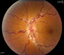
Stasis Retinopathy, Central Retinal Vein Occlusion1256 viewsYoung male who complains of decreased vision in the left eye. VA is 20/20 in the left eye. Fundus photo shows mild central retinal vein occlusion in the left eye.
|
|

Regressed Proliferative Diabetic Retinopathy with Recurrent Vitreous Hemorrhage from Vitreoretinal Traction over a Major Retinal Vessel (Vein)1255 views56-year-old woman has had recurrent vitreous hemorrhage in the left eye from vitreoretinal traction. Diabetes Mellitus for 20 years and excellent response to vitrectomy in the right eye about 1 year ago for recurrent vitreous hemorrhage from traction over a major retinal vessel.
VISUAL ACUITY: Vision OS is 20/200
|
|

Purtchers Retinopathy with Bilateral Central Retinal Artery Occlusions 1252 views50-year-old man traumatic chest compression and leg crushing injury. OD 1/200, OS 3/200
|
|

Retinal Vasculitis due to Lupus (FA)1248 viewsSystemic Lupus Erythematosis - Vasculitis
|
|

Malignant Hypertension - Cotton Wool Spots - Elschnig Spots - Optic Nerve Edema 1244 views
|
|

Preretinal Fibrosis - Tractional Retinal Detachment - Proliferative Diabetic Retinopathy 1233 views33-year-old woman has proliferative diabetic retinopathy in the left eye with fibrosis and tractional retinal detachment. Her vision is hazy in that eye. VISUAL ACUITY: Vision OD is 20/30, OS 20/120
|
|

Diabetic Macular Edema and Hypertensive Retinopathy - Circinate Exudate (Ring Exudate) 1229 views55-year-old woman diabetic for fifteen years and high blood pressure, especially over the last few months. She has had problems with nosebleeds, headaches, and there has been some difficulty bringing her blood pressure down. She said now the blood pressure is under control but it was running 220/120 mmHg for some time. Her vision in the right eye has been poor for four weeks with a spot in the central vision and both eyes have been blurred.
VISUAL ACUITY: Vision OD is 20/160, OS is 20/60
|
|
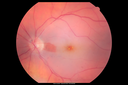
Central Retinal Artery Occlusion with Cilioretinal Sparing - Acute1208 views 77-year-old man was doing fine until midnight of last night when he noticed sudden severe vision loss in the left eye. VISUAL ACUITY: OD 20/20, OS 2/200.
|
|

Mild Proliferative Diabetic Retinopathy1204 viewsRight eye has NVE and left eye has NVD and NVE. VA 20/25 OU.
|
|

A case of Valsalva Retinopathy -11202 views
|
|
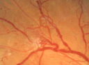
Proliferative Diabetic Retinopathy - Moderate - mild NVD with NVE 1201 viewsVenous beading, vascular loops and NVE are visible on photos
|
|

Disorganized Retinal Inner Layers (DRIL)1201 views72 year old man treated with Avastin for CRVO VA is still 20/200 with absent macular edema (Only 1 month post rx)
|
|

Hemi Central Retinal Vein Occlusion 35 year old non-compliant man1200 views35-year-old man hazy vision in his left eye for about the last three days. OD 20/20, OS 20/30
|
|

Mild Clinically Significant Diabetic Macular Edema1198 views58-year-old woman diabetic for 20 years who has diabetic macular edema in both eyes. She has had waxing and waning edema in the past and I thought it might improve on its own but it did not. She notices her vision is still a little hazy.
VISUAL ACUITY: Vision OD is 20/30, OS is 20/25.
|
|

Ruptured Retinal Arterial Macroaneurysm - Submacular Hemorrhage - Vision 20/400 never recovered - Hypertension 205/941197 views
|
|

Malignant Hypertension - Cotton Wool Spots - Elschnig Spots - Optic Nerve Edema 1196 views6 week fu - CWS are Fading
|
|

Macroaneurysm FP1192 viewsMacroaneurysm with Subretinal Hemorrhage and ME in OS of an Elderly Female
|
|

PDR with heavy PRP laser and worsening macular edema 1191 views51-year-old woman has had extensive laser in both eyes done years ago elsewhere. Her left eye has gradually been declining with macular edema.
VISUAL ACUITY: OD 20/40, OS 20/40
|
|

Retinal Arterial Macroaneurysm - Normal Vision (2 Years before rupture)1190 views78-year-old woman has a retinal arterial macroaneurysm in the right eye. VA 20/25 (pre-rupture photos)
|
|

Regressed Proliferative Diabetic Retinopathy with Recurrent Vitreous Hemorrhage from Vitreoretinal Traction over a Major Retinal Vessel (Vein)1187 views56-year-old woman has had recurrent vitreous hemorrhage in the left eye from vitreoretinal traction. Diabetes Mellitus for 20 years and excellent response to vitrectomy in the right eye about 1 year ago for recurrent vitreous hemorrhage from traction over a major retinal vessel.
VISUAL ACUITY: Vision OS is 20/200
|
|
| 4368 files on 44 page(s) |
1 |
 |
|
|
|
|
|
|
|
|
|


