Most viewed - Retinal Vascular Disease
|

Ischemic CRVO OD and Arterial Macroaneurysm OS510 views83 year old female post PPV, Laser, Ahmed valve for CRVO with vitreous heme and NVG in the right eye. VA was 20/160 3 months following vitrectomy. Left eye has MA which was not treated and involuted post-heme
|
|

Severe PDR with preretinal fibrosis510 views71 year old female untreated for a year following injections because of hurricane Irma. VA 20/30 OD and 5/200 OS. Sudden vision loss 1 month ago left eye was likely an ischemic event. Right eye is being treated with injections and PRP laser.
|
|

Branch Retinal Artery Occlusion - No Plaque510 views74 year old man with 2 days of vision loss. Vision OS is 20/200 but improved to 20/25 in 1 month. He has dysfibrinogen anemia.
|
|

509 views49-year-old woman has central retinal vein occlusion right eye with fluctuating vision for a few month now with OD 20/100. IOP: OD 20
|
|

Branch Retinal Arterial Occlusion 20/20 Vision - Symptoms for 1 Day509 views75-year-old man was seen in the office on 4/6/2010. He noticed a sudden vision change in the right eye yesterday morning. 20/20 Vision both Eyes
|
|

Branch Retinal Arterial Occlusion 20/20 Vision - Symptoms for 1 Day509 views75-year-old man was seen in the office on 4/6/2010. He noticed a sudden vision change in the right eye yesterday morning. 20/20 Vision both Eyes
|
|

Macular Branch Retinal Vein Occlusion and Adult Vitelliform Dystrophy509 views72-year-old woman 2 months post ECCE with poor vision since surgery especially left eye.
VISUAL ACUITY: OD 20/50, OS 20/200
|
|
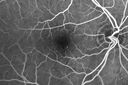
Diabetic Macular Edema - Mild509 views70 year old man with mild edema did well without treatment
|
|

Fibrosis Traction509 viewsYoung female patient with severe diabetes. Patients VA is 20/400 in the right eye. Fundus photos show a lot of fibrosis traction throughout the macula area. Fovea seems flat. Patient will undergo surgery for peeling of traction.
|
|

Proliferative Diabetic Retinopathy - Moderate - mild NVD with NVE 509 viewsFA shows leakage from neovascularization and dark areas in periphery from non-perfusion
|
|

Self Sealed Retinal Arteriol Macroaneurysm- one month after presentation - edema nearly gone509 viewsSD OCT line scan
|
|

PDR NVD left eye and NVE right eye509 views41 year old diabetic woman with PDR in both eyes. High risk in the left eye. She had PRP, Avastin, and vitrectomy x 2 in the left eye with final vision of 20/25. The right eye had eventually PRP laser
|
|

Vascular Remodeling Bilateral BRVO509 views85 year old female 20/25 OU with BRVO 7 years ago in both eyes. She has a change in vision. The FA shows extensive collaterallization and vascular remodeling in each macula.
|
|

Central Retinal Artery Occlusion then Neovascular Glaucoma509 views81 year old male with sudden vision loss. 2 months later developed NVG
|
|

White Retinal Artiole Left eye - Inferotemporal509 views31 year old female with migraines and headaches for the last 12-13 years. Sometimes she gets the visual symptoms with the migraine. When she gets the migraines the pain is on the left side of her head. She gets the problem a few times a month, sometimes more. They usually last 5-6 hours. She has not had a permanent vision change. When she gets a vision change there are spotty dots of blue neon lights in her vision. With her glasses her two eyes are about the same. VA 20/16 in Each Eye
|
|

Twig Old Macular Branch Retinal Vein Occlusion - Venous Venous Collaterals509 views88 year old man with 20/25 vision in his left and only eye. There is vascular remodeling consistent with an old twig BRVO.
|
|

Branch Retinal Artery Occlusion - No Plaque509 views74 year old man with 2 days of vision loss. Vision OS is 20/200 but improved to 20/25 in 1 month. He has dysfibrinogen anemia.
|
|

508 views49-year-old woman has central retinal vein occlusion right eye with fluctuating vision for a few month now with OD 20/100. IOP: OD 20
|
|

AIDS - HIV Retinopathy - Cotton Wool Spots508 views54-year-old woman HIV positive for 18 years. Her CD4 count is currently less than 50 and she was hospitalized for pneumocystis carinii pneumonia 5 months ago. She has noticed just for the last few weeks a few blind spots in the paracentral vision. Predominantly out of her right eye when she closes her left eye, she sees a few temporal blind spots. VA 20/25 in both eyes.
|
|

Post-Op Vitrectomy with Membrane Stripping and Laser508 viewsPatient had surgery to help clear up some vision in the left eye. Pre-op VA was count fingers at 1-ft. Post op VA was 20/200 in the left eye. Patient will return in 3-months for follow-up.
|
|

Ischemic BRVO with non-perfusion (nonperfusion) - recovered from severe vitreous hemorrhage508 views
|
|

Hairy Cell Leukemia - Retinal Hemorrhage and twig Branch Vein Occlusion508 views79 year old man He has had hairy cell leukemia since 2002. He is in remission. His last blood tests were 9/2018. He just moved down here and needs a new leukemia doctor. His vision is fine.
VA OD: Dcc20/25
VA OS: Dcc20/25
IOP: TP: OD:12 OS:12
|
|

Hairy Cell Leukemia - Retinal Hemorrhage and twig Branch Vein Occlusion508 views79 year old man He has had hairy cell leukemia since 2002. He is in remission. His last blood tests were 9/2018. He just moved down here and needs a new leukemia doctor. His vision is fine.
VA OD: Dcc20/25
VA OS: Dcc20/25
IOP: TP: OD:12 OS:12
|
|

Branch Retinal Arterial Occlusion Right Eye - Atheromata - Both Eyes508 views83 year old woman with no symptoms - VA 20/40 in both eyes. Right eye has ST BRAO with silver wiring. Left eye has superotemporal atheromata
|
|

White Retinal Artiole Left eye - Inferotemporal508 views31 year old female with migraines and headaches for the last 12-13 years. Sometimes she gets the visual symptoms with the migraine. When she gets the migraines the pain is on the left side of her head. She gets the problem a few times a month, sometimes more. They usually last 5-6 hours. She has not had a permanent vision change. When she gets a vision change there are spotty dots of blue neon lights in her vision. With her glasses her two eyes are about the same. VA 20/16 in Each Eye
|
|

Moderate PDR508 views43 year old man 20/32 vision in both eyes. Being started on Anti-VEGF injections which will be followed by PRP.
|
|

PDR - Mild Vitreous Hemorrhage and Good Vision507 views58-year-old woman has diabetic retinopathy in both eyes with neovascularization of the optic nerve, worse in the right eye than the left eye. She has had panretinal laser in both eyes and vitreous hemorrhage in the left eye. Her vision, since I saw her last in June, is about the same.
VISUAL ACUITY: Vision OD is 20/25, OS is 20/20.
|
|

Proliferative Diabetic Retinopathy both eyes Type I Diabetic for 25 years507 views47-year-old woman with diabetes for 25 years and decreased vision for 3 months. OD 20/40, OS 20/40
|
|

Diabetic Macular Edema Left Eye - Pre Laser507 views82-year-old woman diabetic for many years, last eye exam 5 years ago with gradual vision loss. OD 20/60, OS 20/70. IOP: OD 16, OS 17.
|
|

Diabetic Macular Edema Left Eye - Pre Laser507 views82-year-old woman diabetic for many years, last eye exam 5 years ago with gradual vision loss. OD 20/60, OS 20/70. IOP: OD 16, OS 17.
|
|
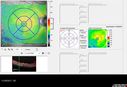
507 views
|
|

Microaneursyms and Macular Edema Not Diabetic507 views85 year old man has been tested about 2-3 times and is always negative for diabetes
|
|

White Retinal Artiole Left eye - Inferotemporal507 views31 year old female with migraines and headaches for the last 12-13 years. Sometimes she gets the visual symptoms with the migraine. When she gets the migraines the pain is on the left side of her head. She gets the problem a few times a month, sometimes more. They usually last 5-6 hours. She has not had a permanent vision change. When she gets a vision change there are spotty dots of blue neon lights in her vision. With her glasses her two eyes are about the same. VA 20/16 in Each Eye
|
|

Vitreous Hemorrhage with no evidence of PDR in the left eye507 views74 year old man with vision loss OS for about a week. The FA shows no PDR in the left eye but the right eye has very mild NVD. Diabetes for 40 years now on insulin.
|
|

PDR and Vitreous Hemorrhage - High Risk Left Eye - Low Risk Right Eye507 views50 year old man with type I diabetes mellitus for 26 years. New Vitreous Hemorrhage in the left eye. Both eyes have NVE. Both also have foveal hypoplasia
|
|

Central Retinal Vein Occlusion - New- and old Macular Scar from AMD506 views87-year-old woman with longstaning macular scars and new vision loss left eye for 2 months. OD: 20/200, OS 20/400.
|
|

Severe Hemorrhage from NVD and NVE Due To PDR506 views60 year old female with Diabetic Retinopathy. VA 20/40 OD HM OS. History of Dot and Blot hemorrhages and cotton-wool spots associated with Diabetes. Severe Hemorrhaging due to NVD and NVE. Pt stated sight decreased very rapidly.
|
|

Fluorescence Angiography of BDR506 viewsPatient is here for eval on diabetic retinopathy. FA photos show significant diabetic retinopathy with hemorrhage inferior temporal to the optic nerve.
|
|

Sickle Retinopathy - Ultra Wide Field Angiogram - Heidelberg506 viewsLeakage from peripheral fronds of NV - HGB SC disease - 28 year old man with vision loss OS from acute vitreous hemorrhage
|
|

HIV Retinopathy506 viewsRetinal edema from microvascular disease
|
|

Acute Branch Retinal Artery Occlusion Left Eye506 viewsThis 84 year old woman had an old BRVO in the right eye and saw a shadow for 2 days in the left eye. She has a superotemporal BRAO. The photos show retinal whitening, the OCT shows inner retinal edema and the FA shows poor circulation.
|
|

Central retinal artery and vein occlusion - Protein C Deficiency 33 year old male506 views33 year old male with vision loss for 3 hours. VA 20/12 right eye, 5/200 left eye. He has a protein C deficiency. The left eye recovered vision in about 1 month to 20/16. FA shows very slow recirculation time.
|
|

PDR NVD left eye and NVE right eye506 views41 year old diabetic woman with PDR in both eyes. High risk in the left eye. She had PRP, Avastin, and vitrectomy x 2 in the left eye with final vision of 20/25. The right eye had eventually PRP laser
|
|

Branch Retinal Arterial Occlusion Right Eye - Atheromata - Both Eyes506 views83 year old woman with no symptoms - VA 20/40 in both eyes. Right eye has ST BRAO with silver wiring. Left eye has superotemporal atheromata
|
|

Proliferative Diabetic Retinopathy - NVD regressed with PRP laser506 views76 year old diabetic man - Presented one year ago with NVD in the left eye. This regressed with laser but then a year later worsened. Additional PRP was done and the NVD regressed again.
|
|

Subtle Branch Retinal Artery Occlusion506 views68 year old man with shadow in the left eye yesterday. IR and OCT show subtle retinal swelling. FA is normal
|
|
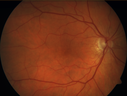
Cilioretinal Artery Occlusion - Nasal (not macular)506 views77 year old female There is a shadow in the top half of her temporal vision in the left eye that came on suddenly yesterday morning. It has not changed since then and the right eye is OK. She has had sinus headaches. She had no focal neurological changes. Â
VA OD: Dcc20/32
VA OS: Dcc20/16-1
IOP: TP: OD:18 OS:19Â
|
|

Central Retinal Vein Occlusion - New- and old Macular Scar from AMD505 views87-year-old woman with longstaning macular scars and new vision loss left eye for 2 months. OD: 20/200, OS 20/400.
|
|

Branch Retinal Arterial Occlusion 20/20 Vision - Symptoms for 1 Day505 views75-year-old man was seen in the office on 4/6/2010. He noticed a sudden vision change in the right eye yesterday morning. 20/20 Vision both Eyes
|
|

Hypertensive Retinopathy505 viewsPatient comes in complaining of spots in vision in both eyes. VA was 20/25 - right eye and 20/20- left eye. Fundus exam reveals little hemorrhages with cotton wool spots due to hypertension and anemia.
|
|
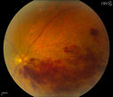
Branch Retinal Vein Occlusion505 viewsPatient's VA was 20/40, right eye. Fundus exam shows branch retinal vein occlusion nasally to the optic nerve in the right eye.
|
|

HIV Retinopathy505 viewsFAF
|
|

Branch Retinal Artery Occlusion505 viewsAcute Inferotemporal Occlusion Right Eye - FA shows delayed circulation, OCT shows edema of superficial layers of retina
|
|

Bilateral Branch Retinal Artery Occlusion - Intraarterial (hollenhorst) plaques visible505 views67 year old with acute visual field loss in each eye. Vision is 20/25 right eye 20/32 left eye. Images show intra-arterial plaques in each eye. The right eye on the optic nerve and the left eye in a branch retinal artery.
|
|

Bilateral Branch Retinal Artery Occlusion - Intraarterial (hollenhorst) plaques visible505 views67 year old with acute visual field loss in each eye. Vision is 20/25 right eye 20/32 left eye. Images show intra-arterial plaques in each eye. The right eye on the optic nerve and the left eye in a branch retinal artery.
|
|

CRVO with Paracentral Acute Middle Maculopath - PAMM505 views56 year old woman with sudden vision loss, hemorrhages in all four quadrants. FA shows good retinal circulation. The OCT shows PAMM lesions in the affected eye. Vision did improve on second visit from 20/160 to 20/60 in about a month
|
|

Intraluminal Plaque Retinal Vessel 505 views69 year old female referred with atypical plaque in the right retina. Her vision is normal. The plaque looks to be adjacent to the blood column visible on the images and the SC OCT shows it to be in the vessel wall.
|
|

Macroaneurysm - Resolved without treatment505 views87 year old female with vision loss OD. Initial FA showed no leakage so no treatment was done and the fluid absorbed over 4 months. Initial VA 20/100, Final VA 20/80
|
|

Grade 4 hypertensive retinopathy505 views55 year old man Hhe did have headaches a few weeks ago and also some nausea and vomiting. He noticed for a few weeks the vision in the left eye was blurred and that the vision in the left eye is dark.
VA OD: Dcc20/20
VA OS: Dcc20/100-1
He was admitted directly to ICU for blood pressure control (290 / 170 mmHg)
3 weeks later his VA increased to 20/20 OD, 20/32 OS
|
|

Central Retinal Vein Occlusion - No macular edema - Pre-foveal opercula505 views62 year old female with 20/20 vision. She had been seeing a spot out of the left eye for 4 years and then noticed splotches in her vision. She never devleoped edema or NV
|
|

Acute Central Retinal Artery Occlusion505 views1 day of vision loss 79 year old man to HM. NVG developed 2 months later.
|
|

Moderate PDR505 views43 year old man 20/32 vision in both eyes. Being started on Anti-VEGF injections which will be followed by PRP.
|
|

Diabetic Macular Ischemia - Enlarged FAZ - OCT-A505 views70 year old female diagnosed with diabetes 2 years ago. Her blood pressure was normal at 116/73. She has vision loss for 6 months in both eyes. VA is 20/100 OU. OCT-A shows macular ischemia.
|
|
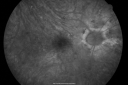
Diabetic Macular Edema with Circinate Exudate Fellow Eye FA504 views74-year-old man has diabetic macular edema in both eyes and background diabetic retinopathy. His vision has blurred over the last few months. His blood sugars have been fluctuating.
VISUAL ACUITY: Vision OD is 20/25, OS is 20/80
|
|

Branch Retinal Arterial Occlusion 20/20 Vision - Symptoms for 1 Day504 views75-year-old man was seen in the office on 4/6/2010. He noticed a sudden vision change in the right eye yesterday morning. 20/20 Vision both Eyes
|
|

Central Retinal Vein Occlusion Recurrent Edema 6 weeks after Lucentis - Nonperfusion Temporally504 views66-year-old woman has a central retinal vein occlusion in the right eye with macular edema. She had intravitreal Lucentis treatment six weeks ago and she has noticed over the last week or two her vision is declining.
VISUAL ACUITY: OD 20/70, OS 20/30.
|
|

Hypertensive Retinopathy504 viewsPatient comes in complaining of spots in vision in both eyes. VA was 20/25 - right eye and 20/20- left eye. Fundus exam reveals little hemorrhages with cotton wool spots due to hypertension and anemia.
|
|

Hairy Cell Leukemia - Retinal Hemorrhage and twig Branch Vein Occlusion504 views79 year old man He has had hairy cell leukemia since 2002. He is in remission. His last blood tests were 9/2018. He just moved down here and needs a new leukemia doctor. His vision is fine.
VA OD: Dcc20/25
VA OS: Dcc20/25
IOP: TP: OD:12 OS:12
|
|

PDR NVD left eye and NVE right eye504 views41 year old diabetic woman with PDR in both eyes. High risk in the left eye. She had PRP, Avastin, and vitrectomy x 2 in the left eye with final vision of 20/25. The right eye had eventually PRP laser
|
|

Branch Retinal Arterial Occlusion Right Eye - Atheromata - Both Eyes504 views83 year old woman with no symptoms - VA 20/40 in both eyes. Right eye has ST BRAO with silver wiring. Left eye has superotemporal atheromata
|
|

Venous Macroaneurysm 6 year old CRVO - treated with focal laser504 views93 year old female with CRVO in the right eye 6 years ago. The visual potential is 20/100 and has recently subjectively and objectively declined. VA is 20/200 and she has new onset macular edema from a venous macroaneurysm that formed at a collateral vessel. This responded nicely to a single laser spot.
|
|

Non-ischemic Central Retinal Vein Occlusion with Macular Edema504 views62 year old man The vision in the right eye started to decline about 10 days ago. Prior to that the two eyes were about the same.
VA OD: Dcc20/125 PH20/50-2
VA OS: Dcc20/20
IOP: TP: OD:21 OS:21
|
|
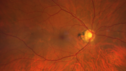
Branch retinal vein occlusion with macular edema504 views71 year old man Starting about 2 weeks ago the left eye was blurred. The right eye is seeing well.
VA OD: sc20/25+2 NccJ1+
VA OS: sc20/63+2 PH20/50-2 Ncc20/400
IOP: TP: OD:19 OS:16
|
|
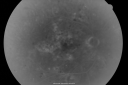
BDR and Diabetic Macular Edema with Fresh Laser Right Eye503 views60-year old man OD 20/60, OS 20/50 worsening vision both eyes
|
|

503 views49-year-old woman has central retinal vein occlusion right eye with fluctuating vision for a few month now with OD 20/100. IOP: OD 20
|
|

Central Retinal Vein Occlusion - New- and old Macular Scar from AMD503 views87-year-old woman with longstaning macular scars and new vision loss left eye for 2 months. OD: 20/200, OS 20/400.
|
|

Branch Retinal Vein Occlusion with Cotton Wool Spot 503 views70-year-old man with blurred vision in the right eye for about one or two months. OD is 20/80, OS is 20/25.
|
|

Panretinal (Scatter) Photocoagulation (Fresh)503 views
|
|

Sickle Retinopathy - Ultra Wide Field Angiogram - Heidelberg503 viewsLeakage from peripheral fronds of NV - HGB SC disease - 28 year old man with vision loss OS from acute vitreous hemorrhage
|
|

Hairy Cell Leukemia - Retinal Hemorrhage and twig Branch Vein Occlusion503 views79 year old man He has had hairy cell leukemia since 2002. He is in remission. His last blood tests were 9/2018. He just moved down here and needs a new leukemia doctor. His vision is fine.
VA OD: Dcc20/25
VA OS: Dcc20/25
IOP: TP: OD:12 OS:12
|
|

Orbital Cellulitis - Optic nerve compression with ocular ischemia503 views81 year old woman with constricted eye movement, orbital cellulitis and severe vision loss in the right eye (LP). She has whitening of the retina and the OCT shows ischemic changes as well as subretinal fluid and blood. The vision in this eye declined to NLP and the eye remained ischemic despite control of her infection.
|
|

Proliferative Diabetic Retinopathy - NVD regressed with PRP laser503 views76 year old diabetic man - Presented one year ago with NVD in the left eye. This regressed with laser but then a year later worsened. Additional PRP was done and the NVD regressed again.
|
|

White Retinal Artiole Left eye - Inferotemporal503 views31 year old female with migraines and headaches for the last 12-13 years. Sometimes she gets the visual symptoms with the migraine. When she gets the migraines the pain is on the left side of her head. She gets the problem a few times a month, sometimes more. They usually last 5-6 hours. She has not had a permanent vision change. When she gets a vision change there are spotty dots of blue neon lights in her vision. With her glasses her two eyes are about the same. VA 20/16 in Each Eye
|
|

Branch Retinal Artery Occlusion - No Plaque503 views74 year old man with 2 days of vision loss. Vision OS is 20/200 but improved to 20/25 in 1 month. He has dysfibrinogen anemia.
|
|

Macroaneurysm - Macular Hemorrhage - Branch Retinal Aterial Occlusion503 views62 year old African American female with chronic hypertension and vision loss for 2 weeks. VA is 20/400
|
|

Macroaneurysm - Macular Hemorrhage - Branch Retinal Aterial Occlusion503 views62 year old African American female with chronic hypertension and vision loss for 2 weeks. VA is 20/400
|
|

Chronic BRAO with Hemorrhages - Sclerosed Vessel503 views95 year old female with blurred vision in both eyes in for a checkup. History of perpapillary CNVM in the left eye 2 years prior not treated. History of stroke with multiple emboli 1 year ago.
Vision 20/32 OD, 20/40 OS
|
|

Cilioretinal Artery Occlusion - Good vision503 views67 year old female - 5 days ago she was getting ready for bed and closed one eye (she had worked for an optometrist for 20 years) and she noticed a spot in the vision in the right eye and came in. She has no focal neurological problems. She once in a while gets some weakness. She has a neurologist and there are some brain lesions. Father had stroke at age 52.
VA OD: Dcc20/20 NccJ1
VA OS: Dcc20/16-2 NccJ1-2
|
|
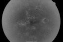
BDR and Diabetic Macular Edema with Fresh Laser Right Eye502 views60-year old man OD 20/60, OS 20/50 worsening vision both eyes
|
|

502 views49-year-old woman has central retinal vein occlusion right eye with fluctuating vision for a few month now with OD 20/100. IOP: OD 20
|
|

Diabetic Macular Edema Left Eye - Pre Laser502 views82-year-old woman diabetic for many years, last eye exam 5 years ago with gradual vision loss. OD 20/60, OS 20/70. IOP: OD 16, OS 17.
|
|

Proliferative Diabetic Retinopathy in patient with Moyamoya Disease502 views28-year-old womandiabetic since age nine, and she also has had multiple strokes. OD is 20/30, OS is 20/30.
|
|

Macular Atrophy - Macular Degeneration and Previous Diabetic Macular Laser 72 Year Old Man502 views72-year-old man has age-related macular degeneration who had macular and peripheral laser done 30 years ago for diabetic retinopathy. He has developed worsening macular atrophy in both eyes
|
|

Proliferative Diabetic Retinopathy Both Eyes - Vitreous Hemorrahge Left Eye502 views67-year-old man has proliferative diabetic retinopathy in both eyes with vitreous hemorrhage in the left eye. He has had pan retinal laser in the left eye. The vitreous hemorrhage has been there for greater than two years and his vision is still hazy. He takes Lumigan in both eyes for his glaucoma.
VISUAL ACUITY: OD 20/40, OS 20/40
|
|
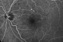
Diabetic Macular Edema - Mild502 views70 year old man with mild edema did well without treatment
|
|

Branch Retinal Vein Occlusion with worse macular edema from hypertension - Photo from 2014 - vision 20/40502 views
|
|

Branch Retinal Vein Occlusion with worse macular edema from hypertension - Photo from 2014 - vision 20/40502 views
|
|

Branch Retinal Vein Occlusion with NVE (Neovascularization Elsewhere)502 views
|
|
502 views
|
|

Branch Retinal Arterial Occlusion Right Eye - Atheromata - Both Eyes502 views83 year old woman with no symptoms - VA 20/40 in both eyes. Right eye has ST BRAO with silver wiring. Left eye has superotemporal atheromata
|
|
| 4416 files on 45 page(s) |
 |
|
|
|
|
20 | |
|
|
|
|


