Most viewed - Retinal Vascular Disease
|

Pathology - Central Retinal Artery Occlusion - 6 weeks old - Inner Retinal Atrophy - Macular Pucker913 viewspatient died about 6 to 8 weeks after the artery occlusion, and he had no history of glaucoma. The inner retina atrophy, which looks very similar to that caused by COAG, is due to recent infarct.
|
|

Diabetic Retinopathy - Foveal Laser Scar Right Eye912 views74-year-old woman has diabetic retinopathy in both eyes. She has had focal laser in her right eye done elsewhere in the mid 90’s and unfortunately the laser was fairly close to the center and as time progressed she developed foveal atrophy around the scars. She has noticed her vision declining since she was here last a year ago. She does take Travatan in both eyes at night for glaucoma.
VISUAL ACUITY: OD 20/200, OS 20/40.
|
|

Central Retinal Artery Occlusion with Cilioretinal Sparing - Acute910 views 77-year-old man was doing fine until midnight of last night when he noticed sudden severe vision loss in the left eye. VISUAL ACUITY: OD 20/20, OS 2/200.
|
|

branch retinal vein occlusion - red free photo907 views70 year old woman with 20/80 vision from a branch retinal vein occlusion and macular edema. She has had laser treatment and intravitreal steroids. Her vision with further laser improved to 20/50.
|
|

Central Retinal Artery Occlusion with Cilioretinal Sparing - Acute - FA Very Slow Filling CRA907 views 77-year-old man was doing fine until midnight of last night when he noticed sudden severe vision loss in the left eye. VISUAL ACUITY: OD 20/20, OS 2/200.
|
|

Retinal Arterial Macroaneurysm - Increased Swelling after Laser 906 views65-year-old woman was seen in the office on January 12, 2011. She has noticed decreased vision in the right eye for the last two or three weeks. She has had some discomfort in both eyes as well. She has a history of low blood pressure, but she does have high cholesterol.
VISUAL ACUITY: OD 20/60
|
|

Neovascularization elsewhere (NVE) Left Eye with Vitreous Hemorrhage904 views
|
|

Macular Branch Retinal Vein Occlusions (2) Left Eye - No Macular Edema (yet) 902 views77-year-old woman with two macular branch retinal vein occlusions in the left eye OD 20/25
|
|

Central Retinal Artery Occlusion with Cilioretinal Sparing - Acute - FA Very Slow Filling CRA900 views 77-year-old man was doing fine until midnight of last night when he noticed sudden severe vision loss in the left eye. VISUAL ACUITY: OD 20/20, OS 2/200.
|
|

Retinal Arterial Macroaneurysm - Ruptured - Vitreous Hemorrhage (Fresh) 5/200 Vision898 views80-year-old woman has a retinal arterial macroaneurysm in the right eye I saw two years ago. She noticed the sudden onset of vision loss last night. OD is 5/200.
|
|

Mild Clinically Significant Diabetic Macular Edema896 views58-year-old woman diabetic for 20 years who has diabetic macular edema in both eyes. She has had waxing and waning edema in the past and I thought it might improve on its own but it did not. She notices her vision is still a little hazy.
VISUAL ACUITY: Vision OD is 20/30, OS is 20/25.
|
|

Serous Retinal Detachment - Hypertensive Retinopathy - 165/120 - Fundus Autofluorescence (FAF)895 views48-year-old woman noticed when she was blinking there was a white flash in the left eye starting about a month ago. She sees it temporally. OD 20/16, OS 20/20. There is a serous retinal detachment superotemporal to the optic nerve falling short of the fovea. Her blood pressure 165/120.
|
|

Ruptured Retinal Arterial Macroaneurysm - Submacular Hemorrhage - Vision 20/400 never recovered - Hypertension 205/94894 views
|
|

Pathology - Central Retinal Artery Occlusion - 6 weeks old - Inner Retinal Atrophy - Macular Pucker894 viewspatient died about 6 to 8 weeks after the artery occlusion, and he had no history of glaucoma. The inner retina atrophy, which looks very similar to that caused by COAG, is due to recent infarct.
|
|
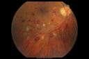
Hairy Cell Leukemia - Retinal Hemorrhage and twig Branch Vein Occlusion893 views79 year old man He has had hairy cell leukemia since 2002. He is in remission. His last blood tests were 9/2018. He just moved down here and needs a new leukemia doctor. His vision is fine.
VA OD: Dcc20/25
VA OS: Dcc20/25
IOP: TP: OD:12 OS:12
|
|

telangiectasia887 views20/20 pt with blurry area in va
|
|

Proliferative Diabetic Retinopathy and Mild Vitreous Hemorrhage885 views58-year-old woman has diabetic retinopathy in both eyes with neovascularization of the optic nerve, worse in the right eye than the left eye. Vision OD is 20/25, OS is 20/20
|
|
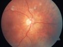
AIDS - HIV Retinopathy - Cotton Wool Spots885 views54-year-old woman HIV positive for 18 years. Her CD4 count is currently less than 50 and she was hospitalized for pneumocystis carinii pneumonia 5 months ago. She has noticed just for the last few weeks a few blind spots in the paracentral vision. Predominantly out of her right eye when she closes her left eye, she sees a few temporal blind spots. VA 20/25 in both eyes.
|
|

Purtscher's Retinopathy884 views19 year old female after pregnancy
|
|

Proliferative Diabetic Retinopathy both eyes Type I Diabetic for 25 years882 views47-year-old woman with diabetes for 25 years and decreased vision for 3 months. OD 20/40, OS 20/40
|
|
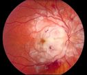
Purtscher's Retinopathy882 views19 year old female after pregnancy
|
|

Branch Retinal Arterial Occlusion 20/20 Vision - Symptoms for 1 Day881 views
|
|

Retinal Arterial Macroaneurysm - Ruptured and self Sealed - Vision Back to 20/80 after 6 Months881 views81-year-old woman vision improving 6 months following rupture of retinal arterial macroaneurysm. Vision OD is 20/80
|
|

Classic BRVO881 viewsClassic branch retinal vein occlusion
|
|

Three years after Injury - Optic Atrophy - Vision is 1/200 in each eye880 views
|
|

Microaneurysms from Ocular Ischemic Syndrome (non-diabetic) Right Eye - Left eye CRAO 1 year ago880 views66-year-old woman lost vision in the left eye from a central retinal artery occlusion 1 year ago. Vision OD is fine. OD 20/25, OS 20/50
|
|

NVD880 viewsDiabetic patient with NVD and NVE
|
|

Cilioretinal artery occlusion and Ophthalmic Artery Occlusion with Relative Sparing of the Central Retinal Artery879 views80-year-old woman who says she was looking in the mirror, covered her right eye and noticed she could not see well out of the left eye. That just happened yesterday. Her last eye check was a few years ago. She also had a carotid endarterectomy on the right side in 2006 shortly after coronary bypass surgery. OD is 20/40, OS is 20/200.
|
|

Retinal Arterial Macroaneurysm - Untreated Hypertension (250/135) - Serous Macular Detachment878 views82 year old asian woman. VA 20/80 Headaches and vision loss for 1 month. Negative medical history but has not been to the doctor for years. (no insurance)
|
|

Macular and Peripheral Retinal Scarring from Diabetic Laser Done 25 years previously877 views72-year-old man had macular and peripheral laser done in the past(1980's)(25 years ago). His vision, although limited, seems about stable to him. He is diabetic for 30 years. OD is 20/160, OS is 20/160.
|
|

Proliferative Diabetic Retinopathy - Moderate - mild NVD with NVE 876 viewsVenous beading, vascular loops and NVE are visible on photos
|
|

Malignant Hypertension - Cotton Wool Spots - Elschnig Spots - Optic Nerve Edema (NFL Scan - Black Line this visit, Gray line 6 weeks ago)874 viewsNerve Swelling is Less
|
|

Purtchers Retinopathy with Bilateral Central Retinal Artery Occlusions 873 views50-year-old man traumatic chest compression and leg crushing injury. OD 1/200, OS 3/200
|
|

Retinal Arterial Macroaneurysm - Increased Swelling after Laser 873 views65-year-old woman was seen in the office on January 12, 2011. She has noticed decreased vision in the right eye for the last two or three weeks. She has had some discomfort in both eyes as well. She has a history of low blood pressure, but she does have high cholesterol.
VISUAL ACUITY: OD 20/60
|
|

Non-Ischemic Central Retinal Vein Occlusion in Young Man (30 years) - Many Cotton Wool Spots873 views30-year-old man with a history of a central retinal vein occlusion for which he was first examined on 2 months ago and his vision then was 4/200. He responded nicely to two Avastin treatments. OD is 20/20, OS is 20/60
|
|

Sectoral Optic Atrophy from an old Branch Retinal Artery Occlusion 873 views74-year-old woman had cataract surgery 3 months ago. Her vision in the right eye has been abnormal since. She notices a shadow in the inferior vision in the right eye. OD is 20/60, OS is 20/50
|
|

Serous Retinal Detachment - Hypertensive Retinopathy - 165/120 - Fluorescein Angiogram872 views48-year-old woman noticed when she was blinking there was a white flash in the left eye starting about a month ago. She sees it temporally. OD 20/16, OS 20/20. There is a serous retinal detachment superotemporal to the optic nerve falling short of the fovea. Her blood pressure 165/120.
|
|

Hemicentral Retinal Vein Occlusion Vision 20/60 871 views84-year-old man has a hemicentral retinal vein occlusion in the right eye. His vision has declined since then.
VISUAL ACUITY: Vision OD is 20/60.
|
|

BRVO arrow showing obstruction point with collateral vessels - red free photo870 views70 year old woman with 20/80 vision from a branch retinal vein occlusion and macular edema. She has had laser treatment and intravitreal steroids. Her vision with further laser improved to 20/50.
|
|

Diabetic Macular Edema and Hypertensive Retinopathy - Circinate Exudate (Ring Exudate) 870 views55-year-old woman diabetic for fifteen years and high blood pressure, especially over the last few months. She has had problems with nosebleeds, headaches, and there has been some difficulty bringing her blood pressure down. She said now the blood pressure is under control but it was running 220/120 mmHg for some time. Her vision in the right eye has been poor for four weeks with a spot in the central vision and both eyes have been blurred.
VISUAL ACUITY: Vision OD is 20/160, OS is 20/60
|
|

Malignant Hypertension - Cotton Wool Spots - Elschnig Spots - Optic Nerve Edema 867 views6 week fu - CWS are Fading
|
|

Central Retinal Artery Occlusion - 2 months old by history863 views77-year-old man was doing fine. His left eye was a little worse than the right eye, and then in the 2 months ago he had sudden severe vision loss in the right eye. Vision OD is 3/200; OS is 20/60. IOP: OD 30, OS 15.
OD: Vertical C/D ratio is 0.4. The macula is white from ischemia. The arteries are narrow.
Photos confirm clinical findings.
OCT SCAN: OCT scan of the right eye shows retinal thickening, consistent with macular edema associated with a central retinal artery occlusion. The left eye has severe inferior retinal thinning, consistent with an old hemicentral retinal artery occlusion in the left eye. The fovea in the left eye is a little bit thin but not too bad.
IMPRESSION: 1. CENTRAL RETINAL ARTERY OCCLUSION RIGHT EYE
2 OLD HEMICENTRAL RETINAL ARTERY OCCLUSION LEFT EYE
3 DIABETES WITHOUT RETINOPATHY
4 ELEVATED INTRAOCULAR PRESSURE RIGHT EYE STATUS POST INJECTION RIGHT EYE A WEEK AGO
DISCUSSION: I explained to the patient that the right eye unfortunately has had a central retinal artery and unfortunately we do not have anything to treat that. His vision will probably improve some over time. He needs to be watched though. It is possible he is developing rubeotic glaucoma, although it is probable the intraocular pressure is high from the injection last week. I asked him to restart the Alphagan, to stop the other two drops, and to return here in three weeks to make sure his pressure is more acceptable.
At this point, having lost almost all the retinal circulation in the right eye and half of the retinal circulation in the left eye, he only has half a retina in the left eye he is working off of. I told him it is very important he keep seeing you and anything that can be done to keep his blood pressure low and his cholesterol reasonable would be helpful to what remains of his retinal circulation.
|
|

Pathology - Central Retinal Artery Occlusion - 6 weeks old - Inner Retinal Atrophy863 viewspatient died about 6 to 8 weeks after the artery occlusion, and he had no history of glaucoma. The inner retina atrophy, which looks very similar to that caused by COAG, is due to recent infarct.
|
|

Regressed Proliferative Diabetic Retinopathy with Recurrent Vitreous Hemorrhage from Vitreoretinal Traction over a Major Retinal Vessel (Vein)863 views56-year-old woman has had recurrent vitreous hemorrhage in the left eye from vitreoretinal traction. Diabetes Mellitus for 20 years and excellent response to vitrectomy in the right eye about 1 year ago for recurrent vitreous hemorrhage from traction over a major retinal vessel.
VISUAL ACUITY: Vision OS is 20/200
|
|

Sectoral Optic Atrophy from an old Branch Retinal Artery Occlusion 860 views74-year-old woman had cataract surgery 3 months ago. Her vision in the right eye has been abnormal since. She notices a shadow in the inferior vision in the right eye. OD is 20/60, OS is 20/50
|
|

Cilioretinal artery occlusion and Ophthalmic Artery Occlusion with Relative Sparing of the Central Retinal Artery859 views80-year-old woman who says she was looking in the mirror, covered her right eye and noticed she could not see well out of the left eye. That just happened yesterday. Her last eye check was a few years ago. She also had a carotid endarterectomy on the right side in 2006 shortly after coronary bypass surgery. OD is 20/40, OS is 20/200.
|
|
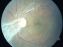
Preretinal Fibrosis - Tractional Retinal Detachment - Proliferative Diabetic Retinopathy 855 views33-year-old woman has proliferative diabetic retinopathy in the left eye with fibrosis and tractional retinal detachment. Her vision is hazy in that eye. VISUAL ACUITY: Vision OD is 20/30, OS 20/120
|
|

Cilioretinal artery occlusion and Ophthalmic Artery Occlusion with Relative Sparing of the Central Retinal Artery854 views80-year-old woman who says she was looking in the mirror, covered her right eye and noticed she could not see well out of the left eye. That just happened yesterday. Her last eye check was a few years ago. She also had a carotid endarterectomy on the right side in 2006 shortly after coronary bypass surgery. OD is 20/40, OS is 20/200.
|
|

Retinal Arterial Macroaneurysm - Recurrent Hemorrhage 6 months post laser853 views6 months post laser: Her vision had improved, but then three weeks ago it worsened again. She is not on any blood thinners. She wasn’t doing any heavy lifting or straining.
VISUAL ACUITY: OD 3/200
|
|
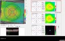
Malignant Hypertension - Cotton Wool Spots - Elschnig Spots - Optic Nerve Edema 853 views6 week fu - CWS are Fading
|
|

Diabetic Retinopathy with Scattered Hemorrhages. 853 viewsDiabetic patient with uncontrolled A1C comes in with blurred vision in the right eye. Fundus photo reveals scattered hemorrhages with cotton wool spots and some edema in the macula. Patient was treated with Avastin for edema.
|
|

Proliferative Diabetic Retinopathy - Moderate - mild NVD with NVE 851 viewsVenous beading, vascular loops and NVE are visible on photos
|
|
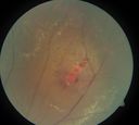
periphery 3. od849 viewsleaking miliary aneurysms
|
|

Ghost vessels with BRVO849 views
|
|
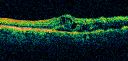
branch retinal vein occlusion - oct line scan847 views70 year old woman with 20/80 vision from a branch retinal vein occlusion and macular edema. She has had laser treatment and intravitreal steroids. Her vision with further laser improved to 20/50.
|
|

Microaneurysms from Ocular Ischemic Syndrome (non-diabetic) Right Eye - Left eye CRAO 1 year ago847 views66-year-old woman lost vision in the left eye from a central retinal artery occlusion 1 year ago. Vision OD is fine. OD 20/25, OS 20/50
|
|

Background Diabetic Retinopathy845 viewsPatient with diabetes for over 11-years comes in with blurred vision. Blood sugar control is very poor. VA 20/50- right eye, 20/25-left eye. Fundus exam shows hard exudates with Circinate Rings and edema. Micro-aneurysms and retinal hemorrhage are present in both eyes. Patient will come back for laser treatment.
|
|
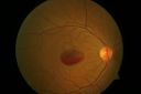
A case of Valsalva Retinopathy -3844 views
|
|

Background Diabetic Retinopathy843 viewsPatient with diabetes for over 11-years comes in with blurred vision. Blood sugar control is very poor. VA 20/50- right eye, 20/25-left eye. Fundus exam shows hard exudates with Circinate Rings and edema. Micro-aneurysms and retinal hemorrhage are present in both eyes. Patient will come back for laser treatment.
|
|

branch retinal vein occlusion - fluorescein angiogram840 views70 year old woman with 20/80 vision from a branch retinal vein occlusion and macular edema. She has had laser treatment and intravitreal steroids. Her vision with further laser improved to 20/50.
|
|

Bilateral Retinal Arteriol Occlusions - Possible Susac Syndrome 840 views80-year-old woman one month ago had vision loss and a vascular occlusion in the right eye. Vision loss occurred in the left eye about 9 years ago with cotton wool spots. Patients has a 30 year history of tinnitis.
|
|

Hemicentral Retinal Vein Occlusion Vision 20/60 840 views84-year-old man has a hemicentral retinal vein occlusion in the right eye. His vision has declined since then.
VISUAL ACUITY: Vision OD is 20/60.
|
|

837 views
|
|

Ruptured Retinal Arterial Macroaneurysm - Submacular Hemorrhage - Vision 20/400 never recovered - Hypertension 205/94836 views
|
|

Retinal Arterial Macroaneurysm - Normal Vision (2 Years before rupture)836 views78-year-old woman has a retinal arterial macroaneurysm in the right eye. VA 20/25 (pre-rupture photos)
|
|

Proliferative Diabetic Retinopathy - Fresh and Old Scatter (Panretinal) Photocoagulation (Laser) - Neovascularization is regressing836 views1 month after initial laser
|
|

Hypertensive Retinopathy with Choroidal Ischemia Right Eye and Vision 8/200 - Blood Pressure 240/120 mmHg IgA Nephropathy835 views39-year-old man two days ago he noticed poor vision in his right eye OD: 8/200; OS: 20/20. BP is 240/120
|
|

Proliferative Sickle Retinopathy - Hemoglobin SC835 views53-year-old woman with a new spider-like floater in the right eye for about six days. She sees one big floater and another little one. She does have hemoglobin SC hemoglobinopathy. VISUAL ACUITY: Vision OD is 20/20; OS is 20/50, PH is 20/20.
|
|

Proliferative Diabetic Retinopathy - Vitreous Hemorrhage and Tractional Retinal Detachment Left Eye834 views52-year-old man has been diabetic for nineteen years developed substantial vision loss over the last month or two. OD 20/70, OS 20/200.
|
|

Malignant Hypertension - Cotton Wool Spots - Elschnig Spots - Optic Nerve Edema 832 viewsMacular Scan Shows Cotton Wool Spots and Nerve Swelling (on Left)
|
|
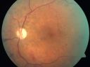
Proliferative Diabetic Retinopathy and Mild Vitreous Hemorrhage831 views58-year-old woman has diabetic retinopathy in both eyes with neovascularization of the optic nerve, worse in the right eye than the left eye. Vision OD is 20/25, OS is 20/20
|
|

Proliferative Diabetic Retinopathy both Eyes Neovascularization of the Disc830 views63-year-old woman has been diabetic for twenty one years who sees new floaters in the right eye. OD 20/40, OS 20/40.
|
|

Hypertensive Retinopathy with Choroidal Ischemia Right Eye and Vision 8/200 - Blood Pressure 240/120 mmHg IgA Nephropathy829 views39-year-old man two days ago he noticed poor vision in his right eye OD: 8/200; OS: 20/20. BP is 240/120
|
|

Central Retinal Artery Occlusion less than 24 hours - 69 year old man VA light perception829 views
|
|

DRIL - Disorganization Retinal Inner Layers829 viewsFollowing treatment for BRVO vision stayed at 3/200 despite resolution of edema
|
|

Proliferative Diabetic Retinopathy both eyes Type I Diabetic for 25 years828 views47-year-old woman with diabetes for 25 years and decreased vision for 3 months. OD 20/40, OS 20/40
|
|

Embolic CRAO color828 views
|
|

Old Branch Retinal Vein Occlusion- Severe CME - Pucker - Diabetic Fundus Photo827 views61-year old man has diabetic retinopathy in the right eye. He also had a macular pucker and macular edema. I did a vitrectomy on September 5th. His vision was initially improving. His vision in the right eye seems to be a little more foggy.
VISUAL ACUITY: OD: 20/200
|
|

FA: CRAO826 viewsCRAO with cilio artery perfusion
|
|

Central Retinal Artery Occlusion with Cilioretinal Sparing - Acute - FA Very Slow Filling CRA825 views 77-year-old man was doing fine until midnight of last night when he noticed sudden severe vision loss in the left eye. VISUAL ACUITY: OD 20/20, OS 2/200.
|
|

Purtchers Retinopathy with Bilateral Central Retinal Artery Occlusions 824 views50-year-old man traumatic chest compression and leg crushing injury. OD 1/200, OS 3/200
|
|

Bilateral Retinal Arteriol Occlusions - Possible Susac Syndrome 823 views80-year-old woman one month ago had vision loss and a vascular occlusion in the right eye. Vision loss occurred in the left eye about 9 years ago with cotton wool spots. Patients has a 30 year history of tinnitis.
|
|

BRVO822 views42 year old female VA 20/25
Branch Retinal Vein Occlusion - Hot Spot on Vein at Occlusion Site
|
|

BRVO with collaterals and cystoid macular edema821 views66 Year Old Man with has had a branch retinal vein occlusion in the right eye with vision loss for the last few months. OD is 20/40, OS is 20/20. IOP: OD 18, OS 19. There is a superotemporal macular branch retinal vein occlusion with edema, hemorrhage, and exudate.
OCT SCAN: The right eye shows edema with a central foveal thickness of 354 microns FA: shows remodeling of the superotemporal vasculature. There are already venous-venous collaterals, suggesting the vein occlusion may be a little more than a few months old. There are multiple microaneurysms superotemporal to the fovea, and there is leakage in the late frames involving the fovea.
|
|

Branch Retinal Vein Occlusion with Macular Edema 3 Months after Intravitreal Kenalog and Grid Laser821 viewsCentral foveal thickness came to normal after laser and intravitreal Kenalog
|
|

Cotton Wool Spots and New Diagnosis of Chronic Lymphocytic leukemia820 views73-year-old man with cotton wool spots diagnosed subsequently with with Chronic Lymphocytice Leukemia
|
|

Hairy Cell Leukemia - Retinal Hemorrhage and twig Branch Vein Occlusion820 views79 year old man He has had hairy cell leukemia since 2002. He is in remission. His last blood tests were 9/2018. He just moved down here and needs a new leukemia doctor. His vision is fine.
VA OD: Dcc20/25
VA OS: Dcc20/25
IOP: TP: OD:12 OS:12
|
|

Retinal Vasculopathy associated with Facioscapulohumeral Muscular Dystrophy 819 views59-year-old woman had a blood pressure spike to 170/90 and was on medications for that. She does have facioscapulohumeral muscular dystrophy, which runs in her family. She has a cousin with the same disorder.
VISUAL ACUITY: Her vision is 20/25 OU. IOP: OD 21, OS 18.
There is no posterior vitreous separation, telangiectatic vessels near the macula. There are patchy retinal hemorrhages. There is no edema or proliferation.
|
|

Diabetic and Pseudophakic Cystoid Macular Edema FA Fellow Eye819 views56-year-old woman has diabetic retinopathy in both eyes and macular edema. She is recently pseudophakic. OD 20/80, OS 20/200.
|
|

Central Retinal Vein Occlusion and Cystoid Macular Edema - Acute - Vision 20/50 819 views68-year-old man six weeks of vision loss right eye from CRVO and CME, vision right eye 20/50
|
|

Preretinal Fibrosis - Tractional Retinal Detachment - Proliferative Diabetic Retinopathy 819 views33-year-old woman has proliferative diabetic retinopathy in the left eye with fibrosis and tractional retinal detachment. Her vision is hazy in that eye. VISUAL ACUITY: Vision OD is 20/30, OS 20/120
|
|

Twig Branch Retinal Vein Occlusion - Recurrent Macular Edema with extended interval Lucentis Therapy after first 6 monthly Injections818 views39-year-old man has a macular branch retinal vein occlusion with macular edema. He had an excellent response to Lucentis for his first 6 treatments. He was last here 6 weeks ago and his vision seems OK to him. OD 20/40.
|
|

Hairy Cell Leukemia - Retinal Hemorrhage and twig Branch Vein Occlusion818 views79 year old man He has had hairy cell leukemia since 2002. He is in remission. His last blood tests were 9/2018. He just moved down here and needs a new leukemia doctor. His vision is fine.
VA OD: Dcc20/25
VA OS: Dcc20/25
IOP: TP: OD:12 OS:12
|
|

Clinically Significant Diabetic Macular Edema both Eyes with Exudates816 views56-year-old woman with diabetes for twelve years. She has noticed blurring of the vision in the right eye for about six months. OD is 20/70, OS is 20/25.
|
|

Old Branch Retinal Vein Occlusion- Severe CME - Pucker - Diabetic Map OCT815 views61-year old man has diabetic retinopathy in the right eye. He also had a macular pucker and macular edema. I did a vitrectomy on September 5th. His vision was initially improving. His vision in the right eye seems to be a little more foggy.
VISUAL ACUITY: OD: 20/200
|
|
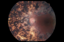
PDR with heavy PRP laser and worsening macular edema 815 views51-year-old woman has had extensive laser in both eyes done years ago elsewhere. Her left eye has gradually been declining with macular edema.
VISUAL ACUITY: OD 20/40, OS 20/40
|
|

Retinal Arterial Atheromata815 views68-year-old woman with records going back 7 years, which clearly document retinal arterial plaques in both eyes.
|
|
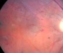
Diabetic and Pseudophakic Cystoid Macular Edema Pretreatment Fundus Photo814 views56-year-old woman has diabetic retinopathy in both eyes and macular edema. She is recently pseudophakic. OD 20/80, OS 20/200.
|
|

813 views
|
|

Branch Retinal Vein Occlusion with NVE (Neovascularization Elsewhere)813 views
|
|
| 4350 files on 44 page(s) |
 |
3 | |
|
|
|
|
|
|
|
|


