Most viewed - Retinal Vascular Disease
|

Ruptured Retinal Arterial Macroaneurysm - Submacular Hemorrhage - Vision 20/400 never recovered - Hypertension 205/94809 views
|
|
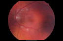
Cotton Wool Spots and New Diagnosis of Chronic Lymphocytic leukemia809 views73-year-old man with cotton wool spots diagnosed subsequently with with Chronic Lymphocytice Leukemia
|
|
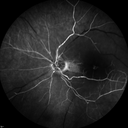
Central Retinal Artery Occlusion with Cilioretinal Sparing - Acute - FA Very Slow Filling CRA809 views 77-year-old man was doing fine until midnight of last night when he noticed sudden severe vision loss in the left eye. VISUAL ACUITY: OD 20/20, OS 2/200.
|
|

BRAO and PAMM808 views
|
|

Retinal Vasculopathy associated with Facioscapulohumeral Muscular Dystrophy early fa807 views59-year-old woman had a blood pressure spike to 170/90 and was on medications for that. She does have facioscapulohumeral muscular dystrophy, which runs in her family. She has a cousin with the same disorder.
VISUAL ACUITY: Her vision is 20/25 OU. IOP: OD 21, OS 18.
There is no posterior vitreous separation, telangiectatic vessels near the macula. There are patchy retinal hemorrhages. There is no edema or proliferation.
|
|

Diabetic Retinopathy807 viewsAnnual diabetic exam. VA 20/30, right eye and 20/50, left eye. Fundus photos shows hemorrhages with cotton wool spots. Exudates are visible around the macula in the right eye. Scattered hemorrhages in the left eye.
|
|

Serous Retinal Detachment - Hypertensive Retinopathy - 165/120 806 views48-year-old woman noticed when she was blinking there was a white flash in the left eye starting about a month ago. She sees it temporally. OD 20/16, OS 20/20. There is a serous retinal detachment superotemporal to the optic nerve falling short of the fovea. Her blood pressure 165/120.
|
|
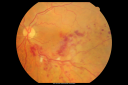
Macular Branch Retinal Vein Occlusion and Adult Vitelliform Dystrophy805 views72-year-old woman 2 months post ECCE with poor vision since surgery especially left eye.
VISUAL ACUITY: OD 20/50, OS 20/200
|
|

Central Retinal Vein Occlusion and Laser Induced Chorioretinal Anastomosis804 views84-year-old woman has age-related macular degeneration in both eyes with foveal atrophy. She also has a central retinal vein occlusion
in the left eye for which she had intravitreal Kenalog and finally chorioretinal anastomosis laser a year
ago. VISUAL ACUITY: Vision OD is 20/160, OS is 7/200
|
|

Sectoral Optic Atrophy from an old Branch Retinal Artery Occlusion 804 views74-year-old woman had cataract surgery 3 months ago. Her vision in the right eye has been abnormal since. She notices a shadow in the inferior vision in the right eye. OD is 20/60, OS is 20/50
|
|

Capillary Non-Perfusion - Severe Non-Proliferative (Background) Diabetic Retinopathy - Type I - 20 Year Old Man Diabetic 16 years803 views20 year old diabetic with poor vision for the past three or four years. He has been diabetic for sixteen years and hemoglobin A1C of 10.0. 20/25 OD, 20/30 OS.
|
|

macula. od803 viewscoalescing exudates
|
|

Clinically Significant Diabetic Macular Edema both Eyes with Exudates802 views56-year-old woman with diabetes for twelve years. She has noticed blurring of the vision in the right eye for about six months. OD is 20/70, OS is 20/25.
|
|

Chronic - Old Diabetic Tractional Retinal Detachment Left Eye802 views77-year-old woman Vision OD is 20/40, OS is 7/200OD: Vertical C/D ratio is 0.3. There is panretinal laser. The macula is flat and attached.
OS: Vertical C/D ratio is 0.3. There is panretinal laser. There is a tractional retinal detachment with preretinal fibrosis nasal to the optic nerves.
|
|
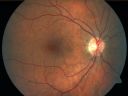
Retinal Vasculopathy associated with Facioscapulohumeral Muscular Dystrophy 801 views59-year-old woman had a blood pressure spike to 170/90 and was on medications for that. She does have facioscapulohumeral muscular dystrophy, which runs in her family. She has a cousin with the same disorder.
VISUAL ACUITY: Her vision is 20/25 OU. IOP: OD 21, OS 18.
There is no posterior vitreous separation, telangiectatic vessels near the macula. There are patchy retinal hemorrhages. There is no edema or proliferation.
59-year-old woman had a blood pressure spike to 170/90 and was on medications for tha
|
|

Clinically Significant Diabetic Macular Edema both Eyes with Exudates801 views56-year-old woman with diabetes for twelve years. She has noticed blurring of the vision in the right eye for about six months. OD is 20/70, OS is 20/25.
|
|
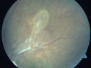
Preretinal Fibrosis - Tractional Retinal Detachment - Proliferative Diabetic Retinopathy 801 views33-year-old woman has proliferative diabetic retinopathy in the left eye with fibrosis and tractional retinal detachment. Her vision is hazy in that eye. VISUAL ACUITY: Vision OD is 20/30, OS 20/120
|
|

BRVO with collaterals and cystoid macular edema800 views66 Year Old Man with has had a branch retinal vein occlusion in the right eye with vision loss for the last few months. OD is 20/40, OS is 20/20. IOP: OD 18, OS 19. There is a superotemporal macular branch retinal vein occlusion with edema, hemorrhage, and exudate.
OCT SCAN: The right eye shows edema with a central foveal thickness of 354 microns FA: shows remodeling of the superotemporal vasculature. There are already venous-venous collaterals, suggesting the vein occlusion may be a little more than a few months old. There are multiple microaneurysms superotemporal to the fovea, and there is leakage in the late frames involving the fovea.
|
|

Serous Retinal Detachment - Hypertensive Retinopathy - 1 month later - less fluid (better blood pressure) - SD OCT800 views48 Year old woman resolving hypertensive serous retinal detachment.
|
|

BRVO with collaterals and cystoid macular edema799 views66 Year Old Man with has had a branch retinal vein occlusion in the right eye with vision loss for the last few months. OD is 20/40, OS is 20/20. IOP: OD 18, OS 19. There is a superotemporal macular branch retinal vein occlusion with edema, hemorrhage, and exudate.
OCT SCAN: The right eye shows edema with a central foveal thickness of 354 microns FA: shows remodeling of the superotemporal vasculature. There are already venous-venous collaterals, suggesting the vein occlusion may be a little more than a few months old. There are multiple microaneurysms superotemporal to the fovea, and there is leakage in the late frames involving the fovea.
|
|
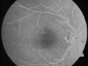
Microaneurysms from Ocular Ischemic Syndrome (non-diabetic) Right Eye - Left eye CRAO 1 year ago799 views66-year-old woman lost vision in the left eye from a central retinal artery occlusion 1 year ago. Vision OD is fine. OD 20/25, OS 20/50
|
|

Twig Branch Retinal Vein Occlusion - Recurrent Macular Edema with extended interval Lucentis Therapy after first 6 monthly Injections799 views39-year-old man has a macular branch retinal vein occlusion with macular edema. He had an excellent response to Lucentis for his first 6 treatments. He was last here 6 weeks ago and his vision seems OK to him. OD 20/40.
|
|

brvo cme with small serous macular detachment 798 views70 year old woman with 20/80 vision from a branch retinal vein occlusion and macular edema. She has had laser treatment and intravitreal steroids. Her vision with further laser improved to 20/50.
|
|

Central Retinal Vein Occlusion and Cystoid Macular Edema - Acute - Vision 20/50 798 views68-year-old man six weeks of vision loss right eye from CRVO and CME, vision right eye 20/50
|
|

Cilioretinal artery occlusion and Ophthalmic Artery Occlusion with Relative Sparing of the Central Retinal Artery798 views80-year-old woman who says she was looking in the mirror, covered her right eye and noticed she could not see well out of the left eye. That just happened yesterday. Her last eye check was a few years ago. She also had a carotid endarterectomy on the right side in 2006 shortly after coronary bypass surgery. OD is 20/40, OS is 20/200.
|
|

Neovascularization elsewhere (NVE) Left Eye with Vitreous Hemorrhage797 views
|
|

Bilateral Retinal Arteriol Occlusions - Possible Susac Syndrome 796 views80-year-old woman one month ago had vision loss and a vascular occlusion in the right eye. Vision loss occurred in the left eye about 9 years ago with cotton wool spots. Patients has a 30 year history of tinnitis.
|
|

Cotton Wool Spots and New Diagnosis of Chronic Lymphocytic leukemia796 views73-year-old man with cotton wool spots diagnosed subsequently with with Chronic Lymphocytice Leukemia
|
|

Central Retinal Artery Occlusion Left Eye 2 Weeks Old - Still Non-Perfusion795 views85-year-old woman who had sudden severevision loss in the left eye two weeks ago. she can see a little more centrally and much more in the periphery, since about the last four days.
VISUAL ACUITY: OD 20/30, OS: Hand motion
|
|

Hypertensive Choroidopathy - Pheochromocytoma - Chronic Very High Blood Pressure 49 YO Male794 views49-year-old man chronic very high blood pressure sometimes the blood pressure will go as high as 260/160, sometimes it is 200/120 and sometimes it is 160/80. He has a pheochromocytoma on the adrenal gland and he has had vision loss in the right eye starting between three and four years ago.
VISUAL ACUITY: OD 20/60, OS 20/25.
|
|

Retinal Arterial Macroaneurysm - Ruptured - Vitreous Hemorrhage (Fresh) 5/200 Vision792 views80-year-old woman has a retinal arterial macroaneurysm in the right eye I saw two years ago. She noticed the sudden onset of vision loss last night. OD is 5/200.
|
|
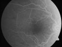
Microaneurysms from Ocular Ischemic Syndrome (non-diabetic) Right Eye - Left eye CRAO 1 year ago792 views66-year-old woman lost vision in the left eye from a central retinal artery occlusion 1 year ago. Vision OD is fine. OD 20/25, OS 20/50
|
|

Diabetic Retinopathy with CME792 viewsSecond opinion for diabetic retinopathy. VA is 20/30, right eye, 20/50, left eye. Patient has mild swelling in the macula of the right eye. No evidence of CME but does have a moderate cataract in the left eye.
|
|

Heavy Panretinal Photocoagulation for proliferative diabetic retinopathy (PRP for PDR)792 views78 year old man who had laser for proliferative diabetic retinopathy in 1981. These photos are from 2020. The left eye also had multiple surgeries for diabetic traction retinal detachment. VA OD 20/32, OS HM
|
|

Proliferative Diabetic Retinopathy both Eyes Neovascularization of the Disc791 views63-year-old woman has been diabetic for twenty one years who sees new floaters in the right eye. OD 20/40, OS 20/40.
|
|

Central Retinal Artery Occlusion Left Eye 2 Weeks Old - Still Non-Perfusion790 views85-year-old woman who had sudden severevision loss in the left eye two weeks ago. she can see a little more centrally and much more in the periphery, since about the last four days.
VISUAL ACUITY: OD 20/30, OS: Hand motion
|
|

Proliferative Diabetic Retinopathy both Eyes Neovascularization of the Disc787 views63-year-old woman has been diabetic for twenty one years who sees new floaters in the right eye. OD 20/40, OS 20/40.
|
|

787 views
|
|

PDR OS with NVE786 views57-year-old man has diabetic retinopathy in both eyes.
Diabetic for 14 years with HgB A1C often over 10.
VISUAL ACUITY: OD 20/30, OS 20/40. PDR OS BDR OD
|
|
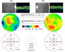
Diabetic Retinopathy - Non-Perfusion - Possible Central Retinal Vein Occlusion785 views68-year-old woman OD 20/70, OS 20/200 CYSTOID MACULAR EDEMA, POSSIBLE EITHER OCULAR ISCHEMIC SYNDROME IN THE RIGHT EYE OR OCCULT CENTRAL RETINAL VEIN OCCLUSION IN THE RIGHT EYE,S EVERE NON-PerfUSION WITH DIABETIC RETINOPATHY IN THE LEFT EYE.
|
|

Proliferative Diabetic Retinopathy both Eyes Neovascularization of the Disc785 views63-year-old woman has been diabetic for twenty one years who sees new floaters in the right eye. OD 20/40, OS 20/40.
|
|

Cilioretinal artery occlusion and Ophthalmic Artery Occlusion with Relative Sparing of the Central Retinal Artery785 views80-year-old woman who says she was looking in the mirror, covered her right eye and noticed she could not see well out of the left eye. That just happened yesterday. Her last eye check was a few years ago. She also had a carotid endarterectomy on the right side in 2006 shortly after coronary bypass surgery. OD is 20/40, OS is 20/200.
|
|

Proliferative Diabetic Retinopathy and Vitreous Hemorrhage - FA shows leakage of dye from NVE783 views
|
|

Macular Branch Retinal Vein Occlusions (2) Left Eye - No Macular Edema (yet) 782 views77-year-old woman with two macular branch retinal vein occlusions in the left eye OD 20/25
|
|

Anemic Retinopathy782 viewsMyelodysplasia - HgB 8 and very low platelet count
|
|

Retinal Arterial Macroaneurysm - Ruptured - Vitreous Hemorrhage (Fresh) 5/200 Vision781 views80-year-old woman has a retinal arterial macroaneurysm in the right eye I saw two years ago. She noticed the sudden onset of vision loss last night. OD is 5/200.
|
|

Diabetic Edema Better - 3 Months Follow-UP - No treatment - SD OCT781 viewsPatient remains asymptomatic and OCT shows less edema
|
|

Malignant Hypertension - Cotton Wool Spots - Elschnig Spots - Optic Nerve Edema 781 views6 week fu - CWS are Fading
|
|
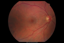
Diabetic Vitreous Hemorrhage Left Eye780 views35-year-old man diabetic for the last 25 years. His hemoglobin A1C has been running between 10 and 11. He was doing fine and then when he woke up from sleeping last night he noticed sudden vision loss in his left eye OD 20/30, OS 20/200.
|
|

Hypertensive Retinopathy with Choroidal Ischemia Right Eye and Vision 8/200 - Blood Pressure 240/120 mmHg IgA Nephropathy780 views39-year-old man two days ago he noticed poor vision in his right eye OD: 8/200; OS: 20/20. BP is 240/120
|
|

Hemicentral Retinal Vein Occlusion Vision 20/60 779 views84-year-old man has a hemicentral retinal vein occlusion in the right eye. His vision has declined since then.
VISUAL ACUITY: Vision OD is 20/60.
|
|

Central Retinal Artery Occlusion Left Eye 2 Weeks Old - Still Non-Perfusion777 views85-year-old woman who had sudden severevision loss in the left eye two weeks ago. she can see a little more centrally and much more in the periphery, since about the last four days.
VISUAL ACUITY: OD 20/30, OS: Hand motion
|
|

Central Retinal Artery Occlusion Left Eye 2 Weeks Old - Still Non-Perfusion776 views85-year-old woman who had sudden severevision loss in the left eye two weeks ago. she can see a little more centrally and much more in the periphery, since about the last four days.
VISUAL ACUITY: OD 20/30, OS: Hand motion
|
|

Preretinal Fibrosis - Tractional Retinal Detachment - Proliferative Diabetic Retinopathy 775 views33-year-old woman has proliferative diabetic retinopathy in the left eye with fibrosis and tractional retinal detachment. Her vision is hazy in that eye. VISUAL ACUITY: Vision OD is 20/30, OS 20/120
|
|
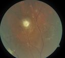
periphery 1. od 775 viewsquiescient/infarcted macroaneurysm
|
|

Proliferative Diabetic Retinopathy - NVD regressed with PRP laser - Animation showing NVD 775 views76 year old diabetic man - Presented one year ago with NVD in the left eye. This regressed with laser but then a year later worsened. Additional PRP was done and the NVD regressed again.
|
|

Branch Retinal Vein Occlusion with Macular Edema774 viewsCentral foveal thickness came to normal after laser and intravitreal Kenalog
|
|

Ruptured Retinal Arterial Macroaneurysm - Submacular Hemorrhage - Vision 20/400 never recovered - Hypertension 205/94774 views
|
|

New BRVO Left Eye Old BRVO Right Eye - Left Eye Shows Twig Superior Vein Occlusion with Edema773 views90-year-old man decreased vision maybe one year since cataract surgery he is no longer able to read. OD 20/200, OS 20/200
|
|
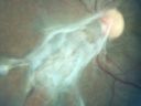
Preretinal Fibrosis - Tractional Retinal Detachment - Proliferative Diabetic Retinopathy 773 views33-year-old woman has proliferative diabetic retinopathy in the left eye with fibrosis and tractional retinal detachment. Her vision is hazy in that eye. VISUAL ACUITY: Vision OD is 20/30, OS 20/120
|
|

Cotton Wool Spots and New Diagnosis of Chronic Lymphocytic leukemia771 views73-year-old man with cotton wool spots diagnosed subsequently with with Chronic Lymphocytice Leukemia
|
|

Diabetic Macular Edema and Hypertensive Retinopathy - Circinate Exudate (Ring Exudate) 771 views55-year-old woman diabetic for fifteen years and high blood pressure, especially over the last few months. She has had problems with nosebleeds, headaches, and there has been some difficulty bringing her blood pressure down. She said now the blood pressure is under control but it was running 220/120 mmHg for some time. Her vision in the right eye has been poor for four weeks with a spot in the central vision and both eyes have been blurred.
VISUAL ACUITY: Vision OD is 20/160, OS is 20/60
|
|

Central Retinal Vein Occlusion Recurrent Edema 6 weeks after Lucentis - Nonperfusion Temporally771 views66-year-old woman has a central retinal vein occlusion in the right eye with macular edema. She had intravitreal Lucentis treatment six weeks ago and she has noticed over the last week or two her vision is declining.
VISUAL ACUITY: OD 20/70, OS 20/30.
|
|

Central Retinal Artery Occlusion with Cilioretinal Sparing - Acute771 views 77-year-old man was doing fine until midnight of last night when he noticed sudden severe vision loss in the left eye. VISUAL ACUITY: OD 20/20, OS 2/200.
|
|

Macular Branch Retinal Vein Occlusions (2) Left Eye - No Macular Edema (yet) 770 views77-year-old woman with two macular branch retinal vein occlusions in the left eye OD 20/25
|
|

769 views63-year-old man diabetic for twenty two years. His central vision is fine.
VISUAL ACUITY: OD 20/25, OS 20/25
|
|
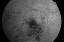
Hypertensive Retinopathy with Choroidal Ischemia Right Eye and Vision 8/200 - Blood Pressure 240/120 mmHg IgA Nephropathy768 views39-year-old man two days ago he noticed poor vision in his right eye OD: 8/200; OS: 20/20. BP is 240/120
|
|

Branch Retinal Arterial Occlusion 20/20 Vision - Symptoms for 1 Day768 views
|
|

Retinal Vasculopathy associated with Facioscapulohumeral Muscular Dystrophy 767 views59-year-old woman had a blood pressure spike to 170/90 and was on medications for that. She does have facioscapulohumeral muscular dystrophy, which runs in her family. She has a cousin with the same disorder.
VISUAL ACUITY: Her vision is 20/25 OU. IOP: OD 21, OS 18.
There is no posterior vitreous separation, telangiectatic vessels near the macula. There are patchy retinal hemorrhages. There is no edema or proliferation.
|
|

New BRVO Left Eye Old BRVO Right Eye - Right Eye Showing Bulls Eye Atrophy from Chronic CME and Non-Perfusion Infero-temporally767 views90-year-old man decreased vision maybe one year since cataract surgery he is no longer able to read. OD 20/200, OS 20/200
|
|

Proliferative Diabetic Retinopathy and Vitreous Hemorrhage - FA shows leakage of dye from NVE767 views
|
|

Twig Branch Retinal Vein Occlusion - Recurrent Macular Edema with extended interval Lucentis Therapy after first 6 monthly Injections766 views39-year-old man has a macular branch retinal vein occlusion with macular edema. He had an excellent response to Lucentis for his first 6 treatments. He was last here 6 weeks ago and his vision seems OK to him. OD 20/40.
|
|

Focal Laser for diabetic macular edema765 views66 year old female with diabetes for 45 years. VA 20/20 OU. Extrafoveal edema in the left eye treated with light focal laser.
|
|

Bilateral Retinal Arteriol Occlusions - Possible Susac Syndrome 765 views80-year-old woman one month ago had vision loss and a vascular occlusion in the right eye. Vision loss occurred in the left eye about 9 years ago with cotton wool spots. Patients has a 30 year history of tinnitis.
|
|

Pathology - Central Retinal Artery Occlusion - 6 weeks old - Inner Retinal Atrophy765 viewspatient died about 6 to 8 weeks after the artery occlusion, and he had no history of glaucoma. The inner retina atrophy, which looks very similar to that caused by COAG, is due to recent infarct.
|
|

765 views
|
|

Retinal Arterial Macroaneurysm - Ruptured and self-sealed after hemorrhage764 views90-year-old woman has vision loss in the right eye for 6 weeks. OD 8/200, OS 20/50. Initial Visit
|
|

Diabetic Macular Edema and Hypertensive Retinopathy - Circinate Exudate (Ring Exudate) 763 views55-year-old woman diabetic for fifteen years and high blood pressure, especially over the last few months. She has had problems with nosebleeds, headaches, and there has been some difficulty bringing her blood pressure down. She said now the blood pressure is under control but it was running 220/120 mmHg for some time. Her vision in the right eye has been poor for four weeks with a spot in the central vision and both eyes have been blurred.
VISUAL ACUITY: Vision OD is 20/160, OS is 20/60
|
|

Proliferative Diabetic Retinopathy both Eyes Neovascularization of the Disc762 views63-year-old woman has been diabetic for twenty one years who sees new floaters in the right eye. OD 20/40, OS 20/40.
|
|

Anemic Retinopathy762 viewsMyelodysplasia - HgB 8 and very low platelet count
|
|

Hypertensive Retinopathy with Choroidal Ischemia Right Eye and Vision 8/200 - Blood Pressure 240/120 mmHg IgA Nephropathy761 views39-year-old man two days ago he noticed poor vision in his right eye OD: 8/200; OS: 20/20. BP is 240/120
|
|

Diabetic Macular Edema and Hypertensive Retinopathy - Circinate Exudate (Ring Exudate) 761 views55-year-old woman diabetic for fifteen years and high blood pressure, especially over the last few months. She has had problems with nosebleeds, headaches, and there has been some difficulty bringing her blood pressure down. She said now the blood pressure is under control but it was running 220/120 mmHg for some time. Her vision in the right eye has been poor for four weeks with a spot in the central vision and both eyes have been blurred.
VISUAL ACUITY: Vision OD is 20/160, OS is 20/60
|
|

Cilioretinal artery occlusion and Ophthalmic Artery Occlusion with Relative Sparing of the Central Retinal Artery761 views80-year-old woman who says she was looking in the mirror, covered her right eye and noticed she could not see well out of the left eye. That just happened yesterday. Her last eye check was a few years ago. She also had a carotid endarterectomy on the right side in 2006 shortly after coronary bypass surgery. OD is 20/40, OS is 20/200.
|
|

Diabetic Retinopathy - Non-Perfusion - Possible Central Retinal Vein Occlusion760 views68-year-old woman OD 20/70, OS 20/200 CYSTOID MACULAR EDEMA, POSSIBLE EITHER OCULAR ISCHEMIC SYNDROME IN THE RIGHT EYE OR OCCULT CENTRAL RETINAL VEIN OCCLUSION IN THE RIGHT EYE,S EVERE NON-PerfUSION WITH DIABETIC RETINOPATHY IN THE LEFT EYE.
|
|

Serous Macular Detachment Left Eye - Possible Hypertensive Choroidopathy v. Central Serous Retinopathy759 views59-year-old woman decreasing vision in the left eye for about two days. She sees a spot in front of her vision. It is a yellow spot which is just straight ahead and moves with her eye. She does have high blood pressure. She checks her blood pressure at home, and even at home her blood pressure runs very high, with the systolic running around 200 and the diastolic around 100, although occasionally it does return to the normal range.
VISUAL ACUITY: Vision OD is 20/60, OS is 20/30
|
|

FA showing NVE, NVD, non- perfusion left eye inferonasally with neovascularization at the margin of the non-perfusion759 views
|
|

Microaneursyms and Macular Edema Not Diabetic759 views85 year old man has been tested about 2-3 times and is always negative for diabetes
|
|

Retinal Vasculopathy associated with Facioscapulohumeral Muscular Dystrophy OCT right eye line scan758 views59-year-old woman had a blood pressure spike to 170/90 and was on medications for that. She does have facioscapulohumeral muscular dystrophy, which runs in her family. She has a cousin with the same disorder.
VISUAL ACUITY: Her vision is 20/25 OU. IOP: OD 21, OS 18.
There is no posterior vitreous separation, telangiectatic vessels near the macula. There are patchy retinal hemorrhages. There is no edema or proliferation.
|
|

Diabetic and Pseudophakic Cystoid Macular Edema Laser Photo - Focal Laser757 views56-year-old woman has diabetic retinopathy in both eyes and macular edema. She is recently pseudophakic. OD 20/80, OS 20/200.
|
|

Diabetic Retinopathy - Non-Perfusion - Possible Central Retinal Vein Occlusion757 views68-year-old woman OD 20/70, OS 20/200 CYSTOID MACULAR EDEMA, POSSIBLE EITHER OCULAR ISCHEMIC SYNDROME IN THE RIGHT EYE OR OCCULT CENTRAL RETINAL VEIN OCCLUSION IN THE RIGHT EYE,S EVERE NON-PerfUSION WITH DIABETIC RETINOPATHY IN THE LEFT EYE.
|
|

Clinically Significant Diabetic Macular Edema both Eyes with Exudates757 views56-year-old woman with diabetes for twelve years. She has noticed blurring of the vision in the right eye for about six months. OD is 20/70, OS is 20/25.
|
|

Central Retinal Vein Occlusion and Laser Induced Chorioretinal Anastomosis756 views84-year-old woman has age-related macular degeneration in both eyes with foveal atrophy. She also has a central retinal vein occlusion
in the left eye for which she had intravitreal Kenalog and finally chorioretinal anastomosis laser a year
ago. VISUAL ACUITY: Vision OD is 20/160, OS is 7/200
|
|

Diabetic Retinopathy - Foveal Laser Scar Right Eye756 views74-year-old woman has diabetic retinopathy in both eyes. She has had focal laser in her right eye done elsewhere in the mid 90’s and unfortunately the laser was fairly close to the center and as time progressed she developed foveal atrophy around the scars. She has noticed her vision declining since she was here last a year ago. She does take Travatan in both eyes at night for glaucoma.
VISUAL ACUITY: OD 20/200, OS 20/40.
|
|

Twig Branch Retinal Vein Occlusion - Recurrent Macular Edema with extended interval Lucentis Therapy after first 6 monthly Injections756 views39-year-old man has a macular branch retinal vein occlusion with macular edema. He had an excellent response to Lucentis for his first 6 treatments. He was last here 6 weeks ago and his vision seems OK to him. OD 20/40.
|
|
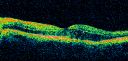
Diabetic Macular Edema and Hypertensive Retinopathy - Circinate Exudate (Ring Exudate) 754 views55-year-old woman diabetic for fifteen years and high blood pressure, especially over the last few months. She has had problems with nosebleeds, headaches, and there has been some difficulty bringing her blood pressure down. She said now the blood pressure is under control but it was running 220/120 mmHg for some time. Her vision in the right eye has been poor for four weeks with a spot in the central vision and both eyes have been blurred.
VISUAL ACUITY: Vision OD is 20/160, OS is 20/60
|
|

Retinal Arterial Macroaneurysm - Ruptured and self-sealed after hemorrhage754 views90-year-old woman has vision loss in the right eye for 6 weeks. OD 8/200, OS 20/50. Initial Visit
|
|

Twig Branch Retinal Vein Occlusion - Recurrent Macular Edema with extended interval Lucentis Therapy after first 6 monthly Injections754 views39-year-old man has a macular branch retinal vein occlusion with macular edema. He had an excellent response to Lucentis for his first 6 treatments. He was last here 6 weeks ago and his vision seems OK to him. OD 20/40.
|
|

Proliferative Diabetic Retinopathy both Eyes Neovascularization of the Disc753 views63-year-old woman has been diabetic for twenty one years who sees new floaters in the right eye. OD 20/40, OS 20/40.
|
|

Branch Retinal Vein Occlusion (BRVO) no macular edema - cotton wool spot753 views85 year old man with asymptomatic BRVO - his vision stayed great without needing any treatment.
|
|

Microaneurysms from Ocular Ischemic Syndrome (non-diabetic) Right Eye - Left eye CRAO 1 year ago752 views66-year-old woman lost vision in the left eye from a central retinal artery occlusion 1 year ago. Vision OD is fine. OD 20/25, OS 20/50
|
|
| 4340 files on 44 page(s) |
 |
|
4 | |
|
|
|
|
|
|
|


