Les plus populaires - Retinal Vascular Disease
|

Proliferative Diabetic Retinopathy both Eyes Neovascularization of the Discvu 712 fois63-year-old woman has been diabetic for twenty one years who sees new floaters in the right eye. OD 20/40, OS 20/40.
|
|

Retinal Arterial Macroaneurysm - Increased Swelling after Laser vu 712 fois65-year-old woman was seen in the office on January 12, 2011. She has noticed decreased vision in the right eye for the last two or three weeks. She has had some discomfort in both eyes as well. She has a history of low blood pressure, but she does have high cholesterol.
VISUAL ACUITY: OD 20/60
|
|

Retinal Arterial Macroaneurysm - Increased Swelling after Laser vu 712 foisVisit 2 - 2 months later:
March 21, 2011. This pleasant 66-year-old woman had laser to retinal arterial microaneurysm in the right eye January 12th. Her vision was getting better, but now it is worse the last few days. She is at home taking care of her demented mother and she and her sister lift her mother a fare amount of the time. I also checked her blood pressure, which was 120/65. VISUAL ACUITY: OD 20/200
|
|

Retinal Vasculopathy associated with Facioscapulohumeral Muscular Dystrophy line osvu 711 fois59-year-old woman had a blood pressure spike to 170/90 and was on medications for that. She does have facioscapulohumeral muscular dystrophy, which runs in her family. She has a cousin with the same disorder.
VISUAL ACUITY: Her vision is 20/25 OU. IOP: OD 21, OS 18.
There is no posterior vitreous separation, telangiectatic vessels near the macula. There are patchy retinal hemorrhages. There is no edema or proliferation.
|
|

Proliferative Sickle Retinopathy - Hemoglobin SCvu 711 fois53-year-old woman with a new spider-like floater in the right eye for about six days. She sees one big floater and another little one. She does have hemoglobin SC hemoglobinopathy. VISUAL ACUITY: Vision OD is 20/20; OS is 20/50, PH is 20/20.
|
|

Central Retinal Vein Occlusion and Laser Induced Chorioretinal Anastomosisvu 711 fois84-year-old woman has age-related macular degeneration in both eyes with foveal atrophy. She also has a central retinal vein occlusion
in the left eye for which she had intravitreal Kenalog and finally chorioretinal anastomosis laser a year
ago. VISUAL ACUITY: Vision OD is 20/160, OS is 7/200
|
|

Diabetic Vitreous Hemorrhage Left Eyevu 710 fois35-year-old man diabetic for the last 25 years. His hemoglobin A1C has been running between 10 and 11. He was doing fine and then when he woke up from sleeping last night he noticed sudden vision loss in his left eye OD 20/30, OS 20/200.
|
|
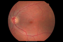
Hypertensive Choroidopathy - Pheochromocytoma - Chronic Very High Blood Pressure 49 YO Malevu 710 fois49-year-old man chronic very high blood pressure sometimes the blood pressure will go as high as 260/160, sometimes it is 200/120 and sometimes it is 160/80. He has a pheochromocytoma on the adrenal gland and he has had vision loss in the right eye starting between three and four years ago.
VISUAL ACUITY: OD 20/60, OS 20/25.
|
|
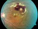
Retinal Arterial Macroaneurysm - Untreated Hypertension (250/135) - Serous Macular Detachmentvu 710 fois82 year old asian woman. VA 20/80 Headaches and vision loss for 1 month. Negative medical history but has not been to the doctor for years. (no insurance)
|
|

Self Sealed Retinal Arteriol Macroaneurysm vu 710 foisFA on presentation showed minimal to no active leakage. Also, there is a small cuff of blood around the MA which is favorable for closure
|
|

Hypertensive Retinopathy with Choroidal Ischemia Right Eye and Vision 8/200 - Blood Pressure 240/120 mmHg IgA Nephropathyvu 709 fois39-year-old man two days ago he noticed poor vision in his right eye OD: 8/200; OS: 20/20. BP is 240/120
|
|

Hemi Central Retinal Vein Occlusion 35 year old non-compliant manvu 708 fois35-year-old man hazy vision in his left eye for about the last three days. OD 20/20, OS 20/30
|
|
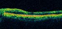
Diabetic and Pseudophakic Cystoid Macular Edema Pre treatment line OSvu 707 fois56-year-old woman has diabetic retinopathy in both eyes and macular edema. She is recently pseudophakic. OD 20/80, OS 20/200.
|
|

Moderate PDRvu 707 fois43 year old man 20/32 vision in both eyes. Being started on Anti-VEGF injections which will be followed by PRP.
|
|

PDR - Mild Vitreous Hemorrhage and Good Visionvu 706 fois58-year-old woman has diabetic retinopathy in both eyes with neovascularization of the optic nerve, worse in the right eye than the left eye. She has had panretinal laser in both eyes and vitreous hemorrhage in the left eye. Her vision, since I saw her last in June, is about the same.
VISUAL ACUITY: Vision OD is 20/25, OS is 20/20.
|
|

vu 706 fois63-year-old man diabetic for twenty two years. His central vision is fine.
VISUAL ACUITY: OD 20/25, OS 20/25
|
|

montage. osvu 706 foisno disease
|
|

Diabetic Vitreous Hemorrhage Left Eyevu 705 fois35-year-old man diabetic for the last 25 years. His hemoglobin A1C has been running between 10 and 11. He was doing fine and then when he woke up from sleeping last night he noticed sudden vision loss in his left eye OD 20/30, OS 20/200.
|
|

Central Retinal Vein Occlusion and Laser Induced Chorioretinal Anastomosisvu 705 fois84-year-old woman has age-related macular degeneration in both eyes with foveal atrophy. She also has a central retinal vein occlusion
in the left eye for which she had intravitreal Kenalog and finally chorioretinal anastomosis laser a year
ago. VISUAL ACUITY: Vision OD is 20/160, OS is 7/200
|
|

Sectoral Optic Atrophy from an old Branch Retinal Artery Occlusion vu 705 fois74-year-old woman had cataract surgery 3 months ago. Her vision in the right eye has been abnormal since. She notices a shadow in the inferior vision in the right eye. OD is 20/60, OS is 20/50
|
|

Hemi Central Retinal Vein Occlusion 35 year old non-compliant manvu 704 fois35-year-old man hazy vision in his left eye for about the last three days. OD 20/20, OS 20/30
|
|

Proliferative Diabetic Retinopathy - Moderate - mild NVD with NVE vu 704 foisVenous beading, vascular loops and NVE are visible on photos
|
|
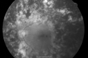
PDR with heavy PRP laser and worsening macular edema red free photovu 703 fois51-year-old woman has had extensive laser in both eyes done years ago elsewhere. Her left eye has gradually been declining with macular edema.
VISUAL ACUITY: OD 20/40, OS 20/40
|
|
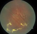
periphery 2. odvu 703 foisleaking miliary aneurysms
|
|

branch retinal vein occlusion - fluorescein angiogramvu 702 fois70 year old woman with 20/80 vision from a branch retinal vein occlusion and macular edema. She has had laser treatment and intravitreal steroids. Her vision with further laser improved to 20/50.
|
|

Retinal Arterial Macroaneurysm - Increased Swelling after Laser vu 702 foisVisit 2 - 2 months later:
March 21, 2011. This pleasant 66-year-old woman had laser to retinal arterial microaneurysm in the right eye January 12th. Her vision was getting better, but now it is worse the last few days. She is at home taking care of her demented mother and she and her sister lift her mother a fare amount of the time. I also checked her blood pressure, which was 120/65. VISUAL ACUITY: OD 20/200
|
|

Bilateral Retinal Arteriol Occlusions - Possible Susac Syndrome vu 701 fois80-year-old woman one month ago had vision loss and a vascular occlusion in the right eye. Vision loss occurred in the left eye about 9 years ago with cotton wool spots. Patients has a 30 year history of tinnitis.
|
|
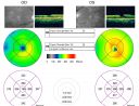
Ocular Ishemic Syndrome - Bilateral Carotid Occlusions - OD 20/60 OS 20/200 Enlarged Foveal Avascular Zone and Temporal Non-perfusionvu 701 fois75-year-old man had laryngeal cancer treated in 15 year ago with radiation. He recently was told that the carotid circulation is closed off on both sides. He does have a patent vertebral artery, which is supplying him pretty well and in addition to that his cardiac output is compromised by poor mitral valves. His consideration is to what can be done about that in the future.
VISUAL ACUITY: OD 20/60, OS 20/200
|
|
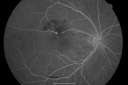
Retinal Arterial Macroaneurysm - Increased Swelling after Laser vu 701 fois65-year-old woman was seen in the office on January 12, 2011. She has noticed decreased vision in the right eye for the last two or three weeks. She has had some discomfort in both eyes as well. She has a history of low blood pressure, but she does have high cholesterol.
VISUAL ACUITY: OD 20/60
|
|

Capillary Non-Perfusion - Severe Non-Proliferative (Background) Diabetic Retinopathy - Type I - 20 Year Old Man Diabetic 16 yearsvu 701 fois20 year old diabetic with poor vision for the past three or four years. He has been diabetic for sixteen years and hemoglobin A1C of 10.0. 20/25 OD, 20/30 OS.
|
|

BRVO with collaterals and cystoid macular edemavu 700 fois66 Year Old Man with has had a branch retinal vein occlusion in the right eye with vision loss for the last few months. OD is 20/40, OS is 20/20. IOP: OD 18, OS 19. There is a superotemporal macular branch retinal vein occlusion with edema, hemorrhage, and exudate.
OCT SCAN: The right eye shows edema with a central foveal thickness of 354 microns FA: shows remodeling of the superotemporal vasculature. There are already venous-venous collaterals, suggesting the vein occlusion may be a little more than a few months old. There are multiple microaneurysms superotemporal to the fovea, and there is leakage in the late frames involving the fovea.
|
|

Hemicentral Retinal Vein Occlusion Vision 20/60 vu 700 fois84-year-old man has a hemicentral retinal vein occlusion in the right eye. His vision has declined since then.
VISUAL ACUITY: Vision OD is 20/60.
|
|

Sectoral Optic Atrophy from an old Branch Retinal Artery Occlusion vu 700 fois74-year-old woman had cataract surgery 3 months ago. Her vision in the right eye has been abnormal since. She notices a shadow in the inferior vision in the right eye. OD is 20/60, OS is 20/50
|
|

Central Retinal Vein Occlusion and Tractional Retinal Detachment for 3 Years (Patient is Diabetic)vu 700 fois67-year-old woman has severe vision loss in the left eye for 3 years OD 20/32, OS 3/200
|
|

Serous Retinal Detachment - Hypertensive Retinopathy - 165/120 - Fundus Autofluorescence (FAF)vu 700 fois48-year-old woman noticed when she was blinking there was a white flash in the left eye starting about a month ago. She sees it temporally. OD 20/16, OS 20/20. There is a serous retinal detachment superotemporal to the optic nerve falling short of the fovea. Her blood pressure 165/120.
|
|

Branch Retinal Vein Occlusion with NVE (Neovascularization Elsewhere)vu 700 fois
|
|
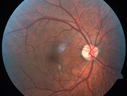
HIV Retinopathyvu 700 foisCentral white spot is an artifact. The small vascular abnormalities are from HIV
|
|

Hypertensive Retinopathy with Choroidal Ischemia Right Eye and Vision 8/200 - Blood Pressure 240/120 mmHg IgA Nephropathyvu 699 fois39-year-old man two days ago he noticed poor vision in his right eye OD: 8/200; OS: 20/20. BP is 240/120
|
|

oct fovea. odvu 699 fois
|
|
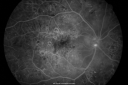
Diabetic and Pseudophakic Cystoid Macular Edema Pretreatment Mid FAvu 698 fois56-year-old woman has diabetic retinopathy in both eyes and macular edema. She is recently pseudophakic. OD 20/80, OS 20/200.
|
|

Central Retinal Vein Occlusion and Cystoid Macular Edema - Acute - Vision 20/50 vu 698 fois68-year-old man six weeks of vision loss right eye from CRVO and CME, vision right eye 20/50
|
|

Retinal Arterial Macroaneurysm - Increased Swelling after Laser vu 697 foisVisit 2 - 2 months later:
March 21, 2011. This pleasant 66-year-old woman had laser to retinal arterial microaneurysm in the right eye January 12th. Her vision was getting better, but now it is worse the last few days. She is at home taking care of her demented mother and she and her sister lift her mother a fare amount of the time. I also checked her blood pressure, which was 120/65. VISUAL ACUITY: OD 20/200
|
|

AIDS - HIV Retinopathy - Cotton Wool Spotsvu 697 fois54-year-old woman HIV positive for 18 years. Her CD4 count is currently less than 50 and she was hospitalized for pneumocystis carinii pneumonia 5 months ago. She has noticed just for the last few weeks a few blind spots in the paracentral vision. Predominantly out of her right eye when she closes her left eye, she sees a few temporal blind spots. VA 20/25 in both eyes.
|
|

Twig Branch Retinal Vein Occlusion - Recurrent Macular Edema with extended interval Lucentis Therapy after first 6 monthly Injectionsvu 697 fois39-year-old man has a macular branch retinal vein occlusion with macular edema. He had an excellent response to Lucentis for his first 6 treatments. He was last here 6 weeks ago and his vision seems OK to him. OD 20/40.
|
|

Trousseau Sign - CRVO and Pancreatic Cancervu 697 foisLeft eye shows CRVO with severe edema. The right eye has multiple small hemorrhages. Some of this is probably anemic retinopathy and some from hypercoaguability of cancer. The patient has pancreatic cancer.
|
|
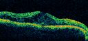
Clinically Significant Diabetic Macular Edema both Eyes with Exudatesvu 696 fois56-year-old woman with diabetes for twelve years. She has noticed blurring of the vision in the right eye for about six months. OD is 20/70, OS is 20/25.
|
|

Retinal Arterial Macroaneurysm - Increased Swelling after Laser vu 696 fois65-year-old woman was seen in the office on January 12, 2011. She has noticed decreased vision in the right eye for the last two or three weeks. She has had some discomfort in both eyes as well. She has a history of low blood pressure, but she does have high cholesterol.
VISUAL ACUITY: OD 20/60
|
|

Focal Laser for diabetic macular edemavu 695 fois66 year old female with diabetes for 45 years. VA 20/20 OU. Extrafoveal edema in the left eye treated with light focal laser.
|
|

Serous Macular Detachment Left Eye - Possible Hypertensive Choroidopathy v. Central Serous Retinopathyvu 695 fois59-year-old woman decreasing vision in the left eye for about two days. She sees a spot in front of her vision. It is a yellow spot which is just straight ahead and moves with her eye. She does have high blood pressure. She checks her blood pressure at home, and even at home her blood pressure runs very high, with the systolic running around 200 and the diastolic around 100, although occasionally it does return to the normal range.
VISUAL ACUITY: Vision OD is 20/60, OS is 20/30
|
|

Proliferative Diabetic Retinopathy and Mild Vitreous Hemorrhagevu 695 fois58-year-old woman has diabetic retinopathy in both eyes with neovascularization of the optic nerve, worse in the right eye than the left eye. Vision OD is 20/25, OS is 20/20
|
|

Central Retinal Vein Occlusion and Laser Induced Chorioretinal Anastomosisvu 695 fois84-year-old woman has age-related macular degeneration in both eyes with foveal atrophy. She also has a central retinal vein occlusion
in the left eye for which she had intravitreal Kenalog and finally chorioretinal anastomosis laser a year
ago. VISUAL ACUITY: Vision OD is 20/160, OS is 7/200
|
|
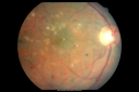
Diabetic and Pseudophakic Cystoid Macular Edema Laser Photo whole Maculavu 694 fois56-year-old woman has diabetic retinopathy in both eyes and macular edema. She is recently pseudophakic. OD 20/80, OS 20/200.
|
|
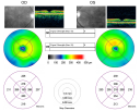
Hypertensive Retinopathy One Year later with good Vision IgA Nephropathyvu 694 foisOD 20/30, OS 20/25 One year following initial episode.
|
|

Hemicentral Retinal Vein Occlusion Vision 20/60 vu 694 fois84-year-old man has a hemicentral retinal vein occlusion in the right eye. His vision has declined since then.
VISUAL ACUITY: Vision OD is 20/60.
|
|

Cilioretinal artery occlusionvu 694 foisCilioretinal artery occlusion with early venous stasis retinopathy
|
|

Proliferative Diabetic Retinopathy - Moderate - mild NVD with NVE vu 694 foisVenous beading, vascular loops and NVE are visible on photos
|
|

Diabetic Patient with Macular Edema and Blood Pressure 200/95vu 693 fois49-year-old decreasing vision over the last year. OD is 20/80, OS 20/80. blood pressure which was 200/95.
|
|

Central Retinal Artery Occlusion Left Eye 2 Weeks Old - Still Non-Perfusionvu 693 fois85-year-old woman who had sudden severevision loss in the left eye two weeks ago. she can see a little more centrally and much more in the periphery, since about the last four days.
VISUAL ACUITY: OD 20/30, OS: Hand motion
|
|

Diabetic Retinopathy - Foveal Laser Scar Right Eyevu 693 fois74-year-old woman has diabetic retinopathy in both eyes. She has had focal laser in her right eye done elsewhere in the mid 90’s and unfortunately the laser was fairly close to the center and as time progressed she developed foveal atrophy around the scars. She has noticed her vision declining since she was here last a year ago. She does take Travatan in both eyes at night for glaucoma.
VISUAL ACUITY: OD 20/200, OS 20/40.
|
|

Capillary Non-Perfusion - Severe Non-Proliferative (Background) Diabetic Retinopathy - Type I - 20 Year Old Man Diabetic 16 yearsvu 693 fois20 year old diabetic with poor vision for the past three or four years. He has been diabetic for sixteen years and hemoglobin A1C of 10.0. 20/25 OD, 20/30 OS.
|
|

A case of Valsalva Retinopathy -2vu 693 fois
|
|

Serous Retinal Detachment - Hypertensive Retinopathy -- 1 month later - less fluid (better blood pressure) SD OCTvu 693 fois48 Year old woman resolving hypertensive serous retinal detachment.
|
|

Serous Retinal Detachment - Hypertensive Retinopathy - 3 months later - Fluid has absorbed (SD OCT VIDEO TAKES TIME TO LOAD)vu 693 fois48 Year old woman resolved hypertensive serous retinal detachment.
|
|

Central Retinal Artery Occlusion Left Eye 2 Weeks Old - Still Non-Perfusionvu 692 fois85-year-old woman who had sudden severevision loss in the left eye two weeks ago. she can see a little more centrally and much more in the periphery, since about the last four days.
VISUAL ACUITY: OD 20/30, OS: Hand motion
|
|

Non-Ischemic Central Retinal Vein Occlusion in Young Man (30 years) - Many Cotton Wool Spotsvu 692 fois30-year-old man with a history of a central retinal vein occlusion for which he was first examined on 2 months ago and his vision then was 4/200. He responded nicely to two Avastin treatments. OD is 20/20, OS is 20/60
|
|
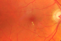
Diabetic Macular Edema - Mildvu 692 fois70 year old man with mild edema did well without treatment
|
|

Retinal Arterial Macroaneurysm - Increased Swelling after Laser vu 691 fois65-year-old woman was seen in the office on January 12, 2011. She has noticed decreased vision in the right eye for the last two or three weeks. She has had some discomfort in both eyes as well. She has a history of low blood pressure, but she does have high cholesterol.
VISUAL ACUITY: OD 20/60
|
|

FA showing NVE, NVD, non- perfusion left eye inferonasally with neovascularization at the margin of the non-perfusion (heavy leakage from NV)vu 691 fois
|
|

Branch Retinal Vein Occlusion with NVE (Neovascularization Elsewhere)vu 691 fois
|
|

Sickle Retinpoathy SC Disease - Vitreous Hemorrhage Left Eye - 29 Year oldvu 691 fois
|
|
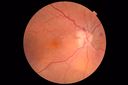
Anemic Retinopathyvu 691 foisMyelodysplasia - HgB 8 and very low platelet count
|
|

Branch Retinal Arterial Occlusion 20/20 Vision - Symptoms for 1 Dayvu 690 fois
|
|

Macular Branch Retinal Vein Occlusions (2) Left Eye - No Macular Edema (yet) vu 690 fois77-year-old woman with two macular branch retinal vein occlusions in the left eye OD 20/25
|
|
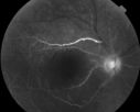
vu 690 fois60 year old male with LP vision due to extensive blood flow loss.
|
|

Montage of Hemi-Central Retinal Artery Occlusionvu 690 fois
|
|
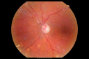
Diabetic Retinopathy - Non-Perfusion - Possible Central Retinal Vein Occlusionvu 689 fois68-year-old woman OD 20/70, OS 20/200 CYSTOID MACULAR EDEMA, POSSIBLE EITHER OCULAR ISCHEMIC SYNDROME IN THE RIGHT EYE OR OCCULT CENTRAL RETINAL VEIN OCCLUSION IN THE RIGHT EYE,S EVERE NON-PerfUSION WITH DIABETIC RETINOPATHY IN THE LEFT EYE.
|
|

Retinal Arterial Macroaneurysm - Increased Swelling after Laser vu 689 fois65-year-old woman was seen in the office on January 12, 2011. She has noticed decreased vision in the right eye for the last two or three weeks. She has had some discomfort in both eyes as well. She has a history of low blood pressure, but she does have high cholesterol.
VISUAL ACUITY: OD 20/60
|
|

Retinal Arterial Macroaneurysm - Increased Swelling after Laser vu 689 foisVisit 2 - 2 months later:
March 21, 2011. This pleasant 66-year-old woman had laser to retinal arterial microaneurysm in the right eye January 12th. Her vision was getting better, but now it is worse the last few days. She is at home taking care of her demented mother and she and her sister lift her mother a fare amount of the time. I also checked her blood pressure, which was 120/65. VISUAL ACUITY: OD 20/200
|
|

BRAO and PAMMvu 689 fois
|
|

Diabetic Macular Ischemia - Enlarged FAZ - OCT-Avu 689 fois70 year old female diagnosed with diabetes 2 years ago. Her blood pressure was normal at 116/73. She has vision loss for 6 months in both eyes. VA is 20/100 OU. OCT-A shows macular ischemia.
|
|

Proliferative Diabetic Retinopathy both eyes Type I Diabetic for 25 yearsvu 688 fois47-year-old woman with diabetes for 25 years and decreased vision for 3 months. OD 20/40, OS 20/40
|
|
vu 688 fois63-year-old man diabetic for twenty two years. His central vision is fine.
VISUAL ACUITY: OD 20/25, OS 20/25
|
|

Serous Retinal Detachment - Hypertensive Retinopathy - 165/120 vu 688 fois48-year-old woman noticed when she was blinking there was a white flash in the left eye starting about a month ago. She sees it temporally. OD 20/16, OS 20/20. There is a serous retinal detachment superotemporal to the optic nerve falling short of the fovea. Her blood pressure 165/120.
|
|
Bilateral Retinal Arteriol Occlusions - Possible Susac Syndrome vu 686 fois80-year-old woman one month ago had vision loss and a vascular occlusion in the right eye. Vision loss occurred in the left eye about 9 years ago with cotton wool spots. Patients has a 30 year history of tinnitis.
|
|

Twig Branch Retinal Vein Occlusion - Recurrent Macular Edema with extended interval Lucentis Therapy after first 6 monthly Injectionsvu 686 fois39-year-old man has a macular branch retinal vein occlusion with macular edema. He had an excellent response to Lucentis for his first 6 treatments. He was last here 6 weeks ago and his vision seems OK to him. OD 20/40.
|
|

wide angle fa late. odvu 686 foisleaking vessels
|
|

Macular Branch Retinal Vein Occlusions (2) Left Eye - No Macular Edema (yet) vu 685 fois77-year-old woman with two macular branch retinal vein occlusions in the left eye OD 20/25
|
|

Chronic Macula-off Tractional Retinal Detachment Left Eye Proliferative Diabetic Retinopathyvu 685 fois81-year-old man with diabetes for 24 years has proliferative diabetic retinopathy in both eyes with a chronic tractional retinal detachment in the left eye 4 years old. OD is 20/40, OS is 2/200
|
|

Capillary Drop Out of the FAZvu 685 foisHigh magnification image of a damaged foveal avascular zone secondary to proliferative diabetic retinopathy. Seen here are multiple laser scars and capillary drop out in FAZ.
|
|

Proliferative Diabetic Retinopathy - Fresh and Old Scatter (Panretinal) Photocoagulation (Laser) - Neovascularization is regressingvu 685 fois1 month after initial laser
|
|

Proliferative Diabetic Retinopathy both eyes Type I Diabetic for 25 yearsvu 684 fois47-year-old woman with diabetes for 25 years and decreased vision for 3 months. OD 20/40, OS 20/40
|
|

Macular Branch Retinal Vein Occlusions (2) Left Eye - No Macular Edema (yet) vu 684 fois77-year-old woman with two macular branch retinal vein occlusions in the left eye OD 20/25
|
|

Cilioretinal artery occlusion and Ophthalmic Artery Occlusion with Relative Sparing of the Central Retinal Arteryvu 684 fois80-year-old woman who says she was looking in the mirror, covered her right eye and noticed she could not see well out of the left eye. That just happened yesterday. Her last eye check was a few years ago. She also had a carotid endarterectomy on the right side in 2006 shortly after coronary bypass surgery. OD is 20/40, OS is 20/200.
|
|

Sickle Retinpoathy SC Disease - Vitreous Hemorrhage Left Eye - 29 Year oldvu 684 foisFrond of NV in the periphery
|
|
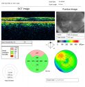
Focal Laser for diabetic macular edemavu 683 fois66 year old female with diabetes for 45 years. VA 20/20 OU. Extrafoveal edema in the left eye treated with light focal laser.
|
|

Sectoral Optic Atrophy from an old Branch Retinal Artery Occlusion vu 683 fois74-year-old woman had cataract surgery 3 months ago. Her vision in the right eye has been abnormal since. She notices a shadow in the inferior vision in the right eye. OD is 20/60, OS is 20/50
|
|

Retinal Arterial Macroaneurysm - Untreated Hypertension (250/135) - Serous Macular Detachmentvu 683 fois82 year old asian woman. VA 20/80 Headaches and vision loss for 1 month. Negative medical history but has not been to the doctor for years. (no insurance)
|
|

branch retinal vein occlusion - fluorescein angiogramvu 682 fois70 year old woman with 20/80 vision from a branch retinal vein occlusion and macular edema. She has had laser treatment and intravitreal steroids. Her vision with further laser improved to 20/50.
|
|

Diabetic Patient with Macular Edema and Blood Pressure 200/95vu 682 fois49-year-old decreasing vision over the last year. OD is 20/80, OS 20/80. blood pressure which was 200/95.
|
|

Branch Retinal Vein Occlusion with Macular Edemavu 681 foisCentral foveal thickness came to normal after laser and intravitreal Kenalog
|
|
| 4350 photos sur 44 page(s) |
 |
|
|
|
6 | |
|
|
|
|
|


