Most viewed - Retinal Vascular Disease
|

Disorganized Retinal Inner Layers (DRIL)662 views72 year old man treated with Avastin for CRVO VA is still 20/200 with absent macular edema (Only 1 month post rx)
|
|

Central Retinal Vein Occlusion - New- and old Macular Scar from AMD661 views87-year-old woman with longstaning macular scars and new vision loss left eye for 2 months. OD: 20/200, OS 20/400.
|
|

Ocular Ishemic Syndrome - Bilateral Carotid Occlusions - OD 20/60 OS 20/200 Enlarged Foveal Avascular Zone and Temporal Non-perfusion661 views75-year-old man had laryngeal cancer treated in 15 year ago with radiation. He recently was told that the carotid circulation is closed off on both sides. He does have a patent vertebral artery, which is supplying him pretty well and in addition to that his cardiac output is compromised by poor mitral valves. His consideration is to what can be done about that in the future.
VISUAL ACUITY: OD 20/60, OS 20/200
|
|

Macular Atrophy - Macular Degeneration and Previous Diabetic Macular Laser 72 Year Old Man661 views72-year-old man has age-related macular degeneration who had macular and peripheral laser done 30 years ago for diabetic retinopathy. He has developed worsening macular atrophy in both eyes
|
|

Retinal Arterial Macroaneurysm - Increased Swelling after Laser 661 viewsVisit 2 - 2 months later:
March 21, 2011. This pleasant 66-year-old woman had laser to retinal arterial microaneurysm in the right eye January 12th. Her vision was getting better, but now it is worse the last few days. She is at home taking care of her demented mother and she and her sister lift her mother a fare amount of the time. I also checked her blood pressure, which was 120/65. VISUAL ACUITY: OD 20/200
|
|

661 views63-year-old man diabetic for twenty two years. His central vision is fine.
VISUAL ACUITY: OD 20/25, OS 20/25
|
|

Proliferative Diabetic Retinopathy - Fresh and Old Scatter (Panretinal) Photocoagulation (Laser) - Neovascularization is regressing661 views1 month after initial laser
|
|

Macular Branch Retinal Vein Occlusions (2) Left Eye - No Macular Edema (yet) 660 views77-year-old woman with two macular branch retinal vein occlusions in the left eye OD 20/25
|
|

BDR OD with Edema659 views57-year-old man has diabetic retinopathy in both eyes.
Diabetic for 14 years with HgB A1C often over 10.
VISUAL ACUITY: OD 20/30, OS 20/40. PDR OS BDR OD
|
|

Cotton Wool Spots Both Eyes after Strep Pneumonia and Cardiac ICU Stay 1 Month Ago 659 views60-year-old man was sick in the hospital in 1 month ago with streptococcal pneumonia. He was in the cardiac intensive care unit and at one point he was in critical condition. He has no visual complaints. OD 20/20, OS 20/25.
|
|

Microaneurysms from Ocular Ischemic Syndrome (non-diabetic) Right Eye - Left eye CRAO 1 year ago659 views66-year-old woman lost vision in the left eye from a central retinal artery occlusion 1 year ago. Vision OD is fine. OD 20/25, OS 20/50
|
|

Central Retinal Vein Occlusion and Tractional Retinal Detachment for 3 Years (Patient is Diabetic)659 views67-year-old woman has severe vision loss in the left eye for 3 years OD 20/32, OS 3/200
|
|

Acute Branch Retinal Artery Occlusion Left Eye659 viewsThis 84 year old woman had an old BRVO in the right eye and saw a shadow for 2 days in the left eye. She has a superotemporal BRAO. The photos show retinal whitening, the OCT shows inner retinal edema and the FA shows poor circulation.
|
|

Hairy Cell Leukemia - Retinal Hemorrhage and twig Branch Vein Occlusion659 views79 year old man He has had hairy cell leukemia since 2002. He is in remission. His last blood tests were 9/2018. He just moved down here and needs a new leukemia doctor. His vision is fine.
VA OD: Dcc20/25
VA OS: Dcc20/25
IOP: TP: OD:12 OS:12
|
|

Wet AMD Small New Hemorrhage - Valsalva658 views79-year-old two weeks ago he was having difficulty seeing out of the right eye. When he covered his left eye things were distorted OD 20/50
|
|
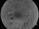
Diabetic Macular Edema and Hypertensive Retinopathy - Circinate Exudate (Ring Exudate) 658 views55-year-old woman diabetic for fifteen years and high blood pressure, especially over the last few months. She has had problems with nosebleeds, headaches, and there has been some difficulty bringing her blood pressure down. She said now the blood pressure is under control but it was running 220/120 mmHg for some time. Her vision in the right eye has been poor for four weeks with a spot in the central vision and both eyes have been blurred.
VISUAL ACUITY: Vision OD is 20/160, OS is 20/60
|
|

Cotton Wool Spots now Gone in Both Eyes657 views60-year-old man was sick in the hospital in 3 month ago with streptococcal pneumonia. He was in the cardiac intensive care unit and at one point he was in critical condition. He has no visual complaints. OD 20/20, OS 20/25. Cotton Wool Spots now GONE
|
|

Diabetic Macular Edema and Hypertensive Retinopathy - Circinate Exudate (Ring Exudate) 657 views55-year-old woman diabetic for fifteen years and high blood pressure, especially over the last few months. She has had problems with nosebleeds, headaches, and there has been some difficulty bringing her blood pressure down. She said now the blood pressure is under control but it was running 220/120 mmHg for some time. Her vision in the right eye has been poor for four weeks with a spot in the central vision and both eyes have been blurred.
VISUAL ACUITY: Vision OD is 20/160, OS is 20/60
|
|

Purtchers Retinopathy with Bilateral Central Retinal Artery Occlusions 656 views50-year-old man traumatic chest compression and leg crushing injury. OD 1/200, OS 3/200
|
|

Serous Retinal Detachment - Hypertensive Retinopathy - 165/120 - Fluorescein Angiogram656 views48-year-old woman noticed when she was blinking there was a white flash in the left eye starting about a month ago. She sees it temporally. OD 20/16, OS 20/20. There is a serous retinal detachment superotemporal to the optic nerve falling short of the fovea. Her blood pressure 165/120.
|
|

Central Retinal Vein Occlusion Recurrent Edema 6 weeks after Lucentis - Nonperfusion Temporally655 views66-year-old woman has a central retinal vein occlusion in the right eye with macular edema. She had intravitreal Lucentis treatment six weeks ago and she has noticed over the last week or two her vision is declining.
VISUAL ACUITY: OD 20/70, OS 20/30.
|
|

FAF Montage of Diabetic Retinopathy654 views70-year old male with severe Diabetic Retinopathy. VA at visit was 20/100 with no improvement with pinhole. Patient had recently undergone cataract surgery to try to regain some of his vison. Panoretinal Photocoagulation scars 360 degrees around the retina...
|
|

Central Retinal Artery Occlusion Left Eye 2 Weeks Old - Still Non-Perfusion653 views85-year-old woman who had sudden severevision loss in the left eye two weeks ago. she can see a little more centrally and much more in the periphery, since about the last four days.
VISUAL ACUITY: OD 20/30, OS: Hand motion
|
|

653 views
|
|

Twig Branch Retinal Vein Occlusion - Recurrent Macular Edema with extended interval Lucentis Therapy after first 6 monthly Injections653 views39-year-old man has a macular branch retinal vein occlusion with macular edema. He had an excellent response to Lucentis for his first 6 treatments. He was last here 6 weeks ago and his vision seems OK to him. OD 20/40.
|
|

Serous Retinal Detachment - Hypertensive Retinopathy - 165/120 - Fluorescein Angiogram653 views48-year-old woman noticed when she was blinking there was a white flash in the left eye starting about a month ago. She sees it temporally. OD 20/16, OS 20/20. There is a serous retinal detachment superotemporal to the optic nerve falling short of the fovea. Her blood pressure 165/120.
|
|

Macular Branch Retinal Vein Occlusions (2) Left Eye - No Macular Edema (yet) 652 views77-year-old woman with two macular branch retinal vein occlusions in the left eye OD 20/25
|
|
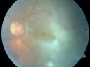
Chronic Macula-off Tractional Retinal Detachment Left Eye Proliferative Diabetic Retinopathy652 views81-year-old man with diabetes for 24 years has proliferative diabetic retinopathy in both eyes with a chronic tractional retinal detachment in the left eye 4 years old. OD is 20/40, OS is 2/200
|
|

PDR - FAF652 views
|
|

Acute Branch Retinal Artery Occlusion Left Eye652 viewsThis 84 year old woman had an old BRVO in the right eye and saw a shadow for 2 days in the left eye. She has a superotemporal BRAO. The photos show retinal whitening, the OCT shows inner retinal edema and the FA shows poor circulation.
|
|

Purtchers Retinopathy with Bilateral Central Retinal Artery Occlusions 651 views50-year-old man traumatic chest compression and leg crushing injury. OD 1/200, OS 3/200
|
|

Retinal Arterial Macroaneurysm - Increased Swelling after Laser 651 views65-year-old woman was seen in the office on January 12, 2011. She has noticed decreased vision in the right eye for the last two or three weeks. She has had some discomfort in both eyes as well. She has a history of low blood pressure, but she does have high cholesterol.
VISUAL ACUITY: OD 20/60
|
|

Pattern Dystrophy - Adult Vitelliform Macular Degeneration651 views72-year-old woman 2 months post ECCE with poor vision since surgery especially left eye.
VISUAL ACUITY: OD 20/50, OS 20/200
|
|
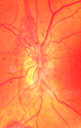
PDR NVD left eye and NVE right eye651 views41 year old diabetic woman with PDR in both eyes. High risk in the left eye. She had PRP, Avastin, and vitrectomy x 2 in the left eye with final vision of 20/25. The right eye had eventually PRP laser
|
|

branch retinal vein occlusion - fluorescein angiogram650 views70 year old woman with 20/80 vision from a branch retinal vein occlusion and macular edema. She has had laser treatment and intravitreal steroids. Her vision with further laser improved to 20/50.
|
|
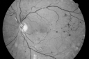
Diabetic Retinopathy - Non-Perfusion - Possible Central Retinal Vein Occlusion650 views68-year-old woman OD 20/70, OS 20/200 CYSTOID MACULAR EDEMA, POSSIBLE EITHER OCULAR ISCHEMIC SYNDROME IN THE RIGHT EYE OR OCCULT CENTRAL RETINAL VEIN OCCLUSION IN THE RIGHT EYE,S EVERE NON-PerfUSION WITH DIABETIC RETINOPATHY IN THE LEFT EYE.
|
|

Bilateral Retinal Arteriol Occlusions - Possible Susac Syndrome 650 views80-year-old woman one month ago had vision loss and a vascular occlusion in the right eye. Vision loss occurred in the left eye about 9 years ago with cotton wool spots. Patients has a 30 year history of tinnitis.
|
|

Diabetic Retinopathy - Foveal Laser Scar Right Eye650 views74-year-old woman has diabetic retinopathy in both eyes. She has had focal laser in her right eye done elsewhere in the mid 90’s and unfortunately the laser was fairly close to the center and as time progressed she developed foveal atrophy around the scars. She has noticed her vision declining since she was here last a year ago. She does take Travatan in both eyes at night for glaucoma.
VISUAL ACUITY: OD 20/200, OS 20/40.
|
|

Capillary Non-Perfusion - Severe Non-Proliferative (Background) Diabetic Retinopathy - Type I - 20 Year Old Man Diabetic 16 years650 views20 year old diabetic with poor vision for the past three or four years. He has been diabetic for sixteen years and hemoglobin A1C of 10.0. 20/25 OD, 20/30 OS.
|
|

Capillary Non-Perfusion - Severe Non-Proliferative (Background) Diabetic Retinopathy - Type I - 20 Year Old Man Diabetic 16 years650 views20 year old diabetic with poor vision for the past three or four years. He has been diabetic for sixteen years and hemoglobin A1C of 10.0. 20/25 OD, 20/30 OS.
|
|

Regressed Proliferative Diabetic Retinopathy with Recurrent Vitreous Hemorrhage from Vitreoretinal Traction over a Major Retinal Vessel (Vein)650 views56-year-old woman has had recurrent vitreous hemorrhage in the left eye from vitreoretinal traction. Diabetes Mellitus for 20 years and excellent response to vitrectomy in the right eye about 1 year ago for recurrent vitreous hemorrhage from traction over a major retinal vessel.
VISUAL ACUITY: Vision OS is 20/200
|
|

Macula Threatening Diabetic Traction Retinal Detachment - Proliferative Diabetic Retinopathy - Vitreous Hemorrhage650 viewsVision 20/50 but tractional retinal detachment supero-temporally is extending toward fovea and vessels are still active on optic nerve.
|
|

Branch Retinal Vein Occlusion with NVE (Neovascularization Elsewhere)650 views
|
|

Vitreo-Traction due to Diabetic Retinopathy650 viewsPatient comes in for follow up on her diabetes. VA is 20/30 in the right eye. Fundus photos shows thick traction nasally to the optic nerve forming a ring around the macula. Will follow up in 6-months.
|
|

Trousseau Sign - CRVO and Pancreatic Cancer650 viewsLeft eye shows CRVO with severe edema. The right eye has multiple small hemorrhages. Some of this is probably anemic retinopathy and some from hypercoaguability of cancer. The patient has pancreatic cancer.
|
|

Bilateral Branch Retinal Artery Occlusion - Intraarterial (hollenhorst) plaques visible650 views67 year old with acute visual field loss in each eye. Vision is 20/25 right eye 20/32 left eye. Images show intra-arterial plaques in each eye. The right eye on the optic nerve and the left eye in a branch retinal artery.
|
|

Retinal Vasculopathy associated with Facioscapulohumeral Muscular Dystrophy late fa649 views59-year-old woman had a blood pressure spike to 170/90 and was on medications for that. She does have facioscapulohumeral muscular dystrophy, which runs in her family. She has a cousin with the same disorder.
VISUAL ACUITY: Her vision is 20/25 OU. IOP: OD 21, OS 18.
There is no posterior vitreous separation, telangiectatic vessels near the macula. There are patchy retinal hemorrhages. There is no edema or proliferation.
|
|

Proliferative Diabetic Retinopathy - Vitreous Hemorrhage and Tractional Retinal Detachment Left Eye649 views52-year-old man has been diabetic for nineteen years developed substantial vision loss over the last month or two. OD 20/70, OS 20/200.
|
|
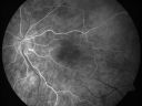
Cilioretinal artery occlusion and Ophthalmic Artery Occlusion with Relative Sparing of the Central Retinal Artery649 views80-year-old woman who says she was looking in the mirror, covered her right eye and noticed she could not see well out of the left eye. That just happened yesterday. Her last eye check was a few years ago. She also had a carotid endarterectomy on the right side in 2006 shortly after coronary bypass surgery. OD is 20/40, OS is 20/200.
|
|
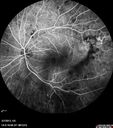
Proliferative Diabetic Retinopathy and Vitreous Hemorrhage - FA shows leakage of dye from NVE649 views
|
|

Hypertensive Retinopathy - BP 240 / 130 mmHg - Grade 4649 views71 year old woman who claims to be asymptomatic with decreased vision, macular edema, cotton wool spots, subretinal fluid and optic nerve edema
|
|
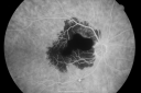
Ruptured Retinal Arterial Macroaneurysm - Submacular Hemorrhage - Vision 20/400 never recovered - Hypertension 205/94648 views
|
|

Central Retinal Vein Occlusion and Laser Induced Chorioretinal Anastomosis648 views84-year-old woman has age-related macular degeneration in both eyes with foveal atrophy. She also has a central retinal vein occlusion
in the left eye for which she had intravitreal Kenalog and finally chorioretinal anastomosis laser a year
ago. VISUAL ACUITY: Vision OD is 20/160, OS is 7/200
|
|

648 views
|
|

Prominent Posterior Hyaloid with Background Diabetic Retinopathy648 viewsPatient comes in for follow up on her Diabetic Retinopathy and glaucoma. Patient's VA was 20/30 in the left eye. Fundus exam presents a Posterior Hyaloid with hemorrhage inferiorily. Patient will be seen again in 6-months for follow up.
|
|

Acute Branch Retinal Artery Occlusion Left Eye648 viewsThis 84 year old woman had an old BRVO in the right eye and saw a shadow for 2 days in the left eye. She has a superotemporal BRAO. The photos show retinal whitening, the OCT shows inner retinal edema and the FA shows poor circulation.
|
|

PDR NVD left eye and NVE right eye648 views41 year old diabetic woman with PDR in both eyes. High risk in the left eye. She had PRP, Avastin, and vitrectomy x 2 in the left eye with final vision of 20/25. The right eye had eventually PRP laser
|
|

Diabetic Vitreous Hemorrhage Left Eye647 views35-year-old man diabetic for the last 25 years. His hemoglobin A1C has been running between 10 and 11. He was doing fine and then when he woke up from sleeping last night he noticed sudden vision loss in his left eye OD 20/30, OS 20/200.
|
|

Diabetic Macular Edema and Hypertensive Retinopathy - Circinate Exudate (Ring Exudate) 647 views55-year-old woman diabetic for fifteen years and high blood pressure, especially over the last few months. She has had problems with nosebleeds, headaches, and there has been some difficulty bringing her blood pressure down. She said now the blood pressure is under control but it was running 220/120 mmHg for some time. Her vision in the right eye has been poor for four weeks with a spot in the central vision and both eyes have been blurred.
VISUAL ACUITY: Vision OD is 20/160, OS is 20/60
|
|

Malignany Hypertension - BP 250/130 - Vision 1/200 in each eye - Diabetic647 views68-year-old woman poor vision in both eyes for the last few years. She is diabetic since 1989. Her blood pressure has been very high. She was in the hospital Friday. Her blood pressure during that visit was 250/130 and her blood pressure in your office she reports was somewhere around the same. I just checked her blood pressure at the beginning of my visit and it was 200/120 in her right arm sitting. Her vision is poor enough that she can barely see to walk around. She has also been feeling nauseous
|
|

Retinal Arterial Macroaneurysm - Ruptured and self-sealed after hemorrhage647 views6 Month Follow-up
91-year-old woman had a retinal arterial macroaneurysm in the right eye 6 Month Follow-up Vision is 20/200.
|
|
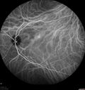
Serous Retinal Detachment - Hypertensive Retinopathy - 165/120 - Indocyanine Green Angiogram647 views48-year-old woman noticed when she was blinking there was a white flash in the left eye starting about a month ago. She sees it temporally. OD 20/16, OS 20/20. There is a serous retinal detachment superotemporal to the optic nerve falling short of the fovea. Her blood pressure 165/120.
|
|

Choroidal Nevus647 views
|
|

Diabetic Retinopathy - Non-Perfusion - Possible Central Retinal Vein Occlusion646 views68-year-old woman OD 20/70, OS 20/200 CYSTOID MACULAR EDEMA, POSSIBLE EITHER OCULAR ISCHEMIC SYNDROME IN THE RIGHT EYE OR OCCULT CENTRAL RETINAL VEIN OCCLUSION IN THE RIGHT EYE,S EVERE NON-PerfUSION WITH DIABETIC RETINOPATHY IN THE LEFT EYE.
|
|

Central Retinal Vein Occlusion - New- and old Macular Scar from AMD646 views87-year-old woman with longstaning macular scars and new vision loss left eye for 2 months. OD: 20/200, OS 20/400.
|
|

Hemi Central Retinal Vein Occlusion 35 year old non-compliant man646 views35-year-old man hazy vision in his left eye for about the last three days. OD 20/20, OS 20/30
|
|

Hypertensive Retinopathy with Choroidal Ischemia Right Eye and Vision 8/200 - Blood Pressure 240/120 mmHg IgA Nephropathy646 views39-year-old man two days ago he noticed poor vision in his right eye OD: 8/200; OS: 20/20. BP is 240/120
|
|

BRAO and PAMM646 views
|
|

Retinal Vasculopathy associated with Facioscapulohumeral Muscular Dystrophy late fa645 views59-year-old woman had a blood pressure spike to 170/90 and was on medications for that. She does have facioscapulohumeral muscular dystrophy, which runs in her family. She has a cousin with the same disorder.
VISUAL ACUITY: Her vision is 20/25 OU. IOP: OD 21, OS 18.
There is no posterior vitreous separation, telangiectatic vessels near the macula. There are patchy retinal hemorrhages. There is no edema or proliferation.
|
|

Hypertensive Retinopathy645 viewsPatient comes in complaining of spots in vision in both eyes. VA was 20/25 - right eye and 20/20- left eye. Fundus exam reveals little hemorrhages with cotton wool spots due to hypertension and anemia.
|
|

Acute Branch Retinal Artery Occlusion Left Eye645 viewsThis 84 year old woman had an old BRVO in the right eye and saw a shadow for 2 days in the left eye. She has a superotemporal BRAO. The photos show retinal whitening, the OCT shows inner retinal edema and the FA shows poor circulation.
|
|

Hairy Cell Leukemia - Retinal Hemorrhage and twig Branch Vein Occlusion645 views79 year old man He has had hairy cell leukemia since 2002. He is in remission. His last blood tests were 9/2018. He just moved down here and needs a new leukemia doctor. His vision is fine.
VA OD: Dcc20/25
VA OS: Dcc20/25
IOP: TP: OD:12 OS:12
|
|

Hypertensive Retinopathy with Choroidal Ischemia Right Eye and Vision 8/200 - Blood Pressure 240/120 mmHg IgA Nephropathy644 views39-year-old man two days ago he noticed poor vision in his right eye OD: 8/200; OS: 20/20. BP is 240/120
|
|

Hemicentral Retinal Vein Occlusion Vision 20/60 644 views84-year-old man has a hemicentral retinal vein occlusion in the right eye. His vision has declined since then.
VISUAL ACUITY: Vision OD is 20/60.
|
|

BDR644 views
|
|

Retinal Vasculopathy associated with Facioscapulohumeral Muscular Dystrophy early fa643 views59-year-old woman had a blood pressure spike to 170/90 and was on medications for that. She does have facioscapulohumeral muscular dystrophy, which runs in her family. She has a cousin with the same disorder.
VISUAL ACUITY: Her vision is 20/25 OU. IOP: OD 21, OS 18.
There is no posterior vitreous separation, telangiectatic vessels near the macula. There are patchy retinal hemorrhages. There is no edema or proliferation.
|
|

Proliferative Diabetic Retinopathy - Vitreous Hemorrhage and Tractional Retinal Detachment Left Eye643 views52-year-old man has been diabetic for nineteen years developed substantial vision loss over the last month or two. OD 20/70, OS 20/200.52-year-old man has been diabetic for nineteen years developed substantial vision loss over the last month or two. OD 20/70, OS 20/200.
|
|

Branch Retinal Arterial Occlusion 20/20 Vision - Symptoms for 1 Day643 views75-year-old man was seen in the office on 4/6/2010. He noticed a sudden vision change in the right eye yesterday morning. 20/20 Vision both Eyes
|
|

NVD - proliferative diabetic retinopathy643 views
|
|

Trousseau Sign - CRVO and Pancreatic Cancer643 viewsLeft eye shows CRVO with severe edema. The right eye has multiple small hemorrhages. Some of this is probably anemic retinopathy and some from hypercoaguability of cancer. The patient has pancreatic cancer.
|
|
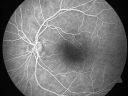
Serous Macular Detachment Left Eye - Possible Hypertensive Choroidopathy v. Central Serous Retinopathy642 views59-year-old woman decreasing vision in the left eye for about two days. She sees a spot in front of her vision. It is a yellow spot which is just straight ahead and moves with her eye. She does have high blood pressure. She checks her blood pressure at home, and even at home her blood pressure runs very high, with the systolic running around 200 and the diastolic around 100, although occasionally it does return to the normal range.
VISUAL ACUITY: Vision OD is 20/60, OS is 20/30
|
|

Proliferative Diabetic Retinopathy - Vitreous Hemorrhage and Tractional Retinal Detachment Left Eye642 views52-year-old man has been diabetic for nineteen years developed substantial vision loss over the last month or two. OD 20/70, OS 20/200.
|
|

Proliferative Diabetic Retinopathy in patient with Moyamoya Disease642 views28-year-old womandiabetic since age nine, and she also has had multiple strokes. OD is 20/30, OS is 20/30.
|
|
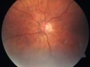
AIDS - HIV Retinopathy - Cotton Wool Spots642 views54-year-old woman HIV positive for 18 years. Her CD4 count is currently less than 50 and she was hospitalized for pneumocystis carinii pneumonia 5 months ago. She has noticed just for the last few weeks a few blind spots in the paracentral vision. Predominantly out of her right eye when she closes her left eye, she sees a few temporal blind spots. VA 20/25 in both eyes.
|
|
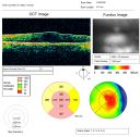
PDR with heavy PRP laser and worsening macular edema map640 views51-year-old woman has had extensive laser in both eyes done years ago elsewhere. Her left eye has gradually been declining with macular edema.
VISUAL ACUITY: OD 20/40, OS 20/40
|
|

New BRVO Left Eye Old BRVO Right Eye - Left Eye Shows Twig Superior Vein Occlusion with Edema640 views90-year-old man decreased vision maybe one year since cataract surgery he is no longer able to read. OD 20/200, OS 20/200
|
|

Central Retinal Artery Occlusion Left Eye 2 Weeks Old - Still Non-Perfusion640 views85-year-old woman who had sudden severevision loss in the left eye two weeks ago. she can see a little more centrally and much more in the periphery, since about the last four days.
VISUAL ACUITY: OD 20/30, OS: Hand motion
|
|

Central Retinal Artery Occlusion Left Eye 2 Weeks Old - Still Non-Perfusion640 views85-year-old woman who had sudden severevision loss in the left eye two weeks ago. she can see a little more centrally and much more in the periphery, since about the last four days.
VISUAL ACUITY: OD 20/30, OS: Hand motion
|
|

Proliferative Sickle Retinopathy - Hemoglobin SC640 views53-year-old woman with a new spider-like floater in the right eye for about six days. She sees one big floater and another little one. She does have hemoglobin SC hemoglobinopathy. VISUAL ACUITY: Vision OD is 20/20; OS is 20/50, PH is 20/20.
|
|

Branch Retinal Vein Occlusion with NVE (Neovascularization Elsewhere)640 viewsOne month post laser
|
|

Trousseau Sign - CRVO and Pancreatic Cancer640 viewsLeft eye shows CRVO with severe edema. The right eye has multiple small hemorrhages. Some of this is probably anemic retinopathy and some from hypercoaguability of cancer. The patient has pancreatic cancer.
|
|

Holenhorst Plaque - Small and asymptomatic640 views72 year old woman with mild dry ARMD. VA 20/40 OU. Asymptomatic plaque noted in the right eye.
|
|

Non-Ischemic Central Retinal Vein Occlusion in Young Man (30 years) - Many Cotton Wool Spots639 views30-year-old man with a history of a central retinal vein occlusion for which he was first examined on 2 months ago and his vision then was 4/200. He responded nicely to two Avastin treatments. OD is 20/20, OS is 20/60
|
|

Trousseau Sign - CRVO and Pancreatic Cancer639 viewsLeft eye shows CRVO with severe edema. The right eye has multiple small hemorrhages. Some of this is probably anemic retinopathy and some from hypercoaguability of cancer. The patient has pancreatic cancer.
|
|
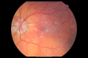
Diabetic Macular Edema with Circinate Exudate 638 views74-year-old man has diabetic macular edema in both eyes and background diabetic retinopathy. His vision has blurred over the last few months. His blood sugars have been fluctuating.
VISUAL ACUITY: Vision OD is 20/25, OS is 20/80
|
|

Central Retinal Artery Occlusion Left Eye 2 Weeks Old - Still Non-Perfusion638 views85-year-old woman who had sudden severevision loss in the left eye two weeks ago. she can see a little more centrally and much more in the periphery, since about the last four days.
VISUAL ACUITY: OD 20/30, OS: Hand motion
|
|

BDR638 views
|
|

Retinal Arterial Macroaneurysm - Untreated Hypertension (250/135) - Serous Macular Detachment638 views82 year old asian woman. VA 20/80 Headaches and vision loss for 1 month. Negative medical history but has not been to the doctor for years. (no insurance)
|
|
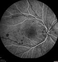
CRVO with Paracentral Acute Middle Maculopath - PAMM638 views56 year old woman with sudden vision loss, hemorrhages in all four quadrants. FA shows good retinal circulation. The OCT shows PAMM lesions in the affected eye. Vision did improve on second visit from 20/160 to 20/60 in about a month
|
|

Lipemia Retinalis638 views31 year old man The patient at the time of imaging had triglycerides of >5680, cholesterol of 1136.
He also does have uncontrolled hypertension and insulin dependent type 2 diabetes, which may be the reason for vitreous extension of the lipids.
|
|
| 4368 files on 44 page(s) |
 |
|
|
|
|
8 | |
|
|
|
|


