Most viewed - Retinal Vascular Disease
|
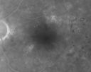
Clinically Significant Diabetic Macular Edema both Eyes with Exudates628 views56-year-old woman with diabetes for twelve years. She has noticed blurring of the vision in the right eye for about six months. OD is 20/70, OS is 20/25.
|
|

Preretinal Fibrosis - Tractional Retinal Detachment - Proliferative Diabetic Retinopathy 628 views33-year-old woman has proliferative diabetic retinopathy in the left eye with fibrosis and tractional retinal detachment. Her vision is hazy in that eye. VISUAL ACUITY: Vision OD is 20/30, OS 20/120
|
|

Intraluminal Plaque Retinal Vessel 628 views69 year old female referred with atypical plaque in the right retina. Her vision is normal. The plaque looks to be adjacent to the blood column visible on the images and the SC OCT shows it to be in the vessel wall.
|
|

Lipemia Retinalis628 views31 year old man The patient at the time of imaging had triglycerides of >5680, cholesterol of 1136.
He also does have uncontrolled hypertension and insulin dependent type 2 diabetes, which may be the reason for vitreous extension of the lipids.
|
|

Diabetic Retinopathy - Non-Perfusion - Possible Central Retinal Vein Occlusion627 views68-year-old woman OD 20/70, OS 20/200 CYSTOID MACULAR EDEMA, POSSIBLE EITHER OCULAR ISCHEMIC SYNDROME IN THE RIGHT EYE OR OCCULT CENTRAL RETINAL VEIN OCCLUSION IN THE RIGHT EYE,S EVERE NON-PerfUSION WITH DIABETIC RETINOPATHY IN THE LEFT EYE.
|
|

Hypertensive Retinopathy with Choroidal Ischemia Right Eye and Vision 8/200 - Blood Pressure 240/120 mmHg IgA Nephropathy627 views39-year-old man two days ago he noticed poor vision in his right eye OD: 8/200; OS: 20/20. BP is 240/120
|
|

Branch Retinal Arterial Occlusion 20/20 Vision - Symptoms for 1 Day627 views
|
|
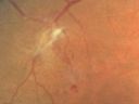
Proliferative Diabetic Retinopathy and Mild Vitreous Hemorrhage627 views58-year-old woman has diabetic retinopathy in both eyes with neovascularization of the optic nerve, worse in the right eye than the left eye. Vision OD is 20/25, OS is 20/20
|
|

branch retinal vein occlusion - fluorescein angiogram626 views70 year old woman with 20/80 vision from a branch retinal vein occlusion and macular edema. She has had laser treatment and intravitreal steroids. Her vision with further laser improved to 20/50.
|
|
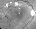
Clinically Significant Diabetic Macular Edema both Eyes with Exudates626 views56-year-old woman with diabetes for twelve years. She has noticed blurring of the vision in the right eye for about six months. OD is 20/70, OS is 20/25.
|
|
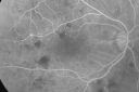
Diabetic Macular Edema and Hypertensive Retinopathy - Circinate Exudate (Ring Exudate) 626 views55-year-old woman diabetic for fifteen years and high blood pressure, especially over the last few months. She has had problems with nosebleeds, headaches, and there has been some difficulty bringing her blood pressure down. She said now the blood pressure is under control but it was running 220/120 mmHg for some time. Her vision in the right eye has been poor for four weeks with a spot in the central vision and both eyes have been blurred.
VISUAL ACUITY: Vision OD is 20/160, OS is 20/60
|
|

NVD - proliferative diabetic retinopathy Heidelberg False Color626 views
|
|

Disorganized Retinal Inner Layers (DRIL)626 views72 year old man treated with Avastin for CRVO VA is still 20/200 with absent macular edema (Only 1 month post rx)
|
|

Diabetic Patient with Macular Edema and Blood Pressure 200/95625 views49-year-old decreasing vision over the last year. OD is 20/80, OS 20/80. blood pressure which was 200/95.
|
|

Chronic - Old Diabetic Tractional Retinal Detachment Left Eye625 views77-year-old woman Vision OD is 20/40, OS is 7/200OD: Vertical C/D ratio is 0.3. There is panretinal laser. The macula is flat and attached.
OS: Vertical C/D ratio is 0.3. There is panretinal laser. There is a tractional retinal detachment with preretinal fibrosis nasal to the optic nerves.
|
|

Holenhorst Plaque - Small and asymptomatic625 views72 year old woman with mild dry ARMD. VA 20/40 OU. Asymptomatic plaque noted in the right eye.
|
|

Panretinal photocoagulation - PRP laser for proliferative diabetic retinopathy625 viewsWide angle image
|
|

Central Retinal Vein Occlusion and Cystoid Macular Edema - Acute - Vision 20/50 624 views68-year-old man six weeks of vision loss right eye from CRVO and CME, vision right eye 20/50
|
|

Diabetic Retinopathy - Non-Perfusion - Possible Central Retinal Vein Occlusion624 views68-year-old woman OD 20/70, OS 20/200 CYSTOID MACULAR EDEMA, POSSIBLE EITHER OCULAR ISCHEMIC SYNDROME IN THE RIGHT EYE OR OCCULT CENTRAL RETINAL VEIN OCCLUSION IN THE RIGHT EYE,S EVERE NON-PerfUSION WITH DIABETIC RETINOPATHY IN THE LEFT EYE.
|
|

Retinal Vasculopathy associated with Facioscapulohumeral Muscular Dystrophy mid fa623 views59-year-old woman had a blood pressure spike to 170/90 and was on medications for that. She does have facioscapulohumeral muscular dystrophy, which runs in her family. She has a cousin with the same disorder.
VISUAL ACUITY: Her vision is 20/25 OU. IOP: OD 21, OS 18.
There is no posterior vitreous separation, telangiectatic vessels near the macula. There are patchy retinal hemorrhages. There is no edema or proliferation.
|
|

Hypertensive Retinopathy One Year later with good Vision IgA Nephropathy623 viewsOD 20/30, OS 20/25 One year following initial episode.
|
|
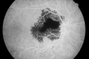
Ruptured Retinal Arterial Macroaneurysm - Submacular Hemorrhage - Vision 20/400 never recovered - Hypertension 205/94623 views
|
|

Acute Branch Retinal Artery Occlusion Left Eye623 viewsThis 84 year old woman had an old BRVO in the right eye and saw a shadow for 2 days in the left eye. She has a superotemporal BRAO. The photos show retinal whitening, the OCT shows inner retinal edema and the FA shows poor circulation.
|
|

Trousseau Sign - CRVO and Pancreatic Cancer623 viewsLeft eye shows CRVO with severe edema. The right eye has multiple small hemorrhages. Some of this is probably anemic retinopathy and some from hypercoaguability of cancer. The patient has pancreatic cancer.
|
|

Proliferative Diabetic Retinopathy - Vitreous Hemorrhage and Tractional Retinal Detachment Left Eye622 views52-year-old man has been diabetic for nineteen years developed substantial vision loss over the last month or two. OD 20/70, OS 20/200.
|
|
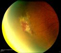
Sickle Cell622 views
|
|

Diabetic Retinopathy - Non-Perfusion - Possible Central Retinal Vein Occlusion621 views68-year-old woman OD 20/70, OS 20/200 CYSTOID MACULAR EDEMA, POSSIBLE EITHER OCULAR ISCHEMIC SYNDROME IN THE RIGHT EYE OR OCCULT CENTRAL RETINAL VEIN OCCLUSION IN THE RIGHT EYE,S EVERE NON-PerfUSION WITH DIABETIC RETINOPATHY IN THE LEFT EYE.
|
|
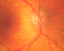
Retinal Arterial Atheromata621 views68-year-old woman with records going back 7 years, which clearly document retinal arterial plaques in both eyes.
|
|

Central Retinal Vein Occlusion and Tractional Retinal Detachment for 3 Years (Patient is Diabetic)621 views67-year-old woman has severe vision loss in the left eye for 3 years OD 20/32, OS 3/200
|
|

BRAO and PAMM621 views
|
|
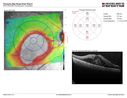
Self Sealed Retinal Arteriol Macroaneurysm621 viewsMAP shows edema and some subretinal fluid
|
|

Diabetic and Pseudophakic Cystoid Macular Edema Late FA - Leakage Macular and Nerve620 views56-year-old woman has diabetic retinopathy in both eyes and macular edema. She is recently pseudophakic. OD 20/80, OS 20/200.
|
|
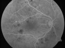
Diabetic Macular Edema and Hypertensive Retinopathy - Circinate Exudate (Ring Exudate) 620 views55-year-old woman diabetic for fifteen years and high blood pressure, especially over the last few months. She has had problems with nosebleeds, headaches, and there has been some difficulty bringing her blood pressure down. She said now the blood pressure is under control but it was running 220/120 mmHg for some time. Her vision in the right eye has been poor for four weeks with a spot in the central vision and both eyes have been blurred.
VISUAL ACUITY: Vision OD is 20/160, OS is 20/60
|
|

Retinal Arterial Macroaneurysm - Untreated Hypertension (250/135) - Serous Macular Detachment620 views82 year old asian woman. VA 20/80 Headaches and vision loss for 1 month. Negative medical history but has not been to the doctor for years. (no insurance)
|
|

Proliferative Diabetic Retinopathy in patient with Moyamoya Disease619 views28-year-old womandiabetic since age nine, and she also has had multiple strokes. OD is 20/30, OS is 20/30.
|
|

Non-Ischemic Central Retinal Vein Occlusion in Young Man (30 years) - Many Cotton Wool Spots619 views30-year-old man with a history of a central retinal vein occlusion for which he was first examined on 2 months ago and his vision then was 4/200. He responded nicely to two Avastin treatments. OD is 20/20, OS is 20/60
|
|

Twig Branch Retinal Vein Occlusion - Recurrent Macular Edema with extended interval Lucentis Therapy after first 6 monthly Injections619 views39-year-old man has a macular branch retinal vein occlusion with macular edema. He had an excellent response to Lucentis for his first 6 treatments. He was last here 6 weeks ago and his vision seems OK to him. OD 20/40.
|
|

PDR - Mild Vitreous Hemorrhage and Good Vision618 views58-year-old woman has diabetic retinopathy in both eyes with neovascularization of the optic nerve, worse in the right eye than the left eye. She has had panretinal laser in both eyes and vitreous hemorrhage in the left eye. Her vision, since I saw her last in June, is about the same.
VISUAL ACUITY: Vision OD is 20/25, OS is 20/20.
|
|

618 views49-year-old woman has central retinal vein occlusion right eye with fluctuating vision for a few month now with OD 20/100. IOP: OD 20
|
|

Retinal Arterial Atheromata618 views68-year-old woman with records going back 7 years, which clearly document retinal arterial plaques in both eyes.
|
|

Bilateral Retinal Arteriol Occlusions - Possible Susac Syndrome 618 views80-year-old woman one month ago had vision loss and a vascular occlusion in the right eye. Vision loss occurred in the left eye about 9 years ago with cotton wool spots. Patients has a 30 year history of tinnitis.
|
|

Orbital Cellulitis - Optic nerve compression with ocular ischemia618 views81 year old woman with constricted eye movement, orbital cellulitis and severe vision loss in the right eye (LP). She has whitening of the retina and the OCT shows ischemic changes as well as subretinal fluid and blood. The vision in this eye declined to NLP and the eye remained ischemic despite control of her infection.
|
|

PDR NVD left eye and NVE right eye618 views41 year old diabetic woman with PDR in both eyes. High risk in the left eye. She had PRP, Avastin, and vitrectomy x 2 in the left eye with final vision of 20/25. The right eye had eventually PRP laser
|
|

Mild Proliferative Diabetic Retinopathy618 viewsRight eye has NVE and left eye has NVD and NVE. VA 20/25 OU.
|
|

Diabetic Macular Edema Left Eye - Post Laser617 views82-year-old woman diabetic for many years, last eye exam 5 years ago with gradual vision loss. OD 20/60, OS 20/70. IOP: OD 16, OS 17.
|
|

Hypertensive Retinopathy with Choroidal Ischemia Right Eye and Vision 8/200 - Blood Pressure 240/120 mmHg IgA Nephropathy617 views39-year-old man two days ago he noticed poor vision in his right eye OD: 8/200; OS: 20/20. BP is 240/120
|
|

Ocular Ishemic Syndrome - Bilateral Carotid Occlusions - OD 20/60 OS 20/200 Enlarged Foveal Avascular Zone and Temporal Non-perfusion617 views75-year-old man had laryngeal cancer treated in 15 year ago with radiation. He recently was told that the carotid circulation is closed off on both sides. He does have a patent vertebral artery, which is supplying him pretty well and in addition to that his cardiac output is compromised by poor mitral valves. His consideration is to what can be done about that in the future.
VISUAL ACUITY: OD 20/60, OS 20/200
|
|

Proliferative Diabetic Retinopathy - Moderate - mild NVD with NVE 617 viewsFAF
|
|

BRAO and PAMM617 views
|
|

Trousseau Sign - CRVO and Pancreatic Cancer617 viewsLeft eye shows CRVO with severe edema. The right eye has multiple small hemorrhages. Some of this is probably anemic retinopathy and some from hypercoaguability of cancer. The patient has pancreatic cancer.
|
|

Cotton Wool Spots now Gone in Both Eyes616 views60-year-old man was sick in the hospital in 3 month ago with streptococcal pneumonia. He was in the cardiac intensive care unit and at one point he was in critical condition. He has no visual complaints. OD 20/20, OS 20/25. Cotton Wool Spots now GONE
|
|
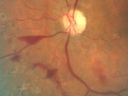
Proliferative Diabetic Retinopathy and Mild Vitreous Hemorrhage616 views58-year-old woman has diabetic retinopathy in both eyes with neovascularization of the optic nerve, worse in the right eye than the left eye. Vision OD is 20/25, OS is 20/20
|
|

Bilateral Retinal Arteriol Occlusions - Possible Susac Syndrome 615 views80-year-old woman one month ago had vision loss and a vascular occlusion in the right eye. Vision loss occurred in the left eye about 9 years ago with cotton wool spots. Patients has a 30 year history of tinnitis.
|
|

Branch Retinal Vein Occlusion with Macular Edema615 viewsCentral foveal thickness came to normal after laser and intravitreal Kenalog
|
|

Branch Retinal Arterial Occlusion 20/20 Vision - Symptoms for 1 Day615 views75-year-old man was seen in the office on 4/6/2010. He noticed a sudden vision change in the right eye yesterday morning. 20/20 Vision both Eyes
|
|

PDR615 views
|
|

Pre-proliferative Diabetic Retinopathy - Peripheral non-perfusion visible on FA615 views
|
|

Chronic BRAO with Hemorrhages - Sclerosed Vessel615 views95 year old female with blurred vision in both eyes in for a checkup. History of perpapillary CNVM in the left eye 2 years prior not treated. History of stroke with multiple emboli 1 year ago.
Vision 20/32 OD, 20/40 OS
|
|

Retinal Arterial Macroaneurysm - Increased Swelling after Laser 614 views65-year-old woman was seen in the office on January 12, 2011. She has noticed decreased vision in the right eye for the last two or three weeks. She has had some discomfort in both eyes as well. She has a history of low blood pressure, but she does have high cholesterol.
VISUAL ACUITY: OD 20/60
|
|

NVE frond614 views
|
|
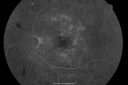
BDR and Diabetic Macular Edema with Fresh Laser Right Eye613 views60-year old man OD 20/60, OS 20/50 worsening vision both eyes
|
|

613 views79-year-old two weeks ago he was having difficulty seeing out of the right eye. When he covered his left eye things were distorted OD 20/50
|
|

Central Retinal Vein Occlusion - New- and old Macular Scar from AMD613 views87-year-old woman with longstaning macular scars and new vision loss left eye for 2 months. OD: 20/200, OS 20/400.
|
|

New BRVO Left Eye Old BRVO Right Eye - Right Eye Showing Bulls Eye Atrophy from Chronic CME and Non-Perfusion Infero-temporally613 views90-year-old man decreased vision maybe one year since cataract surgery he is no longer able to read. OD 20/200, OS 20/200
|
|

Diabetic Macular Edema and Hypertensive Retinopathy - Circinate Exudate (Ring Exudate) 613 views55-year-old woman diabetic for fifteen years and high blood pressure, especially over the last few months. She has had problems with nosebleeds, headaches, and there has been some difficulty bringing her blood pressure down. She said now the blood pressure is under control but it was running 220/120 mmHg for some time. Her vision in the right eye has been poor for four weeks with a spot in the central vision and both eyes have been blurred.
VISUAL ACUITY: Vision OD is 20/160, OS is 20/60
|
|

Proliferative Sickle Retinopathy - Hemoglobin SC613 views53-year-old woman with a new spider-like floater in the right eye for about six days. She sees one big floater and another little one. She does have hemoglobin SC hemoglobinopathy. VISUAL ACUITY: Vision OD is 20/20; OS is 20/50, PH is 20/20.
|
|

BRAO and PAMM613 views
|
|

Acute Branch Retinal Artery Occlusion Left Eye613 viewsThis 84 year old woman had an old BRVO in the right eye and saw a shadow for 2 days in the left eye. She has a superotemporal BRAO. The photos show retinal whitening, the OCT shows inner retinal edema and the FA shows poor circulation.
|
|
612 views
|
|

Serous Macular Detachment Left Eye - Possible Hypertensive Choroidopathy v. Central Serous Retinopathy612 views59-year-old woman decreasing vision in the left eye for about two days. She sees a spot in front of her vision. It is a yellow spot which is just straight ahead and moves with her eye. She does have high blood pressure. She checks her blood pressure at home, and even at home her blood pressure runs very high, with the systolic running around 200 and the diastolic around 100, although occasionally it does return to the normal range.
VISUAL ACUITY: Vision OD is 20/60, OS is 20/30
|
|

Non-Ischemic Central Retinal Vein Occlusion in Young Man (30 years) - Many Cotton Wool Spots612 views30-year-old man with a history of a central retinal vein occlusion for which he was first examined on 2 months ago and his vision then was 4/200. He responded nicely to two Avastin treatments. OD is 20/20, OS is 20/60
|
|

BDR OD with Macular Edema612 views
|
|

Branch Retinal Vein Occlusion with Cotton Wool Spot 611 views70-year-old man with blurred vision in the right eye for about one or two months. OD is 20/80, OS is 20/25.
|
|

Proliferative Diabetic Retinopathy with NVD, NVE, and Pre Retinal Hemorrhage611 viewsJuvenile Onset Diabetic with NVD, NVE and Pre Retinal Hemorrhage.
|
|

Acute Branch Retinal Artery Occlusion Left Eye611 viewsThis 84 year old woman had an old BRVO in the right eye and saw a shadow for 2 days in the left eye. She has a superotemporal BRAO. The photos show retinal whitening, the OCT shows inner retinal edema and the FA shows poor circulation.
|
|

Trousseau Sign - CRVO and Pancreatic Cancer611 viewsLeft eye shows CRVO with severe edema. The right eye has multiple small hemorrhages. Some of this is probably anemic retinopathy and some from hypercoaguability of cancer. The patient has pancreatic cancer.
|
|

Central Retinal Artery Occlusion - Diabetic Retinopathy - NO cherry red spot611 views58 year old man with sudden vision loss in the right eye. The vision had been 20/40 and dropped to 20/200 the day prior to presentation.
VA 20/200 OD, 20/20 OS
FA shows CRAO. No cherry red spot.
|
|

Central Retinal Vein Occlusion and Cystoid Macular Edema - Acute - Vision 20/50 610 views68-year-old man six weeks of vision loss right eye from CRVO and CME, vision right eye 20/50
|
|
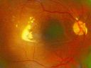
610 views
|
|

Sickle Retinopathy - Ultra Wide Field Angiogram - Heidelberg610 viewsLeakage from peripheral fronds of NV - HGB SC disease - 28 year old man with vision loss OS from acute vitreous hemorrhage
|
|

Hemicentral Vein Occlusion Left Eye610 viewsVision improved from 20/100 - 2 to 20/32 with 5 monthly Avastin treatments.
|
|
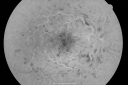
Central Retinal Vein Occlusion and Cystoid Macular Edema - Acute - Vision 20/50 609 views68-year-old man six weeks of vision loss right eye from CRVO and CME, vision right eye 20/50
|
|
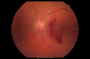
Diabetic Vitreous Hemorrhage Left Eye609 views35-year-old man diabetic for the last 25 years. His hemoglobin A1C has been running between 10 and 11. He was doing fine and then when he woke up from sleeping last night he noticed sudden vision loss in his left eye OD 20/30, OS 20/200.
|
|

Serous Macular Detachment Left Eye - Possible Hypertensive Choroidopathy v. Central Serous Retinopathy609 views59-year-old woman decreasing vision in the left eye for about two days. She sees a spot in front of her vision. It is a yellow spot which is just straight ahead and moves with her eye. She does have high blood pressure. She checks her blood pressure at home, and even at home her blood pressure runs very high, with the systolic running around 200 and the diastolic around 100, although occasionally it does return to the normal range.
VISUAL ACUITY: Vision OD is 20/60, OS is 20/30
|
|

609 views63-year-old man diabetic for twenty two years. His central vision is fine.
VISUAL ACUITY: OD 20/25, OS 20/25
|
|

Neovascularization elsewhere (NVE) Left Eye with Vitreous Hemorrhage609 views
|
|
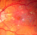
Diabetic Macular Edema with Circinate Exudate Macular Photo Post - Laser (Fresh)608 views74-year-old man has diabetic macular edema in both eyes and background diabetic retinopathy. His vision has blurred over the last few months. His blood sugars have been fluctuating.
VISUAL ACUITY: Vision OD is 20/25, OS is 20/80
|
|

Diabetic and Pseudophakic Cystoid Macular Edema Late FA608 views56-year-old woman has diabetic retinopathy in both eyes and macular edema. She is recently pseudophakic. OD 20/80, OS 20/200.
|
|

Diabetic and Pseudophakic Cystoid Macular Edema Late FA608 views56-year-old woman has diabetic retinopathy in both eyes and macular edema. She is recently pseudophakic. OD 20/80, OS 20/200.
|
|

New BRVO Left Eye Old BRVO Right Eye - Right Eye Showing Bulls Eye Atrophy from Chronic CME and Non-Perfusion Infero-temporally608 views90-year-old man decreased vision maybe one year since cataract surgery he is no longer able to read. OD 20/200, OS 20/200
|
|

Diabetic Macular Edema and Hypertensive Retinopathy - Circinate Exudate (Ring Exudate) 608 views55-year-old woman diabetic for fifteen years and high blood pressure, especially over the last few months. She has had problems with nosebleeds, headaches, and there has been some difficulty bringing her blood pressure down. She said now the blood pressure is under control but it was running 220/120 mmHg for some time. Her vision in the right eye has been poor for four weeks with a spot in the central vision and both eyes have been blurred.
VISUAL ACUITY: Vision OD is 20/160, OS is 20/60
|
|

Cilioretinal artery occlusion and Ophthalmic Artery Occlusion with Relative Sparing of the Central Retinal Artery608 views80-year-old woman who says she was looking in the mirror, covered her right eye and noticed she could not see well out of the left eye. That just happened yesterday. Her last eye check was a few years ago. She also had a carotid endarterectomy on the right side in 2006 shortly after coronary bypass surgery. OD is 20/40, OS is 20/200.
|
|

Serous Retinal Detachment - Hypertensive Retinopathy - 165/120 - Late - ICG shows leakage608 views48-year-old woman noticed when she was blinking there was a white flash in the left eye starting about a month ago. She sees it temporally. OD 20/16, OS 20/20. There is a serous retinal detachment superotemporal to the optic nerve falling short of the fovea. Her blood pressure 165/120.
|
|

Embolic CRAO Early608 views
|
|

Diabetic Retinopathy with CME608 viewsSecond opinion for diabetic retinopathy. VA is 20/30, right eye, 20/50, left eye. Patient has mild swelling in the macula of the right eye. No evidence of CME but does have a moderate cataract in the left eye.
|
|

PDR - Mild Vitreous Hemorrhage and Good Vision607 views58-year-old woman has diabetic retinopathy in both eyes with neovascularization of the optic nerve, worse in the right eye than the left eye. She has had panretinal laser in both eyes and vitreous hemorrhage in the left eye. Her vision, since I saw her last in June, is about the same.
VISUAL ACUITY: Vision OD is 20/25, OS is 20/20.
|
|

Central Retinal Vein Occlusion and Cystoid Macular Edema - Acute - Vision 20/50 607 views68-year-old man six weeks of vision loss right eye from CRVO and CME, vision right eye 20/50
|
|

Wet AMD Small New Hemorrhage - Valsalva607 views79-year-old two weeks ago he was having difficulty seeing out of the right eye. When he covered his left eye things were distorted OD 20/50
|
|
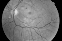
Purtchers Retinopathy with Bilateral Central Retinal Artery Occlusions 607 views50-year-old man traumatic chest compression and leg crushing injury. OD 1/200, OS 3/200
|
|

Serous Retinal Detachment - Hypertensive Retinopathy - 165/120 606 views48-year-old woman noticed when she was blinking there was a white flash in the left eye starting about a month ago. She sees it temporally. OD 20/16, OS 20/20. There is a serous retinal detachment superotemporal to the optic nerve falling short of the fovea. Her blood pressure 165/120.
|
|
| 4345 files on 44 page(s) |
 |
|
|
|
|
9 | |
|
|
|
|


