Most viewed - Retinal Vascular Disease associated with Systemic Disease
|

Hypertensive Retinopathy with Choroidal Ischemia Right Eye and Vision 8/200 - Blood Pressure 240/120 mmHg IgA Nephropathy762 views39-year-old man two days ago he noticed poor vision in his right eye OD: 8/200; OS: 20/20. BP is 240/120
|
|

Serous Macular Detachment Left Eye - Possible Hypertensive Choroidopathy v. Central Serous Retinopathy761 views59-year-old woman decreasing vision in the left eye for about two days. She sees a spot in front of her vision. It is a yellow spot which is just straight ahead and moves with her eye. She does have high blood pressure. She checks her blood pressure at home, and even at home her blood pressure runs very high, with the systolic running around 200 and the diastolic around 100, although occasionally it does return to the normal range.
VISUAL ACUITY: Vision OD is 20/60, OS is 20/30
|
|

Cotton Wool Spots and New Diagnosis of Chronic Lymphocytic leukemia760 views73-year-old man with cotton wool spots diagnosed subsequently with with Chronic Lymphocytice Leukemia
|
|

Bilateral Retinal Arteriol Occlusions - Possible Susac Syndrome 758 views80-year-old woman one month ago had vision loss and a vascular occlusion in the right eye. Vision loss occurred in the left eye about 9 years ago with cotton wool spots. Patients has a 30 year history of tinnitis.
|
|
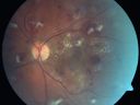
Diabetic Patient with Macular Edema and Blood Pressure 200/95754 views49-year-old decreasing vision over the last year. OD is 20/80, OS 20/80. blood pressure which was 200/95.
|
|

Cotton Wool Spots and New Diagnosis of Chronic Lymphocytic leukemia752 views73-year-old man with cotton wool spots diagnosed subsequently with with Chronic Lymphocytice Leukemia
|
|

Microaneurysms from Ocular Ischemic Syndrome (non-diabetic) Right Eye - Left eye CRAO 1 year ago752 views66-year-old woman lost vision in the left eye from a central retinal artery occlusion 1 year ago. Vision OD is fine. OD 20/25, OS 20/50
|
|

Hypertensive Retinopathy with Choroidal Ischemia Right Eye and Vision 8/200 - Blood Pressure 240/120 mmHg IgA Nephropathy751 views39-year-old man two days ago he noticed poor vision in his right eye OD: 8/200; OS: 20/20. BP is 240/120
|
|

Hypertensive Retinopathy One Year later with good Vision IgA Nephropathy750 viewsOD 20/30, OS 20/25 One year following initial episode.
|
|

Purtchers Retinopathy with Bilateral Central Retinal Artery Occlusions 749 views50-year-old man traumatic chest compression and leg crushing injury. OD 1/200, OS 3/200
|
|

Diabetic Patient with Macular Edema and Blood Pressure 200/95748 views49-year-old decreasing vision over the last year. OD is 20/80, OS 20/80. blood pressure which was 200/95.
|
|

Hypertensive Choroidopathy - Pheochromocytoma - Chronic Very High Blood Pressure 49 YO Male746 views49-year-old man chronic very high blood pressure sometimes the blood pressure will go as high as 260/160, sometimes it is 200/120 and sometimes it is 160/80. He has a pheochromocytoma on the adrenal gland and he has had vision loss in the right eye starting between three and four years ago.
VISUAL ACUITY: OD 20/60, OS 20/25.
|
|

Proliferative Sickle Retinopathy - Hemoglobin SC743 views53-year-old woman with a new spider-like floater in the right eye for about six days. She sees one big floater and another little one. She does have hemoglobin SC hemoglobinopathy. VISUAL ACUITY: Vision OD is 20/20; OS is 20/50, PH is 20/20.
|
|

Bilateral Retinal Arteriol Occlusions - Possible Susac Syndrome 742 views80-year-old woman one month ago had vision loss and a vascular occlusion in the right eye. Vision loss occurred in the left eye about 9 years ago with cotton wool spots. Patients has a 30 year history of tinnitis.
|
|

Hypertensive Choroidopathy - Pheochromocytoma - Chronic Very High Blood Pressure 49 YO Male739 views49-year-old man chronic very high blood pressure sometimes the blood pressure will go as high as 260/160, sometimes it is 200/120 and sometimes it is 160/80. He has a pheochromocytoma on the adrenal gland and he has had vision loss in the right eye starting between three and four years ago.
VISUAL ACUITY: OD 20/60, OS 20/25.
|
|

AIDS - HIV Retinopathy - Cotton Wool Spots738 views54-year-old woman HIV positive for 18 years. Her CD4 count is currently less than 50 and she was hospitalized for pneumocystis carinii pneumonia 5 months ago. She has noticed just for the last few weeks a few blind spots in the paracentral vision. Predominantly out of her right eye when she closes her left eye, she sees a few temporal blind spots. VA 20/25 in both eyes.
|
|
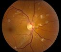
Hypertensive Retinopathy735 viewsPatient comes in complaining of spots in vision in both eyes. VA was 20/25 - right eye and 20/20- left eye. Fundus exam reveals little hemorrhages with cotton wool spots due to hypertension and anemia.
|
|

Diabetic Patient with Macular Edema and Blood Pressure 200/95731 views49-year-old decreasing vision over the last year. OD is 20/80, OS 20/80. blood pressure which was 200/95.
|
|
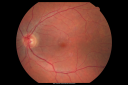
Hypertensive Choroidopathy - Pheochromocytoma - Chronic Very High Blood Pressure 49 YO Male731 views49-year-old man chronic very high blood pressure sometimes the blood pressure will go as high as 260/160, sometimes it is 200/120 and sometimes it is 160/80. He has a pheochromocytoma on the adrenal gland and he has had vision loss in the right eye starting between three and four years ago.
VISUAL ACUITY: OD 20/60, OS 20/25.
|
|

Serous Macular Detachment Left Eye - Possible Hypertensive Choroidopathy v. Central Serous Retinopathy730 views59-year-old woman decreasing vision in the left eye for about two days. She sees a spot in front of her vision. It is a yellow spot which is just straight ahead and moves with her eye. She does have high blood pressure. She checks her blood pressure at home, and even at home her blood pressure runs very high, with the systolic running around 200 and the diastolic around 100, although occasionally it does return to the normal range.
VISUAL ACUITY: Vision OD is 20/60, OS is 20/30
|
|

Hypertensive Retinopathy with Choroidal Ischemia Right Eye and Vision 8/200 - Blood Pressure 240/120 mmHg IgA Nephropathy729 views39-year-old man two days ago he noticed poor vision in his right eye OD: 8/200; OS: 20/20. BP is 240/120
|
|
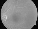
Microaneurysms from Ocular Ischemic Syndrome (non-diabetic) Right Eye - Left eye CRAO 1 year ago727 views66-year-old woman lost vision in the left eye from a central retinal artery occlusion 1 year ago. Vision OD is fine. OD 20/25, OS 20/50
|
|

Retinal Vasculopathy associated with Facioscapulohumeral Muscular Dystrophy line os726 views59-year-old woman had a blood pressure spike to 170/90 and was on medications for that. She does have facioscapulohumeral muscular dystrophy, which runs in her family. She has a cousin with the same disorder.
VISUAL ACUITY: Her vision is 20/25 OU. IOP: OD 21, OS 18.
There is no posterior vitreous separation, telangiectatic vessels near the macula. There are patchy retinal hemorrhages. There is no edema or proliferation.
|
|

Proliferative Sickle Retinopathy - Hemoglobin SC724 views53-year-old woman with a new spider-like floater in the right eye for about six days. She sees one big floater and another little one. She does have hemoglobin SC hemoglobinopathy. VISUAL ACUITY: Vision OD is 20/20; OS is 20/50, PH is 20/20.
|
|

Serous Retinal Detachment - Hypertensive Retinopathy - 165/120 - Fundus Autofluorescence (FAF)724 views48-year-old woman noticed when she was blinking there was a white flash in the left eye starting about a month ago. She sees it temporally. OD 20/16, OS 20/20. There is a serous retinal detachment superotemporal to the optic nerve falling short of the fovea. Her blood pressure 165/120.
|
|

Hypertensive Retinopathy723 viewsPatient comes in complaining of spots in vision in both eyes. VA was 20/25 - right eye and 20/20- left eye. Fundus exam reveals little hemorrhages with cotton wool spots due to hypertension and anemia.
|
|

Serous Retinal Detachment - Hypertensive Retinopathy - 3 months later - Fluid has absorbed (SD OCT VIDEO TAKES TIME TO LOAD)719 views48 Year old woman resolved hypertensive serous retinal detachment.
|
|

Bilateral Retinal Arteriol Occlusions - Possible Susac Syndrome 718 views80-year-old woman one month ago had vision loss and a vascular occlusion in the right eye. Vision loss occurred in the left eye about 9 years ago with cotton wool spots. Patients has a 30 year history of tinnitis.
|
|
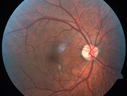
HIV Retinopathy718 viewsCentral white spot is an artifact. The small vascular abnormalities are from HIV
|
|

Hypertensive Retinopathy with Choroidal Ischemia Right Eye and Vision 8/200 - Blood Pressure 240/120 mmHg IgA Nephropathy717 views39-year-old man two days ago he noticed poor vision in his right eye OD: 8/200; OS: 20/20. BP is 240/120
|
|
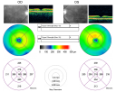
Hypertensive Retinopathy One Year later with good Vision IgA Nephropathy717 viewsOD 20/30, OS 20/25 One year following initial episode.
|
|
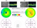
Ocular Ishemic Syndrome - Bilateral Carotid Occlusions - OD 20/60 OS 20/200 Enlarged Foveal Avascular Zone and Temporal Non-perfusion717 views75-year-old man had laryngeal cancer treated in 15 year ago with radiation. He recently was told that the carotid circulation is closed off on both sides. He does have a patent vertebral artery, which is supplying him pretty well and in addition to that his cardiac output is compromised by poor mitral valves. His consideration is to what can be done about that in the future.
VISUAL ACUITY: OD 20/60, OS 20/200
|
|

Serous Retinal Detachment - Hypertensive Retinopathy -- 1 month later - less fluid (better blood pressure) SD OCT713 views48 Year old woman resolving hypertensive serous retinal detachment.
|
|

Serous Macular Detachment Left Eye - Possible Hypertensive Choroidopathy v. Central Serous Retinopathy712 views59-year-old woman decreasing vision in the left eye for about two days. She sees a spot in front of her vision. It is a yellow spot which is just straight ahead and moves with her eye. She does have high blood pressure. She checks her blood pressure at home, and even at home her blood pressure runs very high, with the systolic running around 200 and the diastolic around 100, although occasionally it does return to the normal range.
VISUAL ACUITY: Vision OD is 20/60, OS is 20/30
|
|

Trousseau Sign - CRVO and Pancreatic Cancer712 viewsLeft eye shows CRVO with severe edema. The right eye has multiple small hemorrhages. Some of this is probably anemic retinopathy and some from hypercoaguability of cancer. The patient has pancreatic cancer.
|
|

Diabetic Patient with Macular Edema and Blood Pressure 200/95708 views49-year-old decreasing vision over the last year. OD is 20/80, OS 20/80. blood pressure which was 200/95.
|
|

AIDS - HIV Retinopathy - Cotton Wool Spots708 views54-year-old woman HIV positive for 18 years. Her CD4 count is currently less than 50 and she was hospitalized for pneumocystis carinii pneumonia 5 months ago. She has noticed just for the last few weeks a few blind spots in the paracentral vision. Predominantly out of her right eye when she closes her left eye, she sees a few temporal blind spots. VA 20/25 in both eyes.
|
|
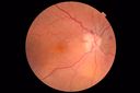
Anemic Retinopathy708 viewsMyelodysplasia - HgB 8 and very low platelet count
|
|

A case of Valsalva Retinopathy -2707 views
|
|
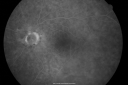
Bilateral Retinal Arteriol Occlusions - Possible Susac Syndrome 704 views80-year-old woman one month ago had vision loss and a vascular occlusion in the right eye. Vision loss occurred in the left eye about 9 years ago with cotton wool spots. Patients has a 30 year history of tinnitis.
|
|

Sickle Retinpoathy SC Disease - Vitreous Hemorrhage Left Eye - 29 Year old704 views
|
|

Hypertensive Choroidopathy - Pheochromocytoma - Chronic Very High Blood Pressure 49 YO Male703 views49-year-old man chronic very high blood pressure sometimes the blood pressure will go as high as 260/160, sometimes it is 200/120 and sometimes it is 160/80. He has a pheochromocytoma on the adrenal gland and he has had vision loss in the right eye starting between three and four years ago.
VISUAL ACUITY: OD 20/60, OS 20/25.
|
|

Serous Retinal Detachment - Hypertensive Retinopathy - 165/120 703 views48-year-old woman noticed when she was blinking there was a white flash in the left eye starting about a month ago. She sees it temporally. OD 20/16, OS 20/20. There is a serous retinal detachment superotemporal to the optic nerve falling short of the fovea. Her blood pressure 165/120.
|
|

Serous Retinal Detachment - Hypertensive Retinopathy - 165/120 - Fluorescein Angiogram703 views48-year-old woman noticed when she was blinking there was a white flash in the left eye starting about a month ago. She sees it temporally. OD 20/16, OS 20/20. There is a serous retinal detachment superotemporal to the optic nerve falling short of the fovea. Her blood pressure 165/120.
|
|

Purtchers Retinopathy with Bilateral Central Retinal Artery Occlusions 702 views50-year-old man traumatic chest compression and leg crushing injury. OD 1/200, OS 3/200
|
|

Sickle Retinpoathy SC Disease - Vitreous Hemorrhage Left Eye - 29 Year old698 viewsFrond of NV in the periphery
|
|

Diabetic Patient with Macular Edema and Blood Pressure 200/95694 views49-year-old decreasing vision over the last year. OD is 20/80, OS 20/80. blood pressure which was 200/95.
|
|

Purtscher's Retinopathy694 views19 year old female after pregnancy
|
|

Bilateral Retinal Arteriol Occlusions - Possible Susac Syndrome 688 views80-year-old woman one month ago had vision loss and a vascular occlusion in the right eye. Vision loss occurred in the left eye about 9 years ago with cotton wool spots. Patients has a 30 year history of tinnitis.
|
|

AIDS - HIV Retinopathy - Cotton Wool Spots688 views54-year-old woman HIV positive for 18 years. Her CD4 count is currently less than 50 and she was hospitalized for pneumocystis carinii pneumonia 5 months ago. She has noticed just for the last few weeks a few blind spots in the paracentral vision. Predominantly out of her right eye when she closes her left eye, she sees a few temporal blind spots. VA 20/25 in both eyes.
|
|

Serous Retinal Detachment - Hypertensive Retinopathy - 165/120 - Fluorescein Angiogram683 views48-year-old woman noticed when she was blinking there was a white flash in the left eye starting about a month ago. She sees it temporally. OD 20/16, OS 20/20. There is a serous retinal detachment superotemporal to the optic nerve falling short of the fovea. Her blood pressure 165/120.
|
|

Retinal Vasculopathy associated with Facioscapulohumeral Muscular Dystrophy 682 views59-year-old woman had a blood pressure spike to 170/90 and was on medications for that. She does have facioscapulohumeral muscular dystrophy, which runs in her family. She has a cousin with the same disorder.
VISUAL ACUITY: Her vision is 20/25 OU. IOP: OD 21, OS 18.
There is no posterior vitreous separation, telangiectatic vessels near the macula. There are patchy retinal hemorrhages. There is no edema or proliferation.
|
|
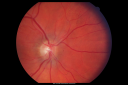
Hypertensive Retinopathy One Year later with good Vision IgA Nephropathy682 viewsOD 20/30, OS 20/25 One year following initial episode.
|
|

Hypertensive Retinopathy682 viewsPatient comes in complaining of spots in vision in both eyes. VA was 20/25 - right eye and 20/20- left eye. Fundus exam reveals little hemorrhages with cotton wool spots due to hypertension and anemia.
|
|
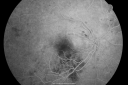
Hypertensive Retinopathy with Choroidal Ischemia Right Eye and Vision 8/200 - Blood Pressure 240/120 mmHg IgA Nephropathy679 views39-year-old man two days ago he noticed poor vision in his right eye OD: 8/200; OS: 20/20. BP is 240/120
|
|

Hypertensive Retinopathy - BP 240 / 130 mmHg - Grade 4679 views71 year old woman who claims to be asymptomatic with decreased vision, macular edema, cotton wool spots, subretinal fluid and optic nerve edema
|
|

Retinal Vasculopathy associated with Facioscapulohumeral Muscular Dystrophy very early fa678 views59-year-old woman had a blood pressure spike to 170/90 and was on medications for that. She does have facioscapulohumeral muscular dystrophy, which runs in her family. She has a cousin with the same disorder.
VISUAL ACUITY: Her vision is 20/25 OU. IOP: OD 21, OS 18.
There is no posterior vitreous separation, telangiectatic vessels near the macula. There are patchy retinal hemorrhages. There is no edema or proliferation.
|
|

Trousseau Sign - CRVO and Pancreatic Cancer677 viewsLeft eye shows CRVO with severe edema. The right eye has multiple small hemorrhages. Some of this is probably anemic retinopathy and some from hypercoaguability of cancer. The patient has pancreatic cancer.
|
|

Serous Retinal Detachment - Hypertensive Retinopathy - 165/120 - Fluorescein Angiogram676 views48-year-old woman noticed when she was blinking there was a white flash in the left eye starting about a month ago. She sees it temporally. OD 20/16, OS 20/20. There is a serous retinal detachment superotemporal to the optic nerve falling short of the fovea. Her blood pressure 165/120.
|
|

Serous Macular Detachment Left Eye - Possible Hypertensive Choroidopathy v. Central Serous Retinopathy674 views59-year-old woman decreasing vision in the left eye for about two days. She sees a spot in front of her vision. It is a yellow spot which is just straight ahead and moves with her eye. She does have high blood pressure. She checks her blood pressure at home, and even at home her blood pressure runs very high, with the systolic running around 200 and the diastolic around 100, although occasionally it does return to the normal range.
VISUAL ACUITY: Vision OD is 20/60, OS is 20/30
|
|

Hypertensive Choroidopathy - Pheochromocytoma - Chronic Very High Blood Pressure 49 YO Male674 views49-year-old man chronic very high blood pressure sometimes the blood pressure will go as high as 260/160, sometimes it is 200/120 and sometimes it is 160/80. He has a pheochromocytoma on the adrenal gland and he has had vision loss in the right eye starting between three and four years ago.
VISUAL ACUITY: OD 20/60, OS 20/25.
|
|
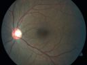
Proliferative Sickle Retinopathy - Hemoglobin SC674 views53-year-old woman with a new spider-like floater in the right eye for about six days. She sees one big floater and another little one. She does have hemoglobin SC hemoglobinopathy. VISUAL ACUITY: Vision OD is 20/20; OS is 20/50, PH is 20/20.
|
|

Wet AMD Small New Hemorrhage - Valsalva673 views79-year-old two weeks ago he was having difficulty seeing out of the right eye. When he covered his left eye things were distorted OD 20/50
|
|

AIDS - HIV Retinopathy - Cotton Wool Spots673 views54-year-old woman HIV positive for 18 years. Her CD4 count is currently less than 50 and she was hospitalized for pneumocystis carinii pneumonia 5 months ago. She has noticed just for the last few weeks a few blind spots in the paracentral vision. Predominantly out of her right eye when she closes her left eye, she sees a few temporal blind spots. VA 20/25 in both eyes.
|
|

Malignany Hypertension - BP 250/130 - Vision 1/200 in each eye - Diabetic673 views68-year-old woman poor vision in both eyes for the last few years. She is diabetic since 1989. Her blood pressure has been very high. She was in the hospital Friday. Her blood pressure during that visit was 250/130 and her blood pressure in your office she reports was somewhere around the same. I just checked her blood pressure at the beginning of my visit and it was 200/120 in her right arm sitting. Her vision is poor enough that she can barely see to walk around. She has also been feeling nauseous
|
|

Malignany Hypertension - BP 250/130 - Vision 1/200 in each eye - Diabetic673 views68-year-old woman poor vision in both eyes for the last few years. She is diabetic since 1989. Her blood pressure has been very high. She was in the hospital Friday. Her blood pressure during that visit was 250/130 and her blood pressure in your office she reports was somewhere around the same. I just checked her blood pressure at the beginning of my visit and it was 200/120 in her right arm sitting. Her vision is poor enough that she can barely see to walk around. She has also been feeling nauseous
|
|

Ocular Ishemic Syndrome - Bilateral Carotid Occlusions - OD 20/60 OS 20/200 Enlarged Foveal Avascular Zone and Temporal Non-perfusion672 views75-year-old man had laryngeal cancer treated in 15 year ago with radiation. He recently was told that the carotid circulation is closed off on both sides. He does have a patent vertebral artery, which is supplying him pretty well and in addition to that his cardiac output is compromised by poor mitral valves. His consideration is to what can be done about that in the future.
VISUAL ACUITY: OD 20/60, OS 20/200
|
|

Purtchers Retinopathy with Bilateral Central Retinal Artery Occlusions 671 views50-year-old man traumatic chest compression and leg crushing injury. OD 1/200, OS 3/200
|
|

Cotton Wool Spots now Gone in Both Eyes671 views60-year-old man was sick in the hospital in 3 month ago with streptococcal pneumonia. He was in the cardiac intensive care unit and at one point he was in critical condition. He has no visual complaints. OD 20/20, OS 20/25. Cotton Wool Spots now GONE
|
|

Bilateral Retinal Arteriol Occlusions - Possible Susac Syndrome 670 views80-year-old woman one month ago had vision loss and a vascular occlusion in the right eye. Vision loss occurred in the left eye about 9 years ago with cotton wool spots. Patients has a 30 year history of tinnitis.
|
|

Ocular Ishemic Syndrome - Bilateral Carotid Occlusions - OD 20/60 OS 20/200 Enlarged Foveal Avascular Zone and Temporal Non-perfusion670 views75-year-old man had laryngeal cancer treated in 15 year ago with radiation. He recently was told that the carotid circulation is closed off on both sides. He does have a patent vertebral artery, which is supplying him pretty well and in addition to that his cardiac output is compromised by poor mitral valves. His consideration is to what can be done about that in the future.
VISUAL ACUITY: OD 20/60, OS 20/200
|
|

Cotton Wool Spots Both Eyes after Strep Pneumonia and Cardiac ICU Stay 1 Month Ago 669 views60-year-old man was sick in the hospital in 1 month ago with streptococcal pneumonia. He was in the cardiac intensive care unit and at one point he was in critical condition. He has no visual complaints. OD 20/20, OS 20/25.
|
|

Microaneurysms from Ocular Ischemic Syndrome (non-diabetic) Right Eye - Left eye CRAO 1 year ago669 views66-year-old woman lost vision in the left eye from a central retinal artery occlusion 1 year ago. Vision OD is fine. OD 20/25, OS 20/50
|
|

Hairy Cell Leukemia - Retinal Hemorrhage and twig Branch Vein Occlusion669 views79 year old man He has had hairy cell leukemia since 2002. He is in remission. His last blood tests were 9/2018. He just moved down here and needs a new leukemia doctor. His vision is fine.
VA OD: Dcc20/25
VA OS: Dcc20/25
IOP: TP: OD:12 OS:12
|
|

Purtchers Retinopathy with Bilateral Central Retinal Artery Occlusions 666 views50-year-old man traumatic chest compression and leg crushing injury. OD 1/200, OS 3/200
|
|

Serous Retinal Detachment - Hypertensive Retinopathy - 165/120 - Fluorescein Angiogram666 views48-year-old woman noticed when she was blinking there was a white flash in the left eye starting about a month ago. She sees it temporally. OD 20/16, OS 20/20. There is a serous retinal detachment superotemporal to the optic nerve falling short of the fovea. Her blood pressure 165/120.
|
|

Serous Retinal Detachment - Hypertensive Retinopathy - 165/120 - Fluorescein Angiogram665 views48-year-old woman noticed when she was blinking there was a white flash in the left eye starting about a month ago. She sees it temporally. OD 20/16, OS 20/20. There is a serous retinal detachment superotemporal to the optic nerve falling short of the fovea. Her blood pressure 165/120.
|
|

Trousseau Sign - CRVO and Pancreatic Cancer663 viewsLeft eye shows CRVO with severe edema. The right eye has multiple small hemorrhages. Some of this is probably anemic retinopathy and some from hypercoaguability of cancer. The patient has pancreatic cancer.
|
|

Hypertensive Retinopathy - BP 240 / 130 mmHg - Grade 4660 views71 year old woman who claims to be asymptomatic with decreased vision, macular edema, cotton wool spots, subretinal fluid and optic nerve edema
|
|

Bilateral Retinal Arteriol Occlusions - Possible Susac Syndrome 658 views80-year-old woman one month ago had vision loss and a vascular occlusion in the right eye. Vision loss occurred in the left eye about 9 years ago with cotton wool spots. Patients has a 30 year history of tinnitis.
|
|

Retinal Vasculopathy associated with Facioscapulohumeral Muscular Dystrophy late fa657 views59-year-old woman had a blood pressure spike to 170/90 and was on medications for that. She does have facioscapulohumeral muscular dystrophy, which runs in her family. She has a cousin with the same disorder.
VISUAL ACUITY: Her vision is 20/25 OU. IOP: OD 21, OS 18.
There is no posterior vitreous separation, telangiectatic vessels near the macula. There are patchy retinal hemorrhages. There is no edema or proliferation.
|
|

Malignany Hypertension - BP 250/130 - Vision 1/200 in each eye - Diabetic656 views68-year-old woman poor vision in both eyes for the last few years. She is diabetic since 1989. Her blood pressure has been very high. She was in the hospital Friday. Her blood pressure during that visit was 250/130 and her blood pressure in your office she reports was somewhere around the same. I just checked her blood pressure at the beginning of my visit and it was 200/120 in her right arm sitting. Her vision is poor enough that she can barely see to walk around. She has also been feeling nauseous
|
|

Retinal Vasculopathy associated with Facioscapulohumeral Muscular Dystrophy late fa655 views59-year-old woman had a blood pressure spike to 170/90 and was on medications for that. She does have facioscapulohumeral muscular dystrophy, which runs in her family. She has a cousin with the same disorder.
VISUAL ACUITY: Her vision is 20/25 OU. IOP: OD 21, OS 18.
There is no posterior vitreous separation, telangiectatic vessels near the macula. There are patchy retinal hemorrhages. There is no edema or proliferation.
|
|

Proliferative Sickle Retinopathy - Hemoglobin SC655 views53-year-old woman with a new spider-like floater in the right eye for about six days. She sees one big floater and another little one. She does have hemoglobin SC hemoglobinopathy. VISUAL ACUITY: Vision OD is 20/20; OS is 20/50, PH is 20/20.
|
|

Lipemia Retinalis655 views31 year old man The patient at the time of imaging had triglycerides of >5680, cholesterol of 1136.
He also does have uncontrolled hypertension and insulin dependent type 2 diabetes, which may be the reason for vitreous extension of the lipids.
|
|
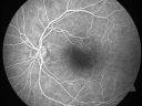
Serous Macular Detachment Left Eye - Possible Hypertensive Choroidopathy v. Central Serous Retinopathy654 views59-year-old woman decreasing vision in the left eye for about two days. She sees a spot in front of her vision. It is a yellow spot which is just straight ahead and moves with her eye. She does have high blood pressure. She checks her blood pressure at home, and even at home her blood pressure runs very high, with the systolic running around 200 and the diastolic around 100, although occasionally it does return to the normal range.
VISUAL ACUITY: Vision OD is 20/60, OS is 20/30
|
|

Hypertensive Retinopathy with Choroidal Ischemia Right Eye and Vision 8/200 - Blood Pressure 240/120 mmHg IgA Nephropathy654 views39-year-old man two days ago he noticed poor vision in his right eye OD: 8/200; OS: 20/20. BP is 240/120
|
|
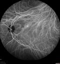
Serous Retinal Detachment - Hypertensive Retinopathy - 165/120 - Indocyanine Green Angiogram654 views48-year-old woman noticed when she was blinking there was a white flash in the left eye starting about a month ago. She sees it temporally. OD 20/16, OS 20/20. There is a serous retinal detachment superotemporal to the optic nerve falling short of the fovea. Her blood pressure 165/120.
|
|

Hairy Cell Leukemia - Retinal Hemorrhage and twig Branch Vein Occlusion654 views79 year old man He has had hairy cell leukemia since 2002. He is in remission. His last blood tests were 9/2018. He just moved down here and needs a new leukemia doctor. His vision is fine.
VA OD: Dcc20/25
VA OS: Dcc20/25
IOP: TP: OD:12 OS:12
|
|

Bilateral Retinal Arteriol Occlusions - Possible Susac Syndrome 653 views80-year-old woman one month ago had vision loss and a vascular occlusion in the right eye. Vision loss occurred in the left eye about 9 years ago with cotton wool spots. Patients has a 30 year history of tinnitis.
|
|

Hypertensive Retinopathy652 viewsPatient comes in complaining of spots in vision in both eyes. VA was 20/25 - right eye and 20/20- left eye. Fundus exam reveals little hemorrhages with cotton wool spots due to hypertension and anemia.
|
|

Retinal Vasculopathy associated with Facioscapulohumeral Muscular Dystrophy early fa651 views59-year-old woman had a blood pressure spike to 170/90 and was on medications for that. She does have facioscapulohumeral muscular dystrophy, which runs in her family. She has a cousin with the same disorder.
VISUAL ACUITY: Her vision is 20/25 OU. IOP: OD 21, OS 18.
There is no posterior vitreous separation, telangiectatic vessels near the macula. There are patchy retinal hemorrhages. There is no edema or proliferation.
|
|

Trousseau Sign - CRVO and Pancreatic Cancer651 viewsLeft eye shows CRVO with severe edema. The right eye has multiple small hemorrhages. Some of this is probably anemic retinopathy and some from hypercoaguability of cancer. The patient has pancreatic cancer.
|
|

Hypertensive Retinopathy with Choroidal Ischemia Right Eye and Vision 8/200 - Blood Pressure 240/120 mmHg IgA Nephropathy650 views39-year-old man two days ago he noticed poor vision in his right eye OD: 8/200; OS: 20/20. BP is 240/120
|
|

Trousseau Sign - CRVO and Pancreatic Cancer649 viewsLeft eye shows CRVO with severe edema. The right eye has multiple small hemorrhages. Some of this is probably anemic retinopathy and some from hypercoaguability of cancer. The patient has pancreatic cancer.
|
|

Trousseau Sign - CRVO and Pancreatic Cancer647 viewsLeft eye shows CRVO with severe edema. The right eye has multiple small hemorrhages. Some of this is probably anemic retinopathy and some from hypercoaguability of cancer. The patient has pancreatic cancer.
|
|
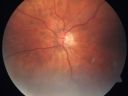
AIDS - HIV Retinopathy - Cotton Wool Spots646 views54-year-old woman HIV positive for 18 years. Her CD4 count is currently less than 50 and she was hospitalized for pneumocystis carinii pneumonia 5 months ago. She has noticed just for the last few weeks a few blind spots in the paracentral vision. Predominantly out of her right eye when she closes her left eye, she sees a few temporal blind spots. VA 20/25 in both eyes.
|
|

Trousseau Sign - CRVO and Pancreatic Cancer645 viewsLeft eye shows CRVO with severe edema. The right eye has multiple small hemorrhages. Some of this is probably anemic retinopathy and some from hypercoaguability of cancer. The patient has pancreatic cancer.
|
|

Central retinal artery and vein occlusion - Protein C Deficiency 33 year old male645 views33 year old male with vision loss for 3 hours. VA 20/12 right eye, 5/200 left eye. He has a protein C deficiency. The left eye recovered vision in about 1 month to 20/16. FA shows very slow recirculation time.
|
|

Diabetic Patient with Macular Edema and Blood Pressure 200/95643 views49-year-old decreasing vision over the last year. OD is 20/80, OS 20/80. blood pressure which was 200/95.
|
|
|
|


